15
Corresponding Author Hadi Akay, MD
Ankara Universitesi Tip Fakültesi Gögüs Cerrahisi Anabilim Dalı İbni Sina Hastanesi, 06100, Ankara, Türkiye Phone : +90 312 3103333 / 3165 Fax : +90 312 3106371 E-mail : [email protected] Received: Oct 27, 2003 • Accepted: May 03, 2004
Ankara Üniversitesi Tıp Fakültesi Mecmuası 2005; 58:15-17 CERRAHİ BİLİMLER / SURGICAL SCIENCES
Olgu Bildirisi / Case Report
Giant bullae of the lung treated successfully by
surgery in a patient with Hyper-IgE Syndrome (Job’s)
Hiper Ig-E sendromu (Job sendromu) tanılı bir olgudaki dev akciğer bülünün cerrahi tadavisi
Batuhan Saçılanateş
1, Figen Doğu
2, Murat Özkan
1, Nisa Akay
3, Aydan İkincioğulları
2, Hadi Akay
11 Department of Thoracic Surgery, Ankara University
School of Medicine, Ankara, Turkey
2 Department of Pediatric Immunology–Allergy, Ankara
University School of Medicine, Ankara, Turkey
3 Department of Dermatology, Ankara University School
of Medicine, Ankara, Turkey
The Hyper-IgE Syndrome (Job’s Syndrome) is clinically characterized with recurrent staphylococ-cal infections of the skin, subcutaneous tissue, upper respiratory tract, lung and bone in early childhood. Herein, we report a ten year-old girl presented with eczema, eosinophilia, elevated serum IgE levels and recurrent lower respiratory tract infections complicated with bulleous for-mation covering almost the whole superior lobe of left lung. Bullae excision via left thoracotomy was successfully performed.
Key words: Job’s syndrome, thoracotomy, bullea excision
Hiperimmünglobulin E sendromu (Job’s Sendromu) erken çocukluk çağında klinik olarak cilt, cil-taltı dokusu, üst solunum yolu, akciğerler ve kemiklerde tekrarlayan stafilokoksik infeksiyonlar ile karakterizedir.Burada egzema, eozinofili, serum IgE düzeyindeki yükseklik ve akciğerin hemen tüm sol üst lobunu kaplayan bül gelişimi ile komplike, tekrarlayan akciğer infeksiyonu ile prezen-te olan 10 yaşındaki bir kız hasta sunulmaktadır. Sol torakotomi yoluyla başarılı bül eksizyonu uygulanmıştır.
Anahtar sözcükler: Job sendromu, torakotomi, bül ekzisyonu
H
yper-IgE Syndrome (Job’s Syndrome) is a rare immunodeficiencydi-sease characterized by recurrent staphylococcal skin abscess, eczema, pneumonia with pneumatocele formation and extremely elevated serum IgE levels (1). The genetic basis is not known and central immunologic defect is largely undefined (2). In this report; a ten year-old girl who was diagnosed as Hyper-IgE Syndrome and had a successful bullae excision by left thoracotomy is presented.
Case report
A 10 year-old girl who was the second child of nonconsanguineous parents was referred to our clinic with a radiologically large pneumatocele in the upper lobe of the left lung. In her past medical history, skin abscess developed at first week of her age and she had a history of recurrent sinopulmonary infections with staphylococcus aureus. She suffered for eczematoid lesions, which were localized in the whole body since her first birthday. In her periodical controls at the age of seven, a giant pneumatocele in the right lung was diagnosed and a pezzer tube was performed into the pneumatocele at another surgical department. One year later, her admission to hospital because of productive cough and fever was eva-luated as emphyema, and tube thoracostomy with systemic anti-staphylococcal antibiotherapy were applied against staphylococcus aureus, which was shown microbiologically in sputum and pleural fluid.
16
Ankara Üniversitesi Tıp Fakültesi Mecmuası 2005; 58(1)
Giant bullae formation treated successfully by surgery in a patient with Hyper-IgE (Job’s) Syndrome
In her admission to our clinic, she had a non-productive cough, dyspnea and fever, which had been continuing for a week. In her physical examination, her weight was 23.5 kg (between 3-10 percentile) and her height was 138.5 cm (between 50-75 percentile). Her face and neck appearances were normal. There were hyperpigmented macular lesions in various parts of the whole body. Hyper-extensible elbow and finger joints were noticed. In the pulmonary ausculta-tion breath sounds were decreased in the upper part of the left chest. Complete blood count yielded hemoglobin level of 11.1 g/dl with significantly decreased mean corpuscular volume (MCV: 59 fl) and increased red cell distribution width (RDW: 16). Her white blood cell, total lymphocy-te, absolute granulocyte and platelet counts were normal
except eosinophilia (total eosinophil count: 820/mm3).
Although blood levels of IgA, IgM and IgG were normal, the level of IgE was extremely elevated to 2500 IU/L. Pe-ripheral blood lymphocyte subset analysis revealed normal T, B lymphocyte and natural killer cell numbers together
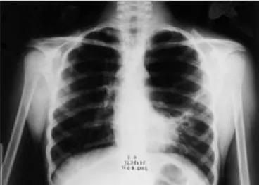
with normal CD4/8 ratio. In vitro lymphoblastic transfor-mation response to PHA and anti-CD3 were found to be normal. Nitroblue tetrazolium test result was found to be normal as well as neutrophile chemotaxis. Hemoglobine electrophoresis showed elevated HbA2 levels (5.8 %) and she was diagnosed as thallasemia minor. In her chest X-ray a large bullae formation at left lung was detected (Figure 1). Pulmonary function tests revealed decreased forced ex-piratory volume in 1 second (FEV1: 1.19 L, 62% of pre-dicted) and forced vital capacity (FVC: 1.02 L, 62 % of predicted). Thorax CT demonstrated bullae formations in both lungs and an approximately 10 cm width bulleous lesion covering nearly the whole superior lobe of left lung (Figure 2). Also there was consolidation area that was lying from hilus to apico-posterior segment of the left lung. This was a considered as pneumonia and staphylococcus aureus was detected in sputulum specimens.
After these evaluations, eventually the patient was di-agnosed as Hyper-IgE Syndrome and she received intrave-nous antibiotics including teicoplanine and amicasine as the anti-staphylococcal treatment, and intravenous
immu-noglobulin (IVIG) prophylaxis. After the 20th day of these
treatments, the patient underwent left thoracotomy. A 9 x 9 x 4 cm sized bullae located in the apicoposterior segment of upper lobe and another bullae located in the anterior segment of upper lobe were explored. Bullae excision and plication were performed for the biggest bullea.
In the postoperative period, air leak was ended at the fifth day, postoperatively. The patient had never had fever or faced with another problem except pain related to tho-racotomy. The prophylactic antibiotic and IVIG treatment continued after the operation and she was discharged at the seventh day, postoperatively. In her follow-up she has done well for 4 months (Figure 3).
Figure 1. Preoperative chest X-ray, showing a large lucent area of the left
lung involving upper middle zones. Figure 3. Postoperative chest X-ray
Figure 2. Preoperative thorax CT, demonstrated the large bullae
Journal of Ankara University Faculty of Medicine 2005; 58(1)
17
B. Sacilanates, F. Dogu, M. Ozkan et al.
Discussion
Hyper-IgE Syndrome in an other words Job’s Syndro-me is characterized with recurrent staphylococcal infecti-ons of skin, subcutaneous tissue, lung and exceptionally high serum IgE levels (>2000 IU /ml) (1). Generally; this syndrome represent as cutaneous infections and staphylo-coccal airway infections include pneumonias, emphyemas with pneumatocele formation in early childhood (3). Be-cause of the lack of surrounding inflammation, the typical purulent abscess formations do not appear in this area so it is called as cold abscess, which is another characteris-tic feature of Job’s Syndrome. Extremely elevated IgE le-vels seem to be the major immunologic problem. Blood and tissue eosinophilia, various chemotactic defects and decreased antibody and T-cell proliferation responses to antigens are the other components of this immunologic
defect. In addition, there are skeletal abnormalities such as hyperextensibility of joints, multiple bone fractures caused by osteopenia, craniosynostosis, dental abnormalities and typical facial appearances can be seen (4). In our patient, the existence of skin abscess, eczema, recurrent sinopulmo-nary infections with multiple pneumatocele formations, hyperextensible joints, eosinophilia and extremely elevated IgE levels revealed the diagnosis of Hyper-IgE Syndrome. She either had the thallassemia minor disease, which is not associated with Hyper-IgE Syndrome. There was clinical and radiological improvement in her clinical course with no dyspnea and fever anymore. In this case, morbidity due to severe lung disease as a result of the recurrent pulmonary infections was succesfully overcomed by surgery and qua-lity of her life was significantly improved.
References
1. Grimbacher G, Holland SM, Gallin JI et al. Hyper- IgE syndrome with recurrent infections- an autosomal dominant multisystem disorder. N Engl J Med 1999;340:692-702.
2. Chehimi J , Elder M , Greene J , et al. Cytokine and chemokine dysregulation in hyper-IgE syndrome. Clin Immunol 2001; 100:49-56.
3. Buckley RH. Hyper Ig E syndrome/ Job’s syndrome. In: Medical Immunology. Mc-Graw Hill. 10th edition. 2001;337.
4. Grimbacher B, Belohradsky BH, Holland SM. Immunoglobulin E in primary immunodeficiency diseases. Allergy 2002;57:995-1007. 5. Hall RA, Salhany EK, Lebel E. Fungal pulmonary abscess in an
adult secondary tohyperimmunoglobulin E (Job’s ) syndrome. Ann Thorac Surg 1995;59:759-761.