Address for Correspondence / Yazışma Adresi: Ümit Akın Dere, MD Başkent University Faculty of Medicine Department of Neurosurgery Bahçelievler, Ankara, Turkey E-mail: [email protected]
©Telif Hakkı 2018 Gazi Üniversitesi Tıp Fakültesi - Makale metnine http://medicaljournal.gazi.edu.tr/ web adresinden ulaşılabilir. ©Copyright 2018 by Gazi University Medical Faculty - Available on-line at web site http://medicaljournal.gazi.edu.tr/
doi:http://dx.doi.org/10.12996/gmj.2018.14
Sacral Spine Eosinophilic Granuloma in an Adult Patient: A Case Report of Unusual Age and
Location
Erişkin Hastada Sakrumun Eozinofilik Granülomu: Ender Yaş ve Lokalizasyonu olan bir Vaka
Ümit Akın Dere
1, Ayfer Aslan
2, A.Memduh Kaymaz
21 Department of Neurosurgery, Başkent University Faculty of Medicine, Ankara, Turkey 2 Department of Neurosurgery, Gazi University Faculty of Medicine, Ankara, Turkey
ABSTRACT
Eosinophilic granuloma one of a triad of diseases under Langerhans’ cell histiocytosis as known as Histiocytosis X, is a rare disorder characterized with uncontrolled proliferation of histiocytes, mostly seen among children and adolescents instead of adults. Although the clinical picture is well defined, there are still unclear aspects on etiology, histology and prognosis. Here we are presenting our experiences on a case of eosinophilic granuloma interestingly in an adult patient. She was admitted with back and hip pain. After her diagnosis of sacral spine osteolytic tumor, the biopsy was performed in our clinic and her histopathology was resulted as eosinophilic granuloma. In this paper we are presenting the clinical and hystopathological features of esonophilic granuloma. We aimed to contribute an additional data to the literature on eosinophilic granuloma presented with an atypical localization and age. According to current literature, there are only few reports on both adults and sacrum affected with eosinophilic granuloma.
Key Words: Eosinophilic granuloma, Langerhans’ cell histiocytosis, histiocytosis X, spine tumor
Received: 05.30.2017 Accepted: 11.23.2017
ÖZET
Eozinofilik granuloma, Langerhans hücreli histiositozis grubundan bir hastalık olan, Histiositozis-X olarak da bilinen, erişkinlere nazaran genellikle çocuk ve adölesanlarda izlenen, kontrolsüz çoğalan histiositlerle karakterize bir rahatsızlıktır. Klinik olarak iyi tanımlanmasına ragmen etyolojik, histolojik ve prognostik faktörler açısından hala netleşmemiş veriler bulunmaktadır. Biz bu olguda eozinofilik granüloması olan erişkin bir hastayı sunuyoruz. Bel ve kalça ağrısı ile kliniğimize başvuran hastaya yapılan incelemelerde sakrumun osteolitik tümörü düşünülerek yapılan cerrahi işlem sonrası alınan doku biyopsisinden yapılan histopatolojik inceleme sonucu eozinofilik granülom olarak gelmiştir. Klinik ve histopatolojik verilerini paylaştığımız bu yazıda amacımız literatürde nadir olarak görülen, erişkin bir hastada rastlanan eozinofilik granülom olgusuyla ilgili klinik tecrübelerimizle literatüere bir katkı sağlamaktır.
Anahtar Sözcükler: Eozinofilik granülom, Langerhans hücreli histiositozis, histiositozis-X, omurga tümörü
Geliş Tarihi: 30.05.2017 Kabul Tarihi: 23.11.2017
INTRODUCTION
Eosinophilic granuloma (under disorders group of Langerhans’ cell histiocytosis as known as Histiocytosis X) is considered as a pediatric a benign osteolytic disease mostly seen in the skull, femur, rib, mandible, pelvis and spine (1, 2). Approximately 80% of cases occur in children younger than 10 years of age (3, 4). Spine involvement has been found in approximately 7-15% of cases (5, 11) with a predilection for the cervical spine (47%), followed by the thoracic (33%) and lumbar (20%) spines in adults (6). Furthermore, there isn’t any report on the frequency of sacral spine involvement in adults. According to our knowledge, in the literature there are only three cases about adult sacral eosinophilic granuloma of which ages were 21, 39 and 41-year-old (2, 4).
Clinical syptoms depend on spinal location and generally concern on severe bone pain which aggravates with palpation (4, 7). For sacral easinophilic granuloma the most common complaints include back pain, tenderness to palpation, bone tumescence, limited range of motion (1, 4, 7). Although peripheral nervous system involvement is uncommon, there are few case reports presented with sciatica due to lytic lesions of the lumbosacral junction (2, 8). Neurological deficits, bowel or bladder dysfunctions are not expected unless presenting of vertebra collapse or peridural spread of tumor (9, 10, 12). Collapse of vertebra is almost only seen in children age group which is called Calve disease (8, 11) . Vertebra plana which exhibits the classic appearance of a vertebral collapse is radiographic manifestation of a bone lesion of eosinophilic granuloma (11, 12). The lytic lesions are all about vertebral body, and the pedicles are almost always preserved except some cases reported (8, 12).
This article describes a case of 37-year-old female with a solitary eosinophilic granuloma on S1 and S2 vertebral body as in the unusual age and location.
CASE REPORT
A 37-year-old female patient had low back and hip pain of 1 year’s duration with onset after parturition. She hadn’t any specific properties in her medical history except of left cervical lymphadenopathy during pregnancy. The biopsy from this location had been resulted non-specific.
When she was admitted to our clinic, the pain was mechanical in nature with tenderness to local pressure on sacrum and resistant to analgesic treatment. She hadn’t any neurological deficit. In her physical examination, there was a mild sacral tumescence and she had bilateral cervical, axillary and inguinal multiple milimetric lymphadenopathies. There weren’t any organomegaly. Laboratory tests showed mild leukocytosis, prolonged activated partial thromboplastin time (aPTT), mild anemia, elevated levels of acute phase proteins as compatible with a moderate inflammation. Standard X-ray radiographic images were normal Upon further evaluation orientated on sacral pain, Magnetic Resonans Imaging (MRI) disclosed a lytic lesion in the sacral vertebral body. On the level of S1 and S2 spine, a soft mass lesion with obliteration of neural foramens, posteriorly narrowing the sacral spinal canal, highly contrast enhancement was detected. The lesion was 4 cm x 8 cm in size and showed extension to the presacral region (Figure 1).
Figure 1: Preoperative lumbosacral MR sagittal and axial images: 4*8 cm sized soft mass lesion in S1 and S2 with highly contrast enhancement, obliteration of neural foramens, posteriorly narrowing the sacral spinal canal and extension to the presacral region.
On Computed Tomography (CT), sacrum was destructed with soft mass lesion elongated through spinal canal toward to presacral region (Figure 2).
Figure 2: Postoperative lumbosacral CT sagittal and axial images: Defective sacral bone with soft mass lesion elongated through spinal canal toward to presacral region.
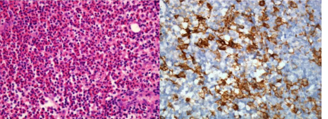
An open biopsy was performed from S1 and S2 vertebral body. Histopthological examinations showed fibrous tissue infiltrated with pleomorphic numerous eosinophils, in-between histiocytic cells, polymorphonuclear leucocytes, lymphocytes and few plasma cells; finally
Langerhans cells with notched marked nucleus and irregular outline. Immunohistochemistry studies were strongly positive for S100 protein and CD1a, poorly positive for Langerin, establishing the diagnosis of eosinophilic granuloma. Pancreatin was negative (Figure 3).
GMJ
2018; 29: 59-61
Dere et al.
Sacral spine eosinophilic granuloma
60
Figure 3: The histopathological images: fibrous tissue infiltrated with pleomorphic numerous eosinophils, histiocytic cells, polymorphonuclear leucocytes, lymphocytes, few plasma cells and Langerhans cells with strongly positive for S100 protein and CD1a, poorly positive for Langerin.
After diagnosis of eosinophilic granuloma, Positron Emission Tomography (PET-CT) was performed for screening of organ involvement and staging the disease. 18F-FDG involvement was detected not only in sacral mass lesion, but
also in left cervical, supraclavicular, mediastinal, abdominal and right inguinal lymph nodes and bone marrow as indicative for lymphoproliferative diseases.
DISCUSSION
Eosinophilic granuloma is an uncommon osteolytic bone disease frequently seen in children and adolescents. However, there are some reports expressing that it is not so less frequent among adults (2). Islenger at all (14) published large retrospective case series of 541 patients of whom 39% were older than 21 years. Although eosinophilic granuloma most frequently effects the skeleton, it constitutes less than 1% of all bone tumors (1, 12). The most common involved bones are skull, ribs, spine, pelvis, and mandible respectively (2, 12, 14). Furthermore, in our literature review there was a case report on eosinophilic granulomas of labial mucosa without bony involvement (13). There are some differences about predilection of eosinophilic granuloma to the spinal parts as cervical, thoracic and lumbar vertebra between pediatrics and adults. In general population, the thoracic vertebra are involved most often (54%) followed by lumbar (35%) and cervical (11%) spine (12, 16). In adults, the cervical vertebra is most often involved (47%) spine bone followed by thoracic (33%) and lumbar (20%) vertebra (1, 6). The rate of sacral vertebra involvement is not mentioned in literature. In our literature review we found only three cases about adult sacral eosinophilic granuloma of which ages were 21, 39 and 41-year-old (2,4).
The diagnosis of eosinophilic granuloma was made with not only histopathology from open biopsy, but also clinical and radiological observation. In histopathologic slides, there are Langerhans cells diffuse positive with S-100 protein and CD1a which point to a group of disorders previously called Histiocytosis-X or currently named Langerhans’ cell histiocytosis which include the triad of Eosinophilic granuloma, Hand-Schüller-Christian syndrome and Letterer-Siwe disease (1, 12). The presence of proliferating Langerhans histiocytes is the histologic hallmark of all three diseases (12). The histopathology shows Histiocytosis X as known as Langerhans’ cell histiocytosis. Further distinguishing features between three conditions are clinical and radiological manifestations. Eosinophilic granuloma constitutes the majority of all cases of histiocytosis X (60%) and characterized with focal bone lesion (12). The intermediate form, called Hand-Schuller-Christian disease, has classical triad of exophthalmos, diabetes insipidus, and bone lesions. The disseminated form called Letterer-Siwe disease is an acute fulminating disorder in usually before the age of three years with diffuse involvement of visceral organs, fever, lymphadenopathy, skin rashes, anemia and thrombocytopenia (1, 7, 12, 13, 15). In our case, the diagnosis of eosinophilic granuloma was made by excluding of the other two condition due to absence of visceral organ involvements, fever, thrombocytopenia, polyuria or polydipsia and exophthalmos.
Radiographic findings of eosinophilic granuloma have generally osteolytic pattern simulating other skeletal lesions such as osteomyelitis, Ewing sarcoma, leukemia, lymphoma, and metastatic lesions (12). To distinguish these widespread radiographic spectrum of diseases, the biopsy may be required and immunohistochemical studies should be performed.
CONCLUSION
This case showed eosinophilic granuloma should be considered in the differential diagnosis of sacral mass lesions in adults besides other lytic bone lesions, although it is rarely seen in adults and sacral spine.
Conflict of interest
No conflict of interest was declared by the authors.
REFERENCES
1. Ueda Y, Murakami H, Demura S, Kawahara N, Tomita K, Tsuchiya H. Eosinophilic granuloma of the lumbar spine in an adult. Orthopedics. 2012 Dec;35:e1818-21.
2. Baillet A1, Grange L, Lafaix PA, Gaudin P, Juvin R. Radiculopathy as
a manifestation of Langerhans' cell histiocytosis. Joint Bone Spine, 74, 2007;190-3
3. Bilge T, Barut S, Yaymaci Y, Alatli C. Solitary eosinophilic granuloma of the lumbar spine in an adult. Case report. Paraplegia. 1995;33:485–7
4. Rimondi E, Mavrogenis AF, Rossi G, Ussia G, Angelini A, Ruggieri P. CT-guided corticosteroid injection for solitary eosinophilic granuloma of the spine. Skeletal Radiol. 2011;40:757-64. 5. Cañadell J, Villas C, Martinez-Denegri J, Azcarate J, Imizcoz A.
Vertebral eosinophilic granuloma. Long-term evolution of a case. Spine (Phila Pa 1976). 1986; 11:767-9.
6. Reddy PK, Vannemreddy PS, Nanda A. Eosinophilic granuloma of spine in adults: a case report and review of literature. Spinal Cord. 2000; 38:766-8.
7. Oberman HA. A clinicopathologic study of 40 cases and review of the literature on eosinophilic granuloma of bone. Hand-SchullerChristian disease and Letterer-Siwe disease. Pediatrics. 1961; 28:307-27.
8. Nakamura H, Nagayama R. Eosinophilic granuloma presenting with local osteolysis in an adult lumbar spine. J Clin Neurosci. 2008; 15:1398-400.
9. Kumar A. Eosinophilic granuloma of the spine with neurological deficit. Orthopedics. 1990;13:1310–2
10. Villas C, Martínez-Peric R, Barrios RH, Beguiristain JL. Eosinophilic granuloma of the spine with and without vertebra plana: long-term follow-up ofsix cases. J Spinal Disord. 1993;6:260-8. 11. Raab P, Hohmann F, Kühl J, Krauspe R. Vertebral remodeling
in eosinophilic granuloma of the spine. A long-term follow-up. Spine (Phila Pa 1976), 1998;23:1351-4.
12. David R, Oria RA, Kumar R, et al. Radiographic features of eosinophilic granuloma of bone. AJR Am J Roentgenol 1989;153:1021–6.
13. V Y, E SK, S N B, A K, Sekhar MS. Solitary extragnathic langerhans cell histiocytosis - a rare case. J Clin Diagn Res. 2015;9:ZD22-3. 14. Islinger RB, Kuklo TR, Owens BD, Horan PJ, Choma TJ, Murphey MD,
et al. Langerhans cell histiocytosis in patients older than 21 years. Clin Orthop 2000;379:231e5
15. Sheila W, Maarten E. Langerhans cell histiocytosis: update for the pediatrician. Curr Opin Pediatr. 2008;1:23-9.
16. Wilner D. Radiology of bone tumors and allied disorders, vol. 2. Philadelphia: Saunders, 1982.