Surgical Interventions for Late Complications
of Arteriovenous Fistulas
Sedat Belli
1, Hakan Yabanoglu
1, Cem Aydogan
2, Alper Parlakgumus
1, Sedat Yildirim
1,
Mehmet Haberal
21
Department of Surgery, Baskent University Faculty of Medicine, Adana, Turkey
2Department of Surgery, Baskent University Faculty of Medicine, Ankara, Turkey
Our aim was to determine the most effective surgical treatment for arteriovenous fistula (AVF) complications after all other methods of salvage have failed. We evaluated 110 patients for 139 complications that occurred after the initial AVF placement and for whom surgical intervention was the last hope for retaining fistula access. Vascular steal syndrome and venous hypertension were the most common complications seen in our patients. The anastomoses of 17 of the vascular steal syndrome cases were narrowed either by stitches or by a polytetrafluoroethylene graft. The second most performed revision surgery was excision of the aneurysm and repair with primary suturing, followed by excision of the aneurysm and interposition grafting. Successful surgical outcomes were achieved in 111 of 139 procedures after revision surgery without constructing a new AVF. AVF salvage surgery is of paramount importance in order to increase the patency rate, which prolongs survival and increases the patient’s quality of life.
Key words: Arteriovenous fistula – Complications – Hemodialysis – Vascular access
B
ecause survival rates in patients with end-stage renal disease have been rising because of developments in treatment procedures, the impor-tance of sustained vascular access for hemodialysis has increased.1Many late complications, such as (1) thrombosis, (2) venous hypertension, (3) aneurysm/pseudoan-eurysm formation, (4) hemorrhage, (5) vascular steal
syndrome (VSS), (6) stenosis, and (7) heart failure may occur following arteriovenous fistula (AVF) surgery.1,2These may cause dysfunction of the AVF
and limit the efficiency of hemodialysis.
Our aim in this retrospective analysis is to present our experience in the surgical treatment of AVF complications in patients for whom all other conservative treatment methods of salvage have
Corresponding author: Sedat Belli, MD, Department of Surgery, Adana Baskent Medical and Research Center, 1250 Adana, Turkey. Tel.:þ90 3223272727; Fax: þ90 3223271276; E-mail: [email protected]
failed. These include radiologic interventions, such as covered stent; coil embolization; application of thrombin or percutaneous transluminal angioplasty balloon dilatation; thromboaspiration; and throm-bolysis. Our study contributes to the literature because data on successful surgical salvage tech-niques for AVFs are limited, despite the relative high frequency of documented AVF complications. Our study focuses on surgical solutions, in particular highlighting minor factors that can increase the success of surgical interventions for compromised AVFs. For this study we examined the clinical presentations and surgical techniques for treating these complications and postintervention outcomes.
Patients and Methods
Patient selection and evaluation
This is a retrospective review of all patients who underwent surgical repair for late complications of their AVFs at our center for 10 years, from January 2001 until December 2010. A total of 1558 AVF-related surgeries were performed in our center during the study period. We evaluated 110 patients for 139 complications that occurred after the initial AVF placement. For the patients in the current study group, surgical intervention was the last hope for retaining fistula access, because all other conserva-tive treatment methods of AVF salvage, as notified above, had failed.
Patient demographic data include age, sex, hyper-tension, diabetes mellitus, coronary artery disease, congestive cardiac failure, smoking, use of anticoag-ulants, region of fistula insertion, the number of fistulas, fistula-related complications, mode of pre-sentation, and surgical technique used for restorative surgery. Furthermore, all of the outcomes were collected from our center’s medical records.
The study was approved by the Baskent Univer-sity Institutional Review Board (project no. KA13/ 27) and by the Baskent University Research Fund.
All patients seen at our center for AVF-related complications are first evaluated by the general surgery department, where the preoperative assess-ment described below is carried out. After this initial assessment, all patients, with the exception of those admitted in critical condition, are referred to the interventional radiology department. The pa-tients selected as cases were subjected to initial procedures in which various endovascular methods were applied; however, there appeared such com-plications as aneurysm thrombosis, and shortness of
the potential cannulation due to partial aneurysm thrombosis.
Once the patients are treated through endovas-cular methods, as a final option the patients return to the general surgery department. As a result, we aimed to perform certain kinds of operations, such as narrowing by nonabsorbable sutures or small-caliber interposition grafts, excision of the aneurysm and repair with primary suturing, narrowing the outflow vein with a tight polytetrafluoroethylene cuff, ligation of the patent distal vein branch, graft interposition, basilic vein transposition, thrombec-tomy, and hemostasis (repair with primary suturing or autogenous veins grafts), which would reach efficient outcomes because there would be no need to create new fistulas. Criteria of AVF complications considered for surgical revision are described in Table 1. Some of these complications are shown in Fig. 1.
Preoperative assessment
The preoperative clinical evaluation includes a color duplex Doppler ultrasound (CDDUS). If there is a suggestion of central stenosis, a fistulogram is performed for further evaluation. This preoperative protocol was carried out on all patients in our study group.
Postoperative follow-up
As is routine in our center for these surgical interventions, heparin (50–100 U/kg body weight) was administered intravenously, and a heparin infusion was continued for 24 hours after the revision surgery. When patients were discharged from the hospital, a low dose of aspirin or dipyridamole was prescribed to be used at home.
Patients were examined for the first time 1 month after discharge, and after this initial follow-up patients were examined within 6 and 12 months.
Table 1 Criteria of AVF complications are considered for surgical revision
a. Exposed graft with overlying skin erosion
b. Signs of local infection (erythematic, purulent discharge, or a draining sinus tract)
c. Aneurysm formation 1. Pain in the area overlying
2. Rapidly expanding aneurysm (.2 cm/6 months) 3. Thrombosis of the aneurysm
d. Ulceration and bleeding
e. Loss of adequate cannulation segment f. Central stenosis
The final follow-up was performed 24 months after the patients were discharged. The patients were examined by the surgery department with CDDUS scans. The corrected AVFs were assessed for functionality, and patency rates were noted.
The postintervention patency status was assessed according to the standards of the Society of Vascular Surgery and the Society of Interventional Radiology. In the follow-up period, a failing AVF was defined as a patent access that was functioning poorly because of hemodynamically significant stenosis (.50%) as documented by CDDUS.3A patent AVF
with normal blood flow may be inefficient for hemodialysis because of other problems, such as outflow vein or in-graft aneurysms without stenosis showing a risk for rupture, infection, or steal syndrome. In these cases, this was also considered as a failing or nonfunctional AVF.3
Statistical methods
Statistical analyses were performed using Statistical Package for the Social Sciences software (version 16.0, SSPS Inc, Chicago, Illinois). Chi-square analysis was used to assess categoric variables. Mann-Whitney U test was used to analyze continuous variables. Categoric variables were expressed as absolute values and percentages. The Kaplan-Meier analysis was used to evaluate patency rates. P , 0.05 was considered statistically significant.
Results
During the study period 110 patients were treated for complications. The demographics and systemic concomitant diseases are detailed in Table 2. The mean duration of renal failure was 49.3 6 40.6 months (range, 1–180 months). A total of 92 (66.2%) of the AVFs with complications had their index surgical procedures performed at other surgical facilities. The mean number of previously per-formed AVFs per patient was 2.1 6 1.2 (range, 1– 7). Most of the complications were detected in AVFs with native vessels and in brachiocephalic fistulas (Table 3).
Fig. 1 (a and b) Photograph of an infected pseudoaneurysm. (c) Giant brachial artery aneurysm in the left arm. (d) Dilation of the cephalic vein with a brachial artery aneurysm in the left upper arm.
Table 2 Patients’ characteristics and demographics
Variable Value
Age, y, mean 6 SD (range) 48.5 6 17.4 (13–78)
.50 y, n (%) 81 (58.3)
Female sex, n (%) 58 (52.7)
Hypertension, n (%) 67 (48.2)
Diabetes mellitus, n (%) 51 (36.7) Coronary artery disease, n (%) 7 (5) Congestive cardiac failure, n (%) 3 (2.1)
Smoking, n (%) 52 (47.2)
Anticoagulant use, n (%)a 46 (41.8) aPatients who have their initial access at our center are
routinely treated with anticoagulants; however, the number is low in our series because of the large number of patients who had their initial access in other centers.
The complications in AVFs performed in the proximal elbow region were more common (Table 4). The surgical procedures performed for each complication type are summarized in Table 4. Although our general strategy was to salvage the AVF, conditions such as life-threatening bleeding, severe ischemia, infection, or ulcerations required the closure of the access (Fig. 1a and 1b).
Severe bleeding and failure of the revised AVF were the most common complications encountered
after salvage surgery (Table 5). A total of 80% of the original AVFs were functional after the revision surgery; in the rest, a new AVF was created during the same session.
The mean follow-up was 16 months, with a range of 1 to 69 months. Postintervention primary paten-cies for all revised AVFs were 80%, 72%, 56%, and 38% at 1, 6, 12, and 24 months, respectively. Demographic characteristics, synthetic graft use, comorbid systemic diseases, complications after
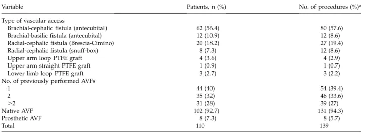
Table 3 Type of vascular access and the number of accesses before the corrective surgery
Variable Patients, n (%) No. of procedures (%)a
Type of vascular access
Brachial-cephalic fistula (antecubital) 62 (56.4) 80 (57.6)
Brachial-basilic fistula (antecubital) 12 (10.9) 12 (8.6)
Radial-cephalic fistula (Brescia-Cimino) 20 (18.2) 27 (19.4)
Radial-cephalic fistula (snuff-box) 8 (7.3) 12 (8.6)
Upper arm loop PTFE graft 4 (3.6) 4 (2.9)
Upper arm straight PTFE graft 1 (0.9) 1 (0.7)
Lower limb loop PTFE graft 3 (2.7) 3 (2.2)
No. of previously performed AVFs
1 44 (40) 54 (39.4) 2 35 (32) 46 (33.6) .2 31 (28) 39 (27) Native AVF 102 (92.7) 131 (94.3) Prosthetic AVF 8 (7.3) 8 (5.7) Total 110 139 PTFE, polytetrafluoroethylene.
aSome patients had more than one procedure.
Table 4 Distribution of complication types due to AVF locations and surgeries performed on AVF complications
Type of complications (n) Site of AVF, n (%) Surgical procedure (n) Proximal (elbow) Distal (wrist) Lower limb
Vascular steal syndrome (35)a 26 (74.3) 9 (25.7) 0 (0) Narrowing by nonabsorbable sutures or small-caliber
interposition grafts (17) Ligation of the fistula (11)
Narrowing the outflow vein with a tight PTFE cuff (7) Venous hypertension (35) 21 (60) 14 (40) 0 (0) Ligation of the patent distal vein branch (31)
Ligation of the fistula (4) Aneurysm/pseudoaneurysm
formation (31)
22 (71) 7 (22.6) 2 (6.4) Excision of the aneurysm and repair with primary suturing (14)
Interposition grafting (12) Ligation of the fistula (5)
Bleeding (13) 12 (92.3) 1 (7.7) 0 (0) Hemostasis (repair with primary suturing or autogenous vein grafts) (8)
Ligation of the fistula (5) Dysfunction of AVF (12) 12 (100) 0 (0) 0 (0) Basilic vein transposition (12) Thrombosis (11) 7 (64) 3 (27) 1 (9) Thrombectomy (11)
Ligation of the fistula (3)
High-output cardiac failure (2) 2 (100) 0 (0) 0 (0) Narrowing the outflow vein with a PTFE graft (2) Total (%) 102 (73.4) 34 (24.5) 3 (2.1) 139 (100%)
PTFE, polytetrafluoroethylene.
surgery, and the complication period had no effects on fistula patency (P . 0.05).
Discussıon
Because of insufficient numbers of kidney donors and difficulties in achieving optimal hygienic conditions for peritoneal dialysis, performing AVFs for periodic hemodialysis has become the first choice of patients with chronic renal failure, performed on more than 65% of patients in 2008 in the United States.4,5 The importance of vascular
access for hemodialysis has increased because of the increasing number of patients surviving longer with chronic renal failure.6Although the use of prosthetic
grafts and the application of central venous cathe-ters are effective, the first choice for vascular access surgeons is the radiocephalic wrist fistula because of its practical use and low complication rates com-pared with other vascular access methods.7–9
Several systemic and/or local complications can occur at various stages after performing AVFs.10–13
The prevalence of these complications has been reported as ranging from 11.4% to 64% in different studies.14–16In this study we have included only the
patients for whom surgical interventions were absolutely necessary; however, many of these complications can generally be treated by radiologic intervention. If the 92 patients whose initial access was performed in other centers were excluded, then the long-term complication rate of the AVFs per-formed in our center was only 3.3%, which is comparably lower than those in the other published studies. Some measures have been found to de-crease AVF-related complications in patients who have end-stage renal disease in our center. These measures include preferring vessels from distal to proximal when evaluating the location for AVFs in the upper extremity, using native vessels, if possible, providing experienced health care professionals in
the hemodialysis units, and education of patients, respectively.
Difficulties in vascular access are one of the most important causes of morbidity and mortality in patients who have chronic renal failure.10 The risk
factors for increased AVF-related complications have been found to include female sex, being of African descent, having diabetes or cardiovascular diseases, having a smoking habit, and being older than 50 years.10,17However, in our patient population
well-established proatherogenic factors did not influence the risk of AVF complications. Furthermore, we found that sex distribution was not a major factor (Table 2). As in the study by Salahi et al,10we found
that complication rates increased with age.
VSS was one of the two most encountered AVF complications in our study. It is likely that the steal syndrome will disappear after the access is aban-doned, yet creation of a new AV access on another extremity is burdened with a high amount risk of recurring peripheral ischemia. Thus, correction of the steal syndrome is to aim at the double goal of preserving the access while improving peripheral arterial circulation. The incidence of ‘‘late’’ VSS is reported to mainly be connected to the location of the AVF. For instance, VSS is reported rarely in AVFs located in the wrist (0.25%), whereas it is more commonly reported in wide brachiocephalic AVFs (10%–20%).12,13We had similar results in our study
with regard to location as a critical factor; moreover, we observed that the onset of VSS was found to occur more readily in fistulas with large anastomo-ses and high flows. In the current study, the percentage of VSSs was 25.1%, all of which occurred in upper extremities. Severe ischemia with nerve damage due to VSS is an emergency and its reported prevalence was 3.9%.18,19 In our group, emergency
surgery due to severe ischemia was necessary in 13 cases (11.8%). The outflow resistance of these AVFs was increased by various methods to direct the blood flow to the distal artery (Table 4).
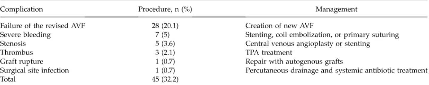
Table 5 Additional complications encountered after management of the initial complication
Complication Procedure, n (%) Management
Failure of the revised AVF 28 (20.1) Creation of new AVF
Severe bleeding 7 (5) Stenting, coil embolization, or primary suturing Stenosis 5 (3.6) Central venous angioplasty or stenting
Thrombus 3 (2.1) TPA treatment
Graft rupture 1 (0.7) Repair with autogenous grafts
Surgical site infection 1 (0.7) Percutaneous drainage and systemic antibiotic treatment
Total 45 (32.2)
The other most common complication was venous hypertension. It usually results from central vein stenosis and/or thrombosis secondary to previous catheterization or valvular incompetence in the AVF outflow vein.20 Another significant cause is
unim-peded flow to the distal venous part of the extremity, which is caused by non-ligation of the distal venous part of the anastomosis. In our center effective treatment was achieved by ligation of the distended vein, which was located in the distal part of the AVF in 31 of the 35 patients (88.6%) who had severe edema in their upper extremities. We observed that all of these patients’ first AVFs had been performed in other centers by the side-to-side technique without the distal venous part ligated. Beigi et al21 reported
that ligation of the distal vein when using the side-to-side technique prevented venous hypertension. In the remaining 4 patients whose index operations were performed by us, additional collateral veins appeared in time, even though the distal vein of the anastomosis was ligated.
Aneurysm/pseudoaneurysm formation is one of the main late-term complications after AVF forma-tion, occurring in from 2% to 10% of patients.10 In
our series the complication ratio in the general population with fistulas was 1.98%, whereas the patient ratio in the complication group was 22.3%. These complications can result in significant life-threatening events, such as rupture, bleeding, or access dysfunction and thrombosis (Fig. 1c and 1d). Traditionally, surgical repair of these aneurysms involves either ligation or resection; however, in this study the surgical aim was to salvage the access.22–25
In a large study including 44 aneurysms involving both AVF and grafts, Georgiadis et al22reported that
the mean primary patency after salvage at 24 months is 32%. Our salvage rate for this type of complication was 83.9% (26 of 31). We used excision of the aneurysm and repair with primary suturing (n ¼ 14) and interposition grafting (n ¼ 12); the long-lasting patency of these 26 salvaged AVFs (44% at 24 months) has encouraged us to continue using these type of interventions rather than ligation.
Bleeding and/or rupture of the AVF from needle access after hemodialysis can be a serious compli-cation that may be lethal if immediate intervention is not initiated. Of all complications requiring surgery, the rate of this complication has been reported as between 1.2% and 4.6%10,12,16 In our
group it was observed in 13 patients (9.3%) and hemostasis was achieved by using primary sutures or autogenous vein grafts in 8 patients (Table 4).
AVFs had to be closed in the remaining 5 patients through ligation.
A dysfunctional AVF can occur when the blood flow is misdirected. Although cephalic vein outflow was targeted, in some patients the flow was directed to the basilic vein, which resulted in inadequate hemodialysis, even though the AVF was technically functioning. In order to use these AVFs, basilic vein transposition was performed on these patients. Hemodialysis was successful in all patients on whom vein transposition was performed; however, in the case of a nonfunctioning cephalic vein, direct brachiobasilic AVFs may be advisable. In their prospective clinical studies Karakayali et al26
rec-ommend that brachiobasilic AVFs and transposition at the same session should have low morbidity, high maturation, and high patency rates, so they should be used as a first-choice solution in situations such as the absence of the cephalic vein.
Thrombosis as a late complication may signifi-cantly decrease the patency rate of AVFs. In patients with thromboses, surgical revision generally results in only 25% to 50% patency.15 In our series the
secondary patency was 70% after thrombectomy during the early postoperative period.
The quality of the initial access as well as the use of the AVF is significant in determining the severity and rate of complications. As is the case with most diseases, prevention is better than treatment. To limit complications, our surgical protocol for initial AVF placement is to use preoperative ultrasono-graphic mapping or venography to choose appro-priate vessels (diameter .2 mm) located in the distal arm for AVF creation.27 The surgeons ensure
that the anastomotic diameter is not greater than 5 mm and distal vein is ligated in order to prevent venous hypertension and high output. This surgical approach combined with trained hemodialysis personnel using the access results in fewer compli-cations, as the data collected from our study showed [our complication rate was 3.3% (47 of 1419) compared with 11.4% to 64% from other studies], and allows the initial AVF to function longer.
Most of the patients included in our study had already undergone surgical operations at different centers, and they were categorized as the patients with severe complications because they had no options in terms of endovascular intervention. Therefore, in this study we have included only the patients on whom endovascular intervention was not an option and surgical interventions were necessary. The retrospective descriptive nature of this study is the main limitation. Because our center
is a reference center and patients’ chronic renal failure duration is longer, such reference rates seem to be higher than the average. Although a good number of patients and procedures were analyzed in the pool, it is hard to isolate a direct causative effect for the occurrence of AVF complications. In addition, the number of patients seems to be sufficient, because they had no record; the patients who had already undergone endovascular interven-tion were not included in the study. Moreover, because a successful radiologic intervention was performed on them, they did not need surgery; therefore, we did not include them in the current study.
Even with the appropriate selection of vessel and anatomic site, where normal anatomy changes permanently, as it does in all AVF surgeries, complications are inevitable. Under these conditions every effort should be made to salvage the func-tioning AVF in order to save virgin vessels for future use. To this end, patients with these complications may be referred to a top-rated tertiary center in the hope that the original AVF has the highest possible chance of being salvaged.
Acknowledgments
We are happy to acknowledge Cagla Sariturk, the statistician of Adana Baskent Medical and Research Center, for the statistical process of this study, and who performed and checked the analysis that made this article possible. The authors declare that they have no competing interests and this study was approved by the Research Fund of Baskent Univer-sity (KA13/27).
References
1. Owen WF. Status of hemodialysis adequacy in the United States: does it account for improved patient survival? Am J Kidney Dis 1998;32(6 suppl 4):S39–S43
2. Murphy GJ, White SA, Nicholson ML. Vascular access for haemodialysis. Br J Surg 2000;87(10):1300–1315
3. Sidawy AN, Gray R, Besarab A, Henry M, Ascher E, Silva M Jr et al. Recommended standards for reports dealing with arteriovenous hemodialysis accesses. J Vasc Surg 2002;35(3): 603–610
4. Jenkins AM, Buist TA, Glover SD. Medium-term follow-up of forty autogenous vein and forty polytetrafluoroethylene (Gore-Tex) grafts for vascular access. Surgery 1980;88(5):667– 672
5. National Institute of Diabetes and Digestive and Kidney Diseases. Kidney and urologic diseases statistics for the United States. NIH Publication No. 11–3895. Available at: http://kidney.niddk.nih.gov/kudiseases/pubs/kustats/ index.aspx. Accessed August 2011
6. Rooijens PP, Burgmans JP, Yo TI, Hop WC, de Smet AA, van den Dorpel MA et al. Autogenous radial-cephalic or prosthetic brachial-antecubital forearm loop AVF in patients with compromised vessels?: a randomized, multicenter study of the patency of primary hemodialysis access. J Vasc Surg 2005; 42(3):481–486; discussions 487
7. III. NKF-K/DOQI Clinical Practice Guidelines for Vascular Access: update 2000. Am J Kidney Dis 2001;37(1 suppl 1):S137– S181
8. Tordoir J, Canaud B, Haage P, Konner K, Basci A, Fouque D et al. EBPG on Vascular Access. Nephrol Dial Transplant 2007; 22(suppl 2):ii88–117
9. Tordoir JH, Mickley V. European guidelines for vascular access: clinical algorithms on vascular access for haemodial-ysis. EDTNA ERCA J 2003;29(3):131–136
10. Salahi H, Fazelzadeh A, Mehdizadeh A, Razmkon A, Malek-Hosseini SA. Complications of arteriovenous fistula in dialysis patients. Transplant Proc 2006;38(5):1261–1264
11. Stern AB, Klemmer PJ. High-output heart failure secondary to arteriovenous fistula. Hemodial Int 2011;15:104–107
12. Vogel KM, Martino MA, O’Brien SP, Kerstein MD. Complica-tions of lower extremity arteriovenous grafts in patients with end-stage renal disease. South Med J 2000;93(6):593–595 13. Malik J, Tuka V, Kasalova Z, Chytilova E, Slavikova M, Clagett
P et al. Understanding the dialysis access steal syndrome: a review of the etiologies, diagnosis, prevention and treatment strategies. J Vasc Access 2008;9(3):155–166
14. Fokou M, Ashuntantang G, Teyang A, Kaze F, Chichom Mefire A et al. Patients characteristics and outcome of 518 arteriove-nous fistulas for hemodialysis in a sub-Saharan African setting. Ann Vasc Surg 2012;26(5):674–679
15. Palder SB, Kirkman RL, Whittemore AD, Hakim RM, Lazarus JM, Tilney NL. Vascular access for hemodialysis: patency rates and results of revision. Ann Surg 1985;202(2):235–239 16. Dix FP, Khan Y, Al-Khaffaf H. The brachial artery-basilic vein
arterio-venous fistula in vascular access for haemodialysis–a review paper. Eur J Vasc Endovasc Surg 2006;31(1):70–79 17. Astor BC, Coresh J, Powe NR, Eustace JA, Klag MJ. Relation
between gender and vascular access complications in hemo-dialysis patients. Am J Kidney Dis 2000;36(6):1126–1134 18. Dunlop MG, Mackinlay JY, Jenkins AM. Vascular access:
experience with the brachiocephalic fistula. Ann R Coll Surg Engl 1986;68(4):203–206
19. Lazarides MK, Staramos DN, Panagopoulos GN, Tzilalis VD, Eleftheriou GJ, Dayantas JN. Indications for surgical treatment of angioaccess-induced arterial ‘‘steal’’. J Am Coll Surg 1998; 187(4):422–426
20. Neville RF, Abularrage CJ, White PW, Sidawy AN. Venous hypertension associated with arteriovenous hemodialysis access. Semin Vasc Surg 2004;17(1):50–56
21. Beigi AA, Masoudpour H, Alavi M. The effect of ligation of the distal vein in snuff-box arteriovenous fistula. Saudi J Kidney Dis Transpl 2009;20(6):1110–1114
22. Georgiadis GS, Lazarides MK, Panagoutsos SA, Kantartzi KM, Lambidis CD, Staramos DN. Surgical revision of complicated false and true vascular access-related aneurysms. J Vasc Surg 2008;47(6):1284–1291
23. Hausegger KA, Tiessenhausen K, Klimpfinger M, Raith J, Hauser H, Tauss J. Aneurysms of hemodialysis access grafts: treatment with covered stents: a report of three cases. Cardiovasc Intervent Radiol 1998;21(4):334–337
24. Wongwanit C, Ruangsetakit C, Sermsathanasawadi N, Chinsakchai K, Mutirangura P. Treatment of iatrogenic pseudoaneurysm of brachial artery with percutaneous
ultra-sonographically guided thrombin injection (PUGTI): a case report and a literature review. J Med Assoc Thai 2007;90(8): 1673–1679
25. Najibi S, Bush RL, Terramani TT, Chaikof EL, Gunnoud AB, Lumsden AB et al. Covered stent exclusion of dialysis access pseudoaneurysms. J Surg Res 2002;106(1):15–19
26. Karakayali FY, Sevmis S, Basaran C, Yabanoglu H, Arat Z, Boyvat F et al. Relationship of preoperative venous and arterial imaging findings to outcomes of brachio-basilic transposition fistulae for hemodialysis: a prospective clinical study. Eur J Vasc Endovasc Surg 2008;35(2):208–213
27. Nursal TZ, Oguzkurt L, Tercan F, Torer N, Noyan T, Karakayali H et al. Is routine preoperative ultrasonographic mapping for arteriovenous fistula creation necessary in patients with favorable physical examination findings?: results of a randomized controlled trial. World J Surg 2006; 30(6):1100–1107