Tension Pneumothorax Associated with Hydatid Cyst Rupture
Ibrahim Can Ku¨rkc¸u¨og˘lu, M.D., Atilla Erog˘lu, M.D., Nurettin Karaog˘lanog˘lu, M.D., and
Pinar Polat, M.D.
Summary: Hydatid disease is common in Turkey. Tension pneumothorax is rare, but it is an important complication of the hydatid cyst with significant morbidity and mortality. Tension hydropneumothorax secondary to the rupture of a hydatid cyst was detected in 5 of 185 spontaneous pneumothorax cases that were treated in the authors’ clinic between 1992 and 1998. All patients were subjected to surgical treatment after urgent tube thoracostomy. No mortality or recurrence was observed at follow-ups of 9 to 24 months.
Key Words: Tension pneumothorax—Hydatid cyst.
Hydatid cyst is a parasitic disease caused by the larval form of Echinococcus granulosus as an intermediary host in humans and seen endemically in Turkey. The inci-dence rate is 1 in 2000, and the lungs are infected in 35% of cases (1). According to data of Turkish Ministry of Health, the incidence is 14 in 100,000 in Turkey (2). Tension pneumothorax is a serious but rare complication of pulmonary hydatid cyst. This complication can be fatal and should be kept in mind in the clinical course of hydatid cyst. The rupture of the hydatid cyst into the pleural cavity must be taken into account in the differ-ential diagnosis of tension pneumothorax. This manu-script presents two of the five cases first treated because of tension pneumothorax and surgically treated after the diagnosis of hydatid cyst.
PATIENTS AND METHODS
All of the patients in this study sought treatment for dyspnea, cyanosis, tachycardia, and hypotension at the authors’ emergency clinic. After chest X ray, tube tho-racostomy was applied because of the diagnosis of ten-sion hydropneumothorax. Tenten-sion hydropneumothorax
and a water-lily sign on intrapleural fluid were detected in two cases. In two other cases, the authors made this diagnosis by detecting a pericystic cavity in the expand-ing lung after tube thoracostomy. Indirect hemagglutina-tion tests were positive in these cases. In the last case, hydatid disease diagnosis was made after posterolateral thoracotomy was performed because of persistent air leak and empyema. The patients who were diagnosed radiologically were subjected to thoracotomy after a few days. In the patient with empyema, thoracotomy was performed after a 3-week tube drainage and antibio-therapy. All patients were treated with albendazole (10/mg/kg/day) for one month postoperatively.
Case 1
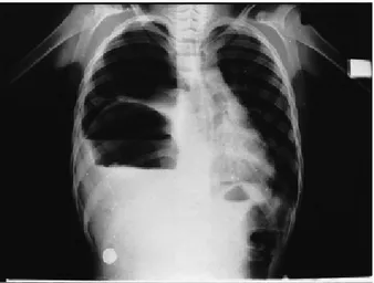
A 10-year-old male whose family earns a living through animal husbandry entered the authors’ emer-gency clinic with complaints of dyspnea and bitter liquid production as a result of coughing. Chest X ray revealed mediastinal displacement, tension pneumothorax, and a water-lily sign floating on the pleural fluid (Fig. 1). Tube thoracostomy was performed because of tension pneu-mothorax. Two-hundred-fifty mL serohemorrhagic fluid was drained. Because of the prediagnosis of a perforated hydatid cyst, the patient was subjected to surgery. Left posterolateral thoracotomy, decortication, cystectomy, and capitonnage were performed. The patient was fol-lowed at monthly intervals. At the end of 9 months of follow-up, the symptoms for which the authors treated him were no longer present.
From the Department of Thoracic Surgery (I.C.K., A.E., N.K.), and the Department of Radiology (P.P.), Atatu¨rk University Medical Fac-ulty, Erzurum, Turkey.
Address correspondence and reprint requests to Dr. Ibrahim Can Ku¨rkc¸u¨og˘lu, Department of Thoracic Surgery Medical Faculty, Atatu¨rk University, 25000 Erzurum, Turkey; e-mail: cankurkcuoglu@ hotmail.com.tr
Journal of Thoracic Imaging
17:78–80 © 2002 Lippincott Williams & Wilkins, Inc., Philadelphia
Case 2:
A 10-year-old boy, whose family lives in a rural area and breeds animals, was hospitalized with complaints of sudden pain in the right side of his chest and dyspnea. On poseteroanterior chest radiography, mediastinal shift, tra-cheal displacement, and lung collapse were detected. There was an appearance of a germinative membrane floating on the intrapleural fluid level (Fig. 2). Eosino-philia was present in peripheral blood. Intradermal Ca-soni and Weinberg complement fixation tests were nega-tive, whereas an indirect hemagglutination test was posi-tive. The patient, who had no other medical problems, was operated on and right posterolateral thoracotomy, cystectomy, and capitonnage were performed. He was discharged on the ninth postoperative day. No recurrence was seen during a 2-year follow up.
DISCUSSION
Pulmonary hydatid cyst and its complications have been known since the time of Hippocrates (3). Pulmo-nary hydatid cysts are asymptomatic until they reach a large size and become complicated. Cough, dyspnea, he-moptysis, and chest pain are the symptoms observed in most patients with pulmonary hydatid cysts. The com-plicated cysts can cause serious clinical results and may mimic various other diseases. In the report of Qian, 356 (42.3%) of 842 patients had complicated hydatid cysts (4). The most frequently seen complications are rupture, abscess formation, and hemoptysis.
The occurrence of pneumothorax because of the rup-ture of the hydatid cyst into the pleural cavity is a rare complication. Few case reports have been presented in the literature (1,5–7). The perforation of a pulmonary
hydatid cyst into the pleural cavity causes symptoms such as chest pain, cough, cyanosis, and choking (3). After the rupture of the cyst, the content of the cyst dissipated to the intrapleural cavity, and the ruptured germinative membrane became collapsed. The parenchy-mal cavity and the bronchopleural fistula together are subject to check-valve mechanism and cause tension pneumothorax. The neighboring lung tissue is com-pressed and the germinative membrane that is floating on the fluid level causes the appearance of a water-lily sign in the pleural cavity.
Cernay reported a rupture into the pleural cavity in 5 of 366 cases (1.3%) (7). Tension pneumothorax was de-tected in 5 of 148 patients (3.37%) who had been oper-ated on for pulmonary hydatid disease during the 6-year period of the present study. In the same period, the num-ber of spontaneous pneumothorax cases was 185. The ratio of tension pneumothorax caused by the rupture of the hydatid cyst was 2.7%. It is thought that the socio-economic and cultural factors of the authors’ region in-crease the rate of complication. It was observed that all spontaneous pneumothorax cases caused by hydatid cyst rupture presented as tension pneumothorax cases. The patient who had empyema was diagnosed by thoracot-omy.
Radiographic signs are generally diagnostic in coun-tries where hydatid disease is common (8). The cyst ap-pears as smoothly outlined, with homogenous-density spherical lesions (3). The radiographic pleural manifes-tations in the acute stage of cyst rupture vary from locu-lated hydropneumothorax to nonloculocu-lated partial, com-plete, or tension hydropneumothorax. A water-lily sign can also be observed in instances of cyst rupture into the pleura (3). Computed tomography and magnetic reso-nance imaging provide important diagnostic clues in
sus-Fig. 2. Chest X ray showing right tension pneumothorax and a
water-lily sign.
FIG. 1. Chest X ray showing left tension pneumothorax and an
in-trapleural water-lily sign.
TENSION PNEUMOTHORAX ASSOCIATED WITH HYDATID CYST RUPTURE 79
pected cases. In 4 patients, pulmonary X ray was very important in the diagnosis of the disease. The determi-nation of a well-defined opaque lesion together with pneumothorax in the lung roentgenograms, and the wa-ter-lily sign in the pleural cavity (encountered in 2 cases), are thought to be pathognomonic findings.
Laboratory methods are useful in the diagnosis of hy-datid disease. Casoni’s intradermal test and Weinberg’s complement fixation test are nonspecific. The false-positive rate of Weinberg’s test is 28%. The indirect hemagglutination test’s superior sensitivity is 66% to 100% and the false-positive rate is 1% to 2% (3). Results of Casoni’s test was positive in 204 patients (91.7%), whereas Weinberg’s complement fixation test was posi-tive in 197 patients (75%) in a study that had been made in the authors’ geographical region (9).
Stewart reported that cough and membrane expecto-ration confirm the diagnosis (6). This symptom was seen only in one of the authors’ patients. Empyema and pleu-ral thickening are the most frequent findings in the au-thors’ cases. Decortication was performed because of pleural thickening in four cases.
Operation is the treatment of choice for pulmonary hydatid cysts (4). Surgical treatment should be per-formed on patients in whom hydatid cyst rupture into the pleural cavity is demonstrated; however, secondary pleu-ral hydatidosis was reported in the literature. To prevent tension pneumothorax complication, tube thoracostomy should be applied preoperatively (1,5–7). The major goal of operation is full excision of cysts and maximum pres-ervation of lung tissue (3). When tension pneumothorax develops in hydatid cyst, urgent intercostal drainage and early thoracotomy is appropriate to prevent extensive complications (6).
Benzimidazole is being used in the medical therapy of hydatid cyst disease. Medical therapy is being used in inoperable cysts, dissemination in difficult localizations, or contraindicated situations to surgery (3). Preoperative antibiotherapy is applied in cases of infected hydatid cyst (3). The absence of recurrence in the follow-up period of the authors’ cases can be explained by the administration of albendazole postoperatively.
The rupture of the hydatid cyst into the pleural cavity results in tension pneumothorax. Hydatid cyst should be remembered in differential diagnosis of tension pneumo-thorax.
REFERENCES
1. Bakir F, al-Omari MM. Echinococcal tension pneumothorax.
Tho-rax 1969;24:547–556.
2. Baris¸ YI., S¸ahin AA, Bilir N. Status of Echinococcosis in Turkey. Publication of Turkish foundation of lung diseases. Ankara: Kent Matbaasi, 1989:28.
3. Symbas PN, Aletras H. Hydatid disease of the lung. In: Shields TW, ed. General Thoracic Surgery, 5th ed. Philadelphia: Lippin-cott Williams & Wilkins, 2000:1113–1122.
4. Qian ZX. Thoracic hydatid Cysts: A report of 842 cases treated over a thirty-year period. Ann Thorac Surg 1988;46:342–346. 5. Connellan SJ, Jowett AW, Wilson RS. Hydatid disease presenting
as tension pneumothorax. Br J Dis Chest 1979;73:405–406. 6. Stewart MP, Cunningham MS. Tension pneumothorax
complicat-ing pulmonary hydatid disease in childhood. Clin Paediatr 1987; 26:422–424.
7. Cernay J, Bensenouci A, Baukhelal H, et al. The rupture of the pleura: a serious complication of the hydatid cyst of the lung in children. Rev Fr Mal Respir 1979;7:45–47.
8. Katz R, Murphy S, Kosloske A. Pulmonary echinococcosis: a pe-diatric disease of the southwestern United States. Pepe-diatrics 1980; 65:1003–1006.
9. Balik AA, Bas¸og˘lu M, C¸ elebi F, et al. Surgical treatment of hydatid disease of the liver: review of 304 cases. Arch Surg 1999;134:166– 169.
I. C. KU¨ RKC¸U¨OG˘LU ET AL. 80