https://doi.org/10.1007/s12012-018-9466-y
The Effects of Lipid Emulsion, Magnesium Sulphate and Metoprolol
in Amitriptyline-Induced Cardiovascular Toxicity in Rats
Saylav Bora1 · Mümin Alper Erdoğan2 · Gürkan Yiğittürk3 · Oytun Erbaş4 · İsmet Parlak5
Published online: 5 June 2018
© Springer Science+Business Media, LLC, part of Springer Nature 2018
Abstract
The aim of this study was to evaluate the effects of metoprolol, lipid emulsion and MgSO4 which can be recommended for prevention of long QT that is one of the lethal consequences of amitriptyline intoxication. Thirty Sprague–Dawley male rats were included. Five groups respectively received the following: saline intraperitoneally (i.p.); amitriptyline (AMT) 100 mg/ kg per os (p.o.) and saline i.p.; AMT 100 mg/kg p.o. and 5 mg/kg metoprolol i.p.; AMT 100 mg/kg p.o. and 20 ml/kg lipid emulsion i.p.; AMT 100 mg/kg p.o. and 75 mg/kg MgSO4 i.p. After 1 h, all groups were analysed by ECG recordings in DII lead; their blood was taken for biochemical examination and euthanasia was performed. For histological examination, cardiac tissues were removed and sections were prepared. QTc was significantly reduced in treatment groups compared to the AMT+saline group. When compared with the AMT+saline, lipid emulsion did not affect pro-BNP and troponin levels in biochemical analysis, but it significantly reduced Caspase 3 expression in histological examination. In the group treated with AMT and metoprolol, there was no significant effect on Caspase 3 expression. In MgSO4-treated group, there was a
significant decrease in troponin, pro-BNP and urea levels biochemically and significant decrease in Caspase 3 expression histologically when compared with the control group. With further studies including clinical studies, MgSO4, lipid emulsion or metoprolol may be used to improve AMT-induced cardiotoxicity. They can possibly become alternative approaches in the future for suicidal or accidental intoxication of tricyclic antidepressant in emergency departments.
Keywords Drug toxicity · QTc · Amitriptyline · Arrhythmia · Electrocardiogram
Introduction
Tricyclic antidepressants (TCAs) are commonly used antide-pressant agents with high intoxication rates. In total, 12,234 cases of TCA toxicity were reported in 2004 by American Association of Poison Control Centers [59].
Amitriptyline (AMT) intoxication is reported to be the most frequent among TCA toxicity, covering 40–58% of TCA overdoses [8, 37]. Of all prescription medication-related toxicities, AMT intoxication is the third most com-mon cause of death, followed by sedative–hypnotic drugs and analgesic drugs [37].
The mortality and morbidity of TCA toxicity are due to direct peripheral α-adrenergic blockage, inhibition of norepi-nephrine reuptake at nerve terminals, a membrane stabiliz-ing or quinidine like effect on myocardium and anticholin-ergic effect [33, 48]. Myocardial sodium channel blockage, ventricular dysrhythmias, myocardial depression, and hypo-tension can be seen in TCA cardiotoxicity [5].
Handling Editor: Rajiv Janardhanan. * Saylav Bora
1 Department of Emergency Medicine, Atatürk Training
and Research Hospital, İzmir Katip Çelebi University, Izmir, Turkey
2 Department of Physiology, Faculty of Medicine, İzmir Katip
Çelebi University, Izmir, Turkey
3 Department of Histology and Embryology, Muğla Sıtkı
Koçman University, Muğla, Turkey
4 Department of Physiology, Faculty of Medicine, Istanbul
Bilim University, Istanbul, Turkey
5 Department of Emergency Medicine, Health Science
University İzmir Bozyaka Research and Training Hospital, Izmir, Turkey
Electrocardiography (ECG) can be used as a predictive marker on cardiac effects of TCA in emergency rooms (ER) [46]. A wide QRS, prolonged QTc and PR interval can be seen due to the prolongation of cardiac action potential and refractory period along with a delay of atrioventricular node (AVN) conduction [11]. The underlying cause is sodium channel blockage leading to TCA toxicity. The effects of TCA toxicity are not related with ingested dosage [40], the prolonged QTc is related to poor prognosis, and this increases the importance of the ECG in the diagnosis and early management of TCA overdose [21, 40]. The prolonga-tion of QT can be seen not only in toxic dosage but also in therapeutic dosages. QT interval is measured from the onset of Q wave and the end of T, and it has to be corrected with heart rate [22]. Generally, Bazett’s equation is used for cor-rection of prolongation of QT [31]. The risk factors for pro-longation of QTc are female sex, advanced age, congenital QT syndrome, electrolyte imbalance such as hypokalemia, endocrine metabolic problems, and so on [54]. It is reported that prolonged QTc increases mortality in TCA toxicity with some diseases as coronary artery disease, syncope, and dia-betes mellitus [4].
The most common, important and lethal consequence of AMT overdose is cardiovascular toxicity, which requires rapid diagnosis and treatment in the ER [37]. Patients with AMT toxicity must be closely monitored. The most lethal cardiovascular adverse effect of AMT overdose is presenta-tion with tachycardia with wide QRS complexes, resulting in a prolonged QT interval. Even though this clinical picture can be identified early and prevented, no prophylactic treat-ment protocol has been established to be used specifically in the ER for this phenomenon.
Metoprolol, a beta blocker, is a class II antiarrhythmic agent. It acts on AVN [17]. These agents are used to control atrial and ventricular rates [50]. The cardiotoxic effects of TCAs such as AMT are powerful and thus result in high mortality rates [18, 46]. Considering this high mortality rate of AMT toxicities, early diagnosis and treatment are very important. The prevention of prolongation of QTc duration can decrease morbidity and mortality in early stages [7].
Magnesium sulphate has been used for decades in the management of eclampsia [49] and pregnancy-induced hypertension [14]. Recent reports on magnesium have emphasized its role in deficiency syndromes [42, 55], includ-ing cardiac arrhythmias.
The evidence for cardiac depression by hypermagnesae-mia is less clear, despite its wide clinical use. Some authors have demonstrated that there is no evidence of myocardial depression at any concentration of serum magnesium [28], whereas others have reported that a single injection of mag-nesium sulphate leads to a decrease in cardiac performance [15]. Although the cardiovascular hemodynamic effects of magnesium sulphate were assessed by some authors in
baboons [28], sheep [44] and humans [16], there have been few reports on magnesium-induced changes in left ventricu-lar diastolic function. The effects of magnesium sulphate on the coronary circulation and myocardial metabolism have not been studied extensively.
Intravenous lipid emulsion is an established, effective treatment for local anesthetic-induced cardiovascular col-lapse [43]. The predominant theory for its mechanism of action is that by creating an expanded, intravascular lipid phase, equilibria are established that drive the offending drug from target tissues into the newly formed ‘lipid sink’. Based on this hypothesis, lipid emulsion has been consid-ered as a candidate for generic reversal of toxicity caused by overdose of any lipophilic drug as AMT [39, 57].
The aim of this study was to explore the most effective, available, practical and adjustable protective agent to be used in the alternative treatment protocol for cardiac toxic-ity caused by AMT overdose within ER settings and whether MgSO4, metoprolol or lipid emulsion would be a more
plau-sible option.
Materials and Methods
Animals and Laboratory Setting
The experimental procedures employed in this study were approved by the Animal Ethics Committee. All experiments were carried out according to the Guide for the Care and Use of Laboratory Animals, as confirmed by National Institute of Health, USA. A total of 30 male Sprague–Dawley rats, each weighing 200–250 g, were used in the study. The rats were kept in a 12-h light/dark cycle (light from 7 am to 7 pm) in quiet rooms with an ambient temperature of 22–24 °C, and were fed standard laboratory food with tap water ad libitum.
Experimental Protocol
The study included 30 Sprague–Dawley male rats, of which 6 were assigned to the control group and were drug-free and the remaining 24 received 100 mg/kg AMT p.o. (Laroxyl 25 mg, Roche, Switzerland). The AMT was administered by initially dissolving the original solid pharmaceutical in tap water and then applying a solution containing 20 mg/ ml AMT via orogastric lavage. Tablets containing 25 mg AMT (Laroxyl, Roche) were suspended in tap water to yield a concentration of 20 mg/ml. According to the weight of each rat, suspended drug solution was completed to 4 ml with tap water. The AMT was given via orogastric tubes. On completion of these processes, these rats were then further randomly divided in four groups. 30 min after AMT admin-istration, we gave the pharmaceuticals to the groups below:
Group A (n:6) received 1 ml/kg 0,9% NaCl saline i.p. (A+S) as placebo
Group B (n:6) received 5 mg/kg metoprolol i.p. (BELOC® 5 mg/5 ml IV AstraZeneca) (A+M)
Group C (n:6) received 20 ml/kg solution of lipid emul-sion i.p. (INTRALIPID 20% Fresenius LE)
(A+LE)
Group D (n:6) received 75 mg/kg MgSO4 i.p. (Magne-sium Sulphate 15% Biopharma) intraperitoneally. At 1 h after drug administration, all rats were applied with ECG on derivation II under anesthesia (Ketamine: 40 mg/ kg, Xylazine: 4 mg/kg). On completion of all assessments, the rats were euthanized and cardiac extraction was made for histological examination.
Assessment of ECG
ECGs were recorded on male Sprague–Dawley rats under anesthesia in the prone position. Electrodes consisted of 26-gauge needles placed subcutaneously for 1 cm. Stand-ard limb leads were constructed from electrodes placed at the paws. Rats were anesthetized by combination of keta-mine hydrochloride at a dose of 40 mg/kg (Alfaketa-mine1, Ege Vet, Alfasan International B.V., Holland) and 4 mg/kg of xylazine hydrochloric (Alfazyne1, Ege Vet, Alfasan Inter-national B.V.) that were administered i.p. Under anesthesia, ketamine (40 mg/kg) and xylazine (4 mg/kg) were admin-istered i.p., ECG was taken in derivation II (BIOPAC MP 150, Goleta, California, USA). Data were evaluated using Biopac Student Lab PRO Version 3.6.7 software (BIOPAC Systems, Inc.), QT interval, T wave duration, and beats per minute (BPM) as the parameters. For the calculation of QTc, Bazett’s formula was used. During the ECG recordings, rec-tal temperatures of the rats were monitored by a recrec-tal probe (HP Viridia 24-C; Hewlett–Packard Company, Palo Alto, California, USA) and the temperature of each rat was kept at approximately 36–37 °C by heating pad.
Histological Examination
Heart tissues were fixed within 10% formol solution. 4-micron sections were obtained, and stained with hae-matoxylin and eosin and Caspase-3 immunohistochemical procedures. Histological examination was applied using an Olympus BX51 microscope.
For immunohistochemistry, sections were incubated with H2O2 (10%) for 30 min to eliminate endogenous per-oxidase activity and then blocked with 10% normal goat serum (Invitrogen) for 1 h at room temperature. After that, sections were incubated in primary antibodies (Caspase-3, SantaCruz Biotechnology; 1/1000) for 24 h at 4 °C. Anti-body detection was carried out with the Histostain-Plus Bulk
kit (Invitrogen) against rabbit IgG and 3,3′-diaminobenzi-dine (DAB) was used to visualize the final product. All sec-tions were washed in PBS and photographed with Olympus C-5050 digital camera mounted on Olympus BX51 micro-scope. Immunoreactivity in the Left Ventricule sections was quantified by using Image J software (National Institutes of Health, Bethesda, MD). To calculate the Caspase-3 immu-noreactivity, Caspase-3 (+) cells were counted at ×40 mag-nification in randomized sections (3–4) for each rat (n = 6) in a blinded fashion.
Biochemical Analyses
Elevated levels of Troponin T and NT-proBNP (N-terminal fragment of NT-proBNP) are significant as their release might indicate cardiac ischemia. These markers are also important to calculate the overall risk for patients with acute coronary syndrome. In this study, blood samples were collected from all rats following the administration of the above-mentioned drugs and the samples were processed at a temperature of − 20 °C. Troponin T and NT-proBNP were then assessed as cardiotoxic markers under elective condi-tions (Thermo Fisher Scientific/Finland Elisa reader, Fine Test Rat Elisa kits).
Na, K, Cl, urea, creatinine and ALT levels were also measured in the collected blood samples (Beckman-Coul-ter AU 2700 PLUS device ve Beckman-Coul(Beckman-Coul-ter commercial kits).
Statistical Analysis
Data are expressed as mean ± SEM. Statistical analyses of the study data were made using the Statistical Package for Social Sciences Windows 15.0 (SPSS 15.0) program. The Shapiro–Wilk test was used to adjust for normal met-ric distribution and discrimination of parametmet-ric and non-parametric variables. The one-way ANOVA test was used to compare parametric variables while the Mann–Whitney U Test was used for the comparison of non-parametric vari-ables. The Tukey method was used in Post hoc analyses for variance homogeneity. For variables that did not show homogenous variance, the Dunnett T3 method was used. A value of p < .05 was accepted as statistically significant.
Results
Electrocardiographic Findings
In Group A (AMT+saline), the QT intervals were signifi-cantly higher than in all the other groups (p < .05). In com-parison with Group A, the QT interval was significantly decreased in all the other groups (p < .05). The measured
and calculated data obtained from the rat ECGs are pre-sented in Table 1.
When control group data were compared to Group A data, it was observed that Group A had both significantly longer QTc and increased T wave duration (p < .005). Compared to Group A, the QTc interval was much more shorter in the other groups (p < .01) (Table 1; Fig. 1).
Biochemical Findings
When Group A was compared to the control group, sig-nificantly higher levels of NT-proBNP were determined in Group A (p < .001). Group D had significantly decreased levels of NT-proBNP compared to Group A (p < .05). There was no significant difference between the other groups in respect of NT-proBNP. The Troponin T and NT-proBNP lev-els in the collected blood samples are presented in Table 2. Compared to the control group measurements, Group A had significantly higher Troponin T levels (p < .05). Group D had significantly decreased Troponin T levels compared to those of Group A (p < .05) (Table 2). There were no significant dif-ferences between the groups in respect of creatinine, ALT, Na+, K+ and Cl− values. Statistically significantly lower
lev-els of urea were measured in Groups B and D, compared to the control group (p < .001). The biochemical measurements obtained from the collected blood samples of all five groups are presented in Table 3.
Immunohistochemical and Apoptotic Process Measures
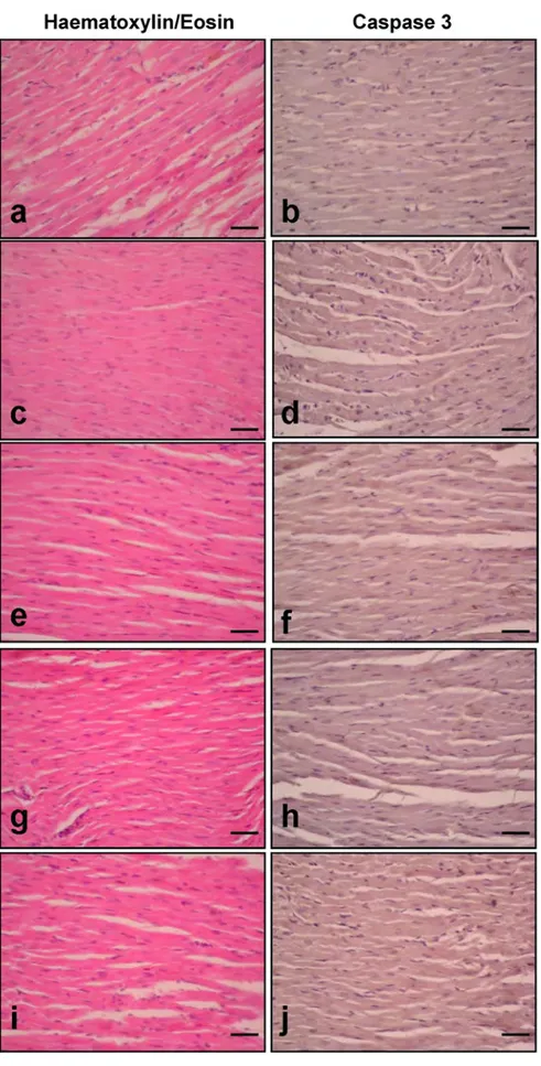
When Group A was compared to the control group, a signifi-cantly higher level of Caspase 3 expression was identified (p < .0001) (Fig. 2c, d). The Caspase 3 expression measure-ments of the histological cardiac examination of the rats are presented in Table 4. Compared to Group A, Groups C, and D had significantly decreased Caspase 3 expression (Fig. 2g–j) (p < .001) (Table 4). There was no significant dif-ference between Group B and Group A in respect of Caspase 3 expression (Fig. 2c–d) (Table 4).
Cardiac sections dyed with haematoxylin and eosin are shown in Fig. 2. While Fig. 2a, b indicate normal micro-scopic structure, Fig. 2c, d shows dilated capillaries and absent or ejected nuclei with necrotic cell groups in the rats that were exposed to AMT. Fig. 2e, f shows similar changes in the group treated with AMT. Fig. 2g–j indicates decreased necrotic areas in addition to dilated capillaries compared to Fig. 2c, d.
Discussion
TCAs are commonly used drugs. Due to widespread use, emergency services administration is frequent due to side effects and toxicity complaints. ECG can be used as a predic-tive marker of the cardiac effects of TCA in the ER [46]. The cardiotoxic effect of AMT TCA is powerful and is responsi-ble for the high mortality rate [18, 46]. In view of this high mortality rate of AMT toxicity, early diagnosis and treat-ment are very important in ER. The prevention of prolonged QTc duration can decrease morbidity and mortality in the early stages [7].
Common ECG abnormalities in AMT toxicity are defined as Na+ channel blockage, PR elongation, prolonged QRS
and QT intervals, alterations on segment ST, T wave altera-tion, right axis deviation and Brugada pattern [53]. The severity of clinical findings does not correlate with serum or urine concentrations and increases the clinical importance of ECG changes [19]. ECG abnormalities have increased importance because arrhythmias and hypotension are late signs of cardiac toxicity. In this study, prolonged QT, QTc and T durations were determined in Group A compared to the control group, which might indicate the presence of car-diotoxicity, independent of the cause. In a previous study conducted in this field, it was reported that AMT did not affect heart rate while causing prolonged QTc [30]. Similar to that study, no change in the heart rate was determined in the current study together with prolonged QTc.
The prolongation of QTc is particularly important in ECG abnormalities. The mechanism which has been sug-gested to be most responsible for QTc prolongation is the Table 1 ECG data
obtained from rats after the administration of treatments
Results are expressed as mean ± SEM (*p < .05, significant when compared to the control group, #p < .05,
significant when compared to Group A, **p < .005, significant when compared to the control group,
##p < .01, significant when compared to Group A)
QT (msn) BPM (beat/min) QTc (msn) T (msn)
Normal (control) 77.4 ± 3.26 249.2 ± 18.1 157.4 ± 9.4 32.4 ± 1.02 Group A (AMT+saline) 98.2 ± 8.07* 330.4 ± 9.85 229.2 ± 17.01** 53.4 ± 3.03** Group B (AMT+metoprolol) 78.2 ± 2.36# 275.4 ± 10.4 167.2 ± 5.3## 34.4 ± 3.45##
Group C (AMT+LE) 76.4 ± 1.92# 292.2 ± 26.2 167.6 ± 9.2## 38.1 ± 1.82##
slowing of depolarization of the cardiac action potential by inhibition of the sodium and potassium channels [10]. It is known that at an elderly age, TCA and higher antipsychotic doses are predictors of significant QTc prolongation [45]. In patients at high risk of cardiac problems, cardiotoxicity may be seen not only at a toxic dosage but also at a therapeutic dosage of AMT [3, 20]. Nose and Barbui reported prolonga-tion of QTc in antidepressant usage and suggested routine
cardiac monitoring for patients prescribed AMT, especially for patients with other risk factors such as cardiovascular comorbidities, and that drug combinations are applied care-fully in consideration of the risk of QTc prolongation [45]. QTc prolongation has been associated with several drugs such as diphenhydramine, ondansetron, pimozide, ziprasi-done, gemfibrozil, amiodarone, citalopram, clarithromy-cin, dofetilide, erythromyclarithromy-cin, ketoconazole, methadone, Fig. 1 The
electrocardiogra-phies of all the five groups. a ECG of control group; b ECG of Group A (AMT+saline); c ECG of Group B
(AMT+metoprolol); d ECG of Group C (AMT+LE); e ECG of Group D (AMT+MgSO4)
a
b
)
ondansetron, haloperidol, pimozide, quetiapine and TCAs [4]. The prolongation of QTc may cause lethal arrhythmias in AMT toxicity [1]. Therefore, early recognition and treat-ment are very important. Despite the mechanism not having yet been fully clarified, some studies in the literature have shown that agents such as adenosine A1 receptor antagonist, theophylline, glucagon, lidocaine or phenytoin have some positive effects on amitriptyline-induced cardiac toxicity [21, 29, 30, 47].
In addition to the presentation of ECG abnormalities, particularly QTc prolongation, the effects of both metopro-lol, lipid emulsion and MgSO4 were also investigated in
this study. The results demonstrated that all shortened QTc and T duration prolongation. To the best of our knowl-edge, there has been no previous study which has inves-tigated the possible effects of metoprolol, lipid emulsion and MgSO4 in AMT toxicity. Beta-blocker treatment for prolonged QTc is a well known, commonly used and valid therapeutic option [2, 7]. The blockade of outward potas-sium currents may be a cause of prolongation of action potential, thereby prolonging the QT interval and T dura-tion. Therefore, drugs such as AMT that prolong QTc may be responsible for the pro-arrhythmic effects through the blockade of outward potassium currents [60]. Metoprolol is known to cause a shortening in action potential due to decreased inward calcium current and increased out-ward potassium currents in myocardial cells [6, 35]. In
particular, the increase in potassium outward from myo-cardial cells triggers a shortening in the action potential, QTc and T durations [60].
In addition to general cardiotoxicity, there are some case reports in the literature which have reported myocar-dial infarct related to AMT overdose [35]. The results of the immunohistochemical examination of cardiac tissue in the current study suggest a significant increase in Caspase 3 expression in Group A specifically, which in turn, could be interpreted as evidence of a possible myocardial infarct playing a role in the pathophysiology of AMT-related car-diac toxicity. Sorodoc et al. [51] reported elevated NT-proBNP and Troponin levels in AMT-related cardiotoxic-ity and postulated that these markers might also be used to detect the severity of toxicity. Similar to these findings [51], significantly elevated NT-proBNP and Troponin lev-els were observed in the affected group as well as similar immunohistochemical changes to the findings of Sorodoc et al. [51] which were reported as histochemical alterations within the left ventricle of subjects in another study.
There have been several clinical studies to date that have recommended using beta blocker agents to treat AMT tox-icity [9]. The basic mechanism of how these agents work could be explained by the action of metoprolol that has been defined as a cardioprotective agent which increases potas-sium flow out of myocardial cells and decreases Ca2+ flow
into the cells thereby triggering a shortened action potential, which in turn leads to a decrease in QTc and T wave dura-tion [60]. As no decline in Caspase 3 activity or histologi-cal recovery on the myocardium was observed in the cur-rent study, it could be suggested that metoprolol could have caused an electrical recovery in respect of cardiotoxicity but was unable to cause a significant alteration in NT-proBNP levels that might underline the inability to prevent apop-tosis and inadequacy in preventing structural myocardial tissue damage. Although there was no decreased Caspase 3 activity in Group B in the current study, in a study of microembolized rats by Su et al. [52], a significant decline was determined in Caspase 3 activity following metoprolol administration. To the best of our knowledge, there has been no clinical or histological study that has shown the effects of metoprolol on AMT intoxication.
Table 2 Troponin T and NT-proBNP levels in blood samples meas-ured by ELISA
Results are expressed as mean ± SEM (*p < .05, significant when compared to the control group, **p < .005, significant when com-pared to the control group, #p < .05, significant when compared to
Group A) NT-proBNP (pg/ml) Troponin T (pg/ml) Control 3.97 ± 0.42 0.86 ± 0.08 Group A (AMT+saline) 12.4 ± 4.17** 2.39 ± 0.9* Group B (AMT+metoprolol) 8.45 ± 1.81 2.06 ± 0.49 Group C (AMT+LE) 14.2 ± 4.28 2.6 ± 0.7 Group D (AMT+MgSO4) 5.6 ± 0.74# 0.9 ± 0.05#
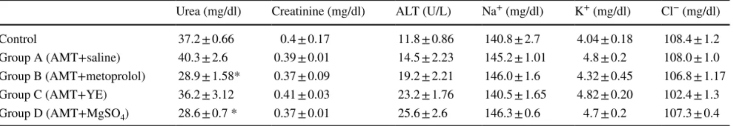
Table 3 The biochemical measurements obtained from the collected blood samples of all groups
Results are expressed as mean ± SEM (*p < .001, significant when compared to the control group)
Urea (mg/dl) Creatinine (mg/dl) ALT (U/L) Na+ (mg/dl) K+ (mg/dl) Cl− (mg/dl)
Control 37.2 ± 0.66 0.4 ± 0.17 11.8 ± 0.86 140.8 ± 2.7 4.04 ± 0.18 108.4 ± 1.2
Group A (AMT+saline) 40.3 ± 2.6 0.39 ± 0.01 14.5 ± 2.23 145.2 ± 1.01 4.8 ± 0.2 108.0 ± 1.0 Group B (AMT+metoprolol) 28.9 ± 1.58* 0.37 ± 0.09 19.2 ± 2.21 146.0 ± 1.6 4.32 ± 0.45 106.8 ± 1.17
Group C (AMT+YE) 36.2 ± 3.12 0.41 ± 0.03 23.2 ± 1.76 140.5 ± 1.65 4.82 ± 0.20 102.4 ± 1.3
Fig. 2 Effects of treatments on haematoxylin/eosin staining and Caspase 3 immunoexpression levels in cardiac tissue sections. Scale bars 50 µm (n = 6/each group). Quantitative analysis of Caspase 3 (+) cell count is given in Table 4. a, b Control, c, d Group A, e, f Group B, g, h Group C, i, j Group D
AMT is a highly lipophylic compound that is very well distributed in lipid-rich contexts [38], making it much more rational therefore to use lipid emulsion in cases of intoxi-cation. Most data on lipid emulsion procedure have been derived from experiences of local anesthetic agent toxicity. There have been reports that all lipophylic agents including lipid emulsion would be useful in experimental studies [23,
61]. With several clinical trials, it has been shown that suc-cessful results could be obtained by using the lipid emulsion method in AMT toxicity [11, 24]. In the current study, as a result of lipid emulsion, QTc and T wave duration shortened while a decrease in Caspase 3 activity was observed. In con-trast to the study hypothesis, lipid emulsion did not provide any alteration in NT-proBNP or Troponin levels. The lipid emulsion administered group also showed cellular recovery at a histological level. These findings support evidence indi-cating that the lipid emulsion method is based on the lipid sink theory, which suggests that lipid emulsion makes com-partments in plasma and provides sequestration of lipophylic agents, thereby reducing free drug concentration in plasma, and reducing the toxic effects of drugs on target tissues while acting through the same mechanism against cardiotoxicity [41, 57]. This effect has been shown for lipophylic agents such as bupivacaine and cocaine in particular [12, 57, 58]. In a trial by Weinberg et al. [57], asystole was generated in rats with the use of bupivacaine and then lipid emulsion was administered to one group. From examination of the cardiac biopsy materials, the group that had been given lipid emul-sion was determined to have significantly lower bupivacaine levels [57]. In another study by Carreiro et al. [12], prior to generation of cocaine toxicity, rats in one group were premedicated with lipid emulsion while the control group was given normal saline. Lower mortality rates were seen in the group that had received lipid emulsion compared to the control group [12].
Tzivoni et al. [56] reported that MgSO4 prevented the
emergence of Torsades de Pointes without improving pro-longed QTc interval. It was similarly observed in the current
study that MgSO4 caused significantly shorter QTc and T waves. MgSO4 also seemed to be more effective on
immuno-histochemical levels, compared to metoprolol or lipid emul-sion, causing a decline in NT-proBNP and Troponin levels. MgSO4 administration also resulted in significant decreases in Caspase 3 activity, possibly through the Mg natural role of modulating calcium ions, reflecting its cardioprotective efficacy [13, 34]. In a study of 40 rabbits, Jalal et al. [27] generated myocardial infarction on the rabbits and in three groups, pioglitazone, MgSO4 and omega-3 fatty acid were
administered separately to each group. MgSO4 was seen to
be able to reduce the infarct area and decreased the number of inflammatory cells such as polymorphonuclear (PMN) or mononuclear (MN) in the cardiac tissue. This process was interpreted as having been caused by the coronary vasodila-tor, immunomodulatory, anti-aggregatory and free oxygen radical cleanser effects of MgSO4 [27, 32]).
In the current study, AMT toxicity was created in rats and a significant decrease was seen in Caspase 3 expression in the rat group that was given MgSO4. The reduced volume of the infarct area identified in the study by Jalal et al. [27] might be related to MgSO4 acting through a mechanism that
possibly decreased the expression of Caspase 3 in rats that were treated with the molecule. Studies that have reported reduced myocardial infarct areas and less myocyte loss related to reperfusion damage support this perspective [25,
26]. In the current study, QTc and T wave duration decreased in the AMT toxicity group administered with MgSO4. How-ever, in another trial that was conducted using imipramine, which is another form of TCA, Mg administration was seen to increase cardiotoxicity and caused further adversities in cardiac conductivity abnormalities [36].
There are many experimental studies and case reports related to AMT toxicity in literature, although, to the best of our knowledge, this is the first study evaluating and compar-ing the electrophysiological, immunohistochemical and bio-chemical effects of metoprolol, lipid emulsion and MgSO4 on the cardiotoxic effects of AMT. In this study, the benefi-cial effects of these treatments were demonstrated on the cardiotoxic effect of AMT intoxication. The results of this study showed that MgSO4 was more effective on shortness of QTc prolongation and immunohistochemical/biochemical effects than the other treatments in AMT toxic rats.
Conclusion
In conclusion, AMT has strong cardiotoxic effects and it presents with ECG abnormalities such as prolongation of QTc duration, which is significant in cardiotoxicity and can lead to increased mortality. The results of this study indicate that MgSO4, lipid emulsion, and metoprolol are all effec-tive treatment methods which can be applied to prevent the Table 4 Evaluation of Caspase 3 immunoexpression in cardiac tissue
extracted from the animals after administration of agents
Results are expressed as mean ± SEM (*p < .0001, significant when compared to the control group, #p < .001, significant when compared
to Group A) Caspase 3 immu-noexpression levels Control 10.3 ± 2.6 Group A (AMT+saline) 48.7 ± 5.3* Group B (AMT+metoprolol) 43.8 ± 6.9 Group C (AMT+LE) 25.2 ± 3.3# Group D (AMT+MgSO4) 26.9 ± 4.8#
clinical emergence of elongated QTc. Efficacy of MgSO4 administration in Torsades de Pointes has been proven and well established. There is considerable value in the current study since it has shown these benefits at the histological, electrophysiological and biochemical levels.
To conclude, it could be hypothesized that it would be beneficial to administer MgSO4 or lipid emulsion as
prophy-lactic agents in AMT toxicity prior to symptomatic cardiac dysfunction. Replication of these findings in preclinical and clinical human populations might provide huge innovations and directions to classical treatment options that target the prevention of cardiotoxicity in AMT intoxication. With fur-ther investigations including clinical studies, MgSO4, lipid emulsion or metoprolol may be used to ameliorate AMT-induced cardiotoxicity. They could also be used in patients who are in a cardiac risk group and take amitriptyline treat-ment regularly.
Compliance with Ethical Standards
Conflict of interest The authors declare that they have no conflict of interest.
References
1. Akgun, A., Kalkan, S., Hocaoglu, N., Gidener, S., & Tuncok, Y. (2008). Effects of adenosine receptor antagonists on amitriptyline-induced QRS prolongation in isolated rat hearts. Clinical Toxicol-ogy (Philadelphia), 46(7), 677–685.
2. Akman, T., Erbas, O., Akman, L., & Yilmaz, A. U. (2014). Prevention of pazopanib-prevention induced prolonged cardiac repolarization and proarrhytmic effects. Arquivos Brasileiros de Cardiologia, 103(5), 403–409.
3. Alvarez, P. A., & Pahissa, J. (2010). QT alterations in psychophar-macology: Proven candidates and suspects. Current Drug Safety, 5(1), 97–104.
4. Balasubramaniyam, N., Palaniswamy, C., Aronow, W. S., Khera, S., Balasubramanian, G., Harikrishnan, P., et al. (2013). Associa-tion of corrected QT interval with long-term mortality in patients with syncope. Archives of Medical Science, 9(6), 1049–1054. 5. Barber, M. J., Starmer, C. F., & Grant, A. O. (1991). Blockade
of cardiac sodium channels by amitriptyline and diphenylhydan-toin. Evidence for two use-dependent binding sites. Circulation Research, 69(3), 677–696.
6. Barrington, P. L., & Ten Eick, R. E. (1990). Characterization of the electrophysiological effects of metoprolol on isolated feline ventricular myocytes. Journal of Pharmacology and Experimental Therapeutics, 252(3), 1043–1052.
7. Basol, N., & Erbas, O. (2016). The effects of diltiazem and meto-prolol in QTc prolongation due to amitriptyline intoxication. Human & Experimental Toxicology, 35(1), 29–34.
8. Bateman, D. N., Chick, J., Good, A. M., Kelly, C. A., & Mas-terton, G. (2004). Are selective serotonin re-uptake inhibitors associated with an increased risk of self-harm by antidepressant overdose? European Journal of Clinical Pharmacology, 60, 2214. 9. Baysal, T., Oran, B., Doğan, M., Cimen, D., Elmas, S., &
Karaaslan, S. (2007). Beta-blocker treatment in an adolescent with amitriptyline intoxication. The Anatolian Journal of Cardiology, 7(3), 324–325.
10. Beach, S. R., Celano, C. M., Noseworthy, P. A., Januzzi, J. L., & Huffman, J. C. (2013). QTc prolongation, torsades de pointes, and psychotropic medications. Psychosomatics, 54(1), 1–13. 11. Blaber, M. S., Khan, J. N., Nrebner, J. A., & McColm, R. (2012).
“Lipid rescue” for tricyclic antidepressant cardiotoxicity. The Journal of Emergency Medicine, 43(3), 465–467.
12. Carreiro, S., Blum, J., & Hack, J. B. (2014). Pretreatment with intravenous lipid emulsion reduces mortality from cocaine toxicity in a rat model. Annals of Emergency Medicine, 64(1), 32–37. 13. Celebi, O., Diker, E., & Aydogdu, S. (2008). Clinical importance
of cardiac troponins. Archives of the Turkish Society of Cardiol-ogy, 36(4), 269–277.
14. Cotton, D. B., Gonik, B., & Dorman, K. F. (1984). Cardiovascu-lar alterations in severe pregnancy-induced hypertension: Acute effects of intravenous magnesium sulphate. American Journal of Obstetrics and Gynecology, 148, 162–165.
15. Critelli, G., Ferro, G., Peschle, C., Perticone, F. R., Rengo, F. R., & Condorelli, M. (1977). Myocardial contractility after injection or prolonged infusion of magnesium sulphate. Acta Cardiologica, 32, 65–73.
16. Dandavino, A., Woods, J. R., Murayama, K., Brinkman, C. R., & Assali, N. S. (1977). Circulatory effects of magnesium sulphate in normotensive and renal hypertensive pregnant sheep. American Journal of Obstetrics and Gynecology, 127, 769–774.
17. Demircan, C., Cikriklar, H. I., Engindeniz, Z., Cebicci, H., Atar, N., Guler, V., et al. (2005). Comparison of the effectiveness of intravenous diltiazem and metoprolol in the management of rapid ventricular rate in atrial fibrillation. Emergency Medicine Journal, 22(6), 411–414.
18. Dianat, S., Zarei, M. R., Hassanian-Moghaddam, H., Rashidi-Ranjbar, N., Rahimian, R., & Rasouli, M. R. (2011). Tricyclic antidepressants intoxication in Tehran, Iran: Epidemiology and associated factors. Human & Experimental Toxicology, 30(4), 283–288.
19. Dinleyici, E. C., Kilic, Z., Sahin, S., Tutuncu-Toker, R., Eren, M., Yargic, Z. A., et al. (2013). Heart rate variability in children with tricyclic antidepressant intoxication. Cardiology Research and Practice, 2013, 196506.
20. Erbas, O., & Yilmaz, M. (2015). Metoprolol and diltiazem ame-liorate ziprasidone-induced prolonged corrected QT interval in rats. Toxicology and Industrial Health, 31(12), 1152–1157. 21. Foianini, A., Joseph Wiegand, T., & Benowitz, N. (2010). What
is the role of lidocaine or phenytoin in tricyclic antidepressant-induced cardiotoxicity? Clinical Toxicology, 48(4), 325–330. 22. Fossa, A. A., Zhou, M., Brennan, N., Round, P., & Ford, J. (2014).
Use of continuous ECG for improvements in assessing the stand-ing response as a positive control for QT prolongation. Annals of Noninvasive Electrocardiology, 19(1), 82–89.
23. Harvey, M., & Cave, G. (2007). Intralipid outperforms sodium bicarbonate in a rabbit model of clomipramine toxicity. Annals of Emergency Medicine, 49(2), 178–185.
24. Harvey, M., & Cave, G. (2012). Case report: Successful lipid resuscitation in multi-drug overdose with predominant tricyclic antidepressant toxidrome. International Journal of Emergency Medicine, 5(1), 8.
25. Holly, T. A., Drincic, A., Byun, Y., Nakamura, S., Harris, K., Klocke, F. J., et al. (1999). Caspase inhibition reduces myo-cyte cell death induced by myocardial ischemia and reperfu-sion in vivo. Journal of Molecular and Cellular Cardiology, 31, 1709–1715.
26. Hussain, A., Gharanei, A. M., Nagra, A. S., & Maddock, H. L. (2014). Caspase inhibition Via A3 adenosine receptors: A new cardioprotective mechanism against myocardial infarction. Car-diovascular Drugs and Therapy, 28, 19–32.
27. Jalal, A. N., Yasseri, K., & Kadhim, H. A. (2009). Histopathologi-cal monitorring of cardioprotective effects of MgSO4, pioglitazone
and omega-3 fatty acids in rabbits induced with myocardial infarc-tion. Kufa Medical Journal, 12(1), 476–481.
28. James, M. F. M., Cork, R. C., & Dennett, J. E. (1987). Cardiovas-cular effects of magnesium sulphate in the baboon. Magnesium, 6, 314–324.
29. Kalkan, S., Hocaoglu, N., Oransay, K., Buyukdeligoz, M., & Tuncok, Y. (2012). Adenosine mediated cardiovascular toxicity in amitriptyline poisoned rats. Drug and Chemical Toxicology, 35(4), 423–431.
30. Kaplan, Y. C., Hocaoglu, N., Oransay, K., Kalkan, S., & Tuncok, Y. (2008). Effect of glucagon on amitriptyline-induced cardiovas-cular toxicity in rats. Human & Experimental Toxicology, 27(4), 321–325.
31. Karmakar, S., Padman, A., Mane, N. S., & Sen, T. (2013). Hypokalemia: A potent risk for QTc prolongation in clarithro-mycin treated rats. European Journal of Pharmacology, 709(1–3), 80–84.
32. Kemp, P. A., Gardiner, S. M., March, J. E., Bennett, T., & Rubin, P. C. (1994). Effects of NG-nitro-L-arginine methyl ester on regional hemomodynamic responses to MgSO4 in conscious rats.
British Journal of Pharmacology, 111(1), 325–331.
33. Kerr, G. W., McGuffie, A. C., & Wilkie, S. (2001). Tricyclic anti-depressant overdose: A review. Emergency Medicine Journal, 18(4), 236–241.
34. Kharb, S., & Singh, V. (2000). Magnesium deficiency potentiates free radical production associated with myocardial infarction. The Journal of the Association of Physicians of India, 48, 484–485. 35. Kiyan, S., Aksay, E., Yanturali, S., Atilla, R., & Ersel, M. (2006).
Acute myocardial infarction associated with amitripthyline over-dose. Basic & Clinical Pharmacology & Toxicology, 98, 462–466. 36. Kline, J. A., DeStefano, A. A., Schroeder, J. D., & Raymond,
R. M. (1994). Magnesium potantiates imipramine toxicity in the isolated rat heart. Annals of Emergency Medicine, 24, 224–232. 37. Legome, E. (2006). Toxicity, antidepressant. Emergency medicine
online textbook. Retrieved September 26, from http://www.emedi cine.com.
38. Levine, M., Brooks, D. E., Franken, A., & Graham, R. (2012). Delayed-onset seizure and cardiac arrest after amitriptyline over-dose, treated with intravenous lipid emulsion therapy. Pediatrics, 130(2), E432–E438.
39. Li, J., Iorga, A., Sharma, S., Youn, J. Y., Partow-Navid, R., Umar, S., et al. (2012). Intralipid, a clinically safe compound, protects the heart against ischemia-reperfusion injury more efficiently than cyclosporine-A. Anesthesiology: The Journal of the American Society of Anesthesiologists, 117(4), 836–846.
40. Liebelt, E. L. (2011). Cyclic antidepressants. In L. S. Nelson, N. A. Lewin, M. A. Howland, R. S. Hoffman, L. R. Goldfrank, & N. E. Flomenbaum (Eds.), Goldfrank’s toxicologic emergencies (9th ed., pp. 1049–1057). New York: Mcgraw Hill Companies. 41. Lou, P., Lucchinetti, E., Zhang, L., Affolter, A., Schaub, M. C.,
Gandhi, M., et al. (2014). The mechanism of intralipid®-mediated cardioprotection complex IV inhibition by the active metabolite, palmitoylcarnitine, generates reactive oxygen species and acti-vates reperfusion injury salvage kinases. PLoS ONE. https ://doi. org/10.1371/journ al.pone.00872 05.
42. Martin, B. J., Black, J., & McLelland, A. S. (1991). Hipomagne-semi in elderly hospital admissions: A study of clinical signifi-cance. QJM: An International Journal of Medicine, 78, 177–184. 43. Mazoit, J. X., Le Guen, R., Beloeil, H., & Benhamou, D. (2009).
Binding of long-lasting local anesthetics to lipid emulsions. Anes-thesiology, 110, 380–386.
44. Mroczek, W. J., Lee, W. R., & Davidov, M. E. (1970). Effect of magnesium sulphate on cardiovascular hemomodynamics. Angiol-ogy, 10, 720–724.
45. Nose`, M., & Barbui, C. (2014). Do antidepressants prolong the QT interval? Epidemiology and Psychiatric Sciences, 23(1), 19–20.
46. Olgun, H., Yildirim, Z. K., Karacan, M., & Ceviz, N. (2009). Clinical, electrocardiographic, and laboratory findings in children with amitriptyline intoxication. Pediatric Emergency Care, 25(3), 170–173.
47. Oransay, K., Kalkan, S., Hocaoglu, N., Arici, A., & Tuncok, Y. (2011). An alternative antidote therapy in amitriptyline-induced rat toxicity model: Theophylline. Drug and Chemical Toxicology, 34(1), 53–60.
48. Potter, W. Z., & Hollister, L. E. (2004). Antidepressant agents. In B. G. Katzung (Ed.), Basic and clinical pharmacology. A LANGE medical book (pp. 482–496). New York: McGraw-Hill.
49. Pritchard, J. A., & Pritchard, S. A. (1975). Standardized treat-ment of 154 consecutive cases of eclampsia. American Journal of Obstetrics and Gynecology, 123, 543–552.
50. Shantsila, E., Watson, T., & Lip, G. Y. (2007). Drug-induced QT-interval prolongation and proarrhythmic risk in the treatment of atrial arrhythmias. Europace, 9(Suppl 4), iv37–i44.
51. Sorodoc, V., Sorodoc, L., Ungureanu, D., Sava, A., & Jaba, I. M. (2013). Troponin T and NT-proBNP as biomarkers of early myocardial damage amitriptyline-induced cardiovascular toxicity in rats. International Journal of Toxicology, 32(5), 351–357. 52. Su, Q., Li, L., Liu, Y. C., Zhou, Y., Lu, Y. G., & Wen, W. M.
(2013). Effect of metoprolol on myocardial apoptosis and cas-pase-9 activation after coronary microembolization in rats. Exper-imental & Clinical Cardiology, 18(2), 161–165.
53. Thanacoody, H. K., & Thomas, S. H. (2005). Tricyclic antidepres-sant poisoning: Cardiovascular toxicity. Toxicological Reviews, 24(3), 205–214.
54. Trinkley, K. E., Lee Page, R., Lien, H., Yamanouye, K., & Tisdale, J. E. (2013). QT interval prolongation and the risk of torsades de pointes: Essentials for clinicians. Current Medical Research and Opinion, 29(12), 1719–1726.
55. Turlapaty, P. D. M. V., & Altura, B. M. (1980). Magnesium defi-ciency produces spasms of coronary arteries: Relationship to etiology of sudden death ischemic heart disease. Science, 208, 198–200.
56. Tzivoni, D., Banai, S., Schuger, C., Benhorin, J., Keren, A., Got-tlieb, S., et al. (1988). Treatment of torsade de pointes with mag-nesium sulfate. Circulation, 77, 392–397.
57. Weinberg, G. L., Ripper, R., Murphy, P., Edelman, L. B., Hoff-man, W., Strichartz, G., et al. (2006). Lipid infusion accelerates removal of bupivacaine and recovery from bupivacaine toxicity in the isolated rat heart. Regional Anesthesia and Pain Medicine, 31, 296–303.
58. Weinberg, G. L., VadeBoncouer, T., Ramaraju, G. A., Garcia-Amaro, M. F., & Cwik, M. J. (1998). Pretreatment or resuscita-tion with a lipid infusion shifts the dose-response to bupivacaine-induced asystole. Anesthesiology, 88, 1071–1075.
59. Woolf, A. D., Erdman, A. R., Nelson, L. S., Caravati, E. M., Cobaugh, D. J., Booze, L. L., et al. (2007). Tricyclic antidepres-sant poisoning: An evidence-based consensus guideline for out-of-hospital management. Clinical Toxicology, 45(3), 203–233. 60. Yap, Y. G., & Camm, A. J. (2003). Drug induced QT prolongation
and torsades de pointes. Heart, 89(11), 1363–1372.
61. Yoav, G., Odelia, G., & Shaltiel, C. (2002). A lipid emulsion reduces mortality from clomipramine overdose in rats. Veterinary and Human Toxicology, 44(1), 30–30.