https://doi.org/10.1007/s11033-019-04620-1
ORIGINAL ARTICLE
The anticancer activity of visnagin, isolated from Ammi visnaga L.,
against the human malignant melanoma cell lines, HT 144
Fatma Aydoğmuş‑Öztürk1,2,3 · Humera Jahan3 · Neslihan Beyazit4 · Keriman Günaydın1 ·
Muhammad Iqbal Choudhary3,5,6
Received: 14 November 2018 / Accepted: 18 January 2019 / Published online: 29 January 2019 © Springer Nature B.V. 2019
Abstract
Melanoma is a cancer of melanocyte cells and has the highest global incidence. There is a need to develop new drugs for the treatment of this deadly cancer, which is resistant to currently used treatment modalities. We investigated the anticancer activity of visnagin, a natural furanochromone derivative, isolated from Ammi visnaga L., against malignant melanoma (HT 144) cell lines. The singlet oxygen production capacity of visnagin was determined by the RNO bleaching method while cytotoxic activity by the MTT assay. Further, HT 144 cells treated with visnagin were also exposed to visible light (λ ≥ 400 nm) for 25 min to examine the illumination cytotoxic activity. The apoptosis was measured by flow cytometry with annexin V/PI dual staining technique. The effect of TNF-α secretion on apoptosis was also investigated. In standard MTT assay, vis-nagin (100 µg/mL) exhibited 80.93% inhibitory activity against HT 144 cancer cell lines, while in illuminated MTT assay at same concentration it showed lesser inhibitory activity (63.19%). Visnagin was induced apoptosis due to the intracellular generation of reactive oxygen species (ROS) and showed an apoptotic effect against HT 144 cell lines by 25.88%. However, it has no effect on TNF-α secretion. Our study indicates that visnagin can inhibit the proliferation of malignant melanoma, apparently by inducing the intracellular oxidative stress.
Keywords Visnagin · Ammi visnaga L. · Malignant melanoma · Apoptosis
Introduction
Melanoma is a malignant type of cancer of the melanocytes [1, 2]. The skin cancer is a common disease among the can-cer types diagnosed in the world [3, 4]. Melanoma accounts for about 4–5% among the skin cancers. But it causes 75% of skin cancer related deaths [5]. Unfortunately, the incidence and mortality rate of malignant melanoma has been increas-ing worldwide [6, 7]. Melanoma is the most aggressive form of skin cancer. Much of the genetic, functional, and bio-chemical studies revealed that melanoma cells are highly resistant to chemotherapeutic drugs. This resistance is due to the opposition to apoptosis and reprogramming prolifera-tion, and survival pathways along the course of melanoma progression [8]. Therefore, it is necessary to develop new drugs, which can overcome the resistance against chemo-therapy, and destroy the tumor cells.
Plant-derived secondary metabolites are relatively less toxic and possess lead compounds for modern drug devel-opment. In this context, Ammi visnaga L. is one of the most promising plants because of its chemical composition * Keriman Günaydın
1 Department of Molecular Biology and Genetics, Faculty of Science, Istanbul University, 34134 Istanbul, Turkey 2 Köyceğiz Vocational School, Muğla Sıtkı Koçman
University, Köyceğiz, 48800 Muğla, Turkey
3 Dr. Panjwani Center for Molecular Medicine and Drug Research, International Center for Chemical and Biological Science, University of Karachi, Karachi 75270, Pakistan 4 Department of Chemistry, Faculty of Arts and Sciences,
Mustafa Kemal University, 31060 Hatay, Turkey
5 H.E.J. Research Institute of Chemistry, International Center for Chemical and Biological Science, University of Karachi, Karachi 75270, Pakistan
6 Department of Biochemistry, Faculty of Science, King Abdulaziz University, 21412 Jeddah, Saudi Arabia
consisting of mainly furanochromone derivatives [9]. Furan-ochromones are well known oxygen-containing heterocy-clic compounds which perform critical biological functions in nature. Ammi visnaga L. is an annual or perennial plant native to the Mediterranean flora, and is a species of flower-ing plant belongs to the Umbelliferae family. Its fruits have been used for centuries in Egyptian and Middle Eastern folk medicine as diuretic infusions, and against the kidney and bladder stones. In addition to its folk uses, it is cur-rently utilized by pharmaceutical companies as a source of furanochromones (khellin, and visnagin). The active biologi-cal entities of A. visnaga, khellin, and visnagin have been widely employed for their vasodilating and antispasmodic activities [10]. Apart from this, several studies have also been conducted to evaluate the photo-biological properties of khellin and visnagin. Furanochromones (khellin, and visnagin) have an isomeric backbone with that of the pso-ralens exhibiting broad range of photo-biological activities [11]. The structural resemblance between furanochromones and psoralens (furanocoumarins) has led to many studies in which the photo-biological, photosensitizing, and phototoxic properties of khellin and visnagin were investigated [11–15].
The current study aims to evaluate the anticancer activ-ity of visnagin against HT 144 cell lines for the possible treatment of melanoma. In this study, we determined the singlet oxygen (1O
2) production capacity and the cytotoxic
activity of visnagin against HT 144 cancerous and 3T3 mouse fibroblast cell lines. The apoptosis, the impact on the generation of reactive oxygen species (ROS), and the release of tumor necrosis factor-alpha (TNF)-α in an HT 144 human melanoma cell model in vitro was also investigated. This study on visnagin will lead to further studies against malignant melanoma.
Materials and methods
Plant material
Ammi visnaga L. was harvested in September 2014 near
Hatay, Turkey, and a voucher specimen have been depos-ited in the Herbarium of the Faculty of Pharmacy of Istanbul University (Herbarium number: ISTE83916).
Isolation of visnagin
Visnagin was isolated from the fruits of Ammi visnaga L. by a rapid and easy technique. Briefly, the dry, ripe, and grounded fruits (1 kg) were extracted with 2 L of hot water for 48 h, and the extract was concentrated to 1/8 volume by rotary evaporator. It was then extracted many times with 150 mL n-hexane. The collected hexane fraction was dried by using Na2SO4, filtered, and the solvent was removed
under reduced pressure to afford the crude product, which was subjected to silica gel 60 column chromatography. The elution with dichloromethane/ethyl acetate (9:1) yielded 1.1 g of visnagin (m.p. 142–143 °C) [9].
Determination of singlet oxygen
The assay media was prepared by passing oxygen from dem-ineralize water for 2 h. Sequentially, 20 µL of the 2 mg/ mL visnagin dissolved in methanol (final concentration 200 µg/mL), 20 µL 0.05 M phosphate buffer (pH 7.4), and 80 µL 25 µM RNO was added to 96-well plate, and then the reaction was initiated by 80 µL 10 mM imidazole. The 12 cm away placed wells were illuminated with a 150 W halogen lamp by putting above 6 mm plexiglass filters. The meas-urements were taken every 2 min at 440 nm and terminated after 24 min. The resulting absorbance was plotted versus time. Methanol was used as a control. Rose Bengal (32 µM), singlet oxygen inducer, was used as a standard [16]. Metha-nol, RNO, imidazole, and Rose Bengal were purchased from Sigma (Saint Louis, Missouri, USA).
Cell lines and culture conditions
HT 144 (human malign melanoma) (ATCC; Manassas, VA, USA), and 3T3 (normal mouse fibroblast) cell lines were provided by the Biobank of PCMD, ICCBS (International Center for Chemical and Biological Science, University of Karachi, Pakistan). HT 144 cells were cultured in McCoy’s 5A (Gibco, ThermoFisherScientific, USA), and 3T3 cells were cultured in DMEM(Gibco, ThermoFisherScientific, USA), supplemented with 10% fetal bovine serum (PAA; PAA Laboratory GmbH, Austria). The cultures were incu-bated in a 5% CO2 humidified atmosphere at 37 °C.
MTT cytotoxicity assay
The cytotoxic activity of the visnagin was tested by MTT assay [17]. HT 144 (7 × 10⁴ cells/well), and 3T3
(6 × 10⁴ cells/well) cells were seeded in 96-well culture
plates and grown overnight. The cells were treated with vis-nagin at different concentrations (12.5, 25, 50, 100 µg/mL) for 24 h. After incubation, media were replaced with 50 µL MTT (0.2 mg/mL) and incubated in a 5% CO2 incubator at
37 °C for 3 h. HT 144 cells were also treated with visible light (λ ≥ 400 nm) in illuminated assay, and the illumina-tion cytotoxic activity of visnagin was also tested by MTT assay. In illumination cytotoxicity assay, the cells were exposed to visible light under a 6 mm plexiglass filter with a 150 W halogen lamp for 30 min. At the end of the incuba-tion, the media were removed from 96-well plates, and the formazan crystals were dissolved with 100 µL DMSO. Dox-orubicin was used as a positive control. Absorbances were
measured at 570 and 540 nm, respectively, by using Spectra Max spectrophotometer (Applied Biosystems, CA, USA).
ROS assay
HT 144 cells were seeded in 96-well black fluorescent cell culture treated plate at a density of 1 × 10⁵ cells/mL and
incubated for 24 h. HT 144 cell lines seeded on 96-well plate was pre-treated with the 2′,7′-dichloro-dihydro-fluores-cein diacetate (DCFH-DA) probe at 37 °C for 45 min. After removal of the probe, the cells were washed with 1X PBS and treated with 100 µg/mL of visnagin for 24 h. 0.5% H2O2 was used as a positive control. At the end of incubation, fluorescence measurements were made by using Spectra Max spectrophotometer (Applied Biosystems, CA, USA) at the wavelengths of 495 (excitation) and 520 nm (emission) [18, 19].
Apoptosis assay
Apoptosis analysis was performed by using the FITC Annexin V Apoptosis Detection Kit II (BD Pharmingen, BD Biosciences, San Jose, CA, USA). HT 144 cell lines were seeded in a 24-well plate with a density of 5 × 10⁴ cells/
mL and incubated for 24 h. 100 µL of visnagin (100 µg/mL) was added to a final volume of 1000 µL. Incubated for 24 h at 37 °C. After incubation, 10 × 100 µL binding buffer were added. 1 µL propidium iodide (PI), and 2 µL FITC Annexin V were added and incubated for 15 min in the dark. As a positive control untreated cells and as a negative control DMSO was used. The data were analyzed by CellQuest Pro software, using a BD FACSCalibur instrument (BD Pharmingen, BD Biosciences, San Jose, CA, USA) [20].
Measurement of TNF‑α secretion
The TNF-α concentration in the supernatant was deter-mined by performing DuoSet ELISA Human TNF-α DY 210 (RandD Systems, Minneapolis, MN, USA). Briefly, HT 144 cells were seeded in a 48-well cell culture plate at a density of 2 × 10⁶ cells/mL. At the end of incubation, 50
µL stop solution was added, and measured at 450 nm, using Spectra Max spectrophotometer (Molecular Devices, San Jose, CA, USA) [21]. Untreated cells were used as a posi-tive control while doxorubicin in four concentrations as a negative control.
Statistical analyses
All data on singlet oxygen production, cytotoxicity, illumi-nation cytotoxicity, ROS, apoptosis, and TNF-α secretion assays are the average of three independent experiments. The data were analyzed via One-way ANOVA and were expressed as mean ± SEM, p-values < 0.05 were considered significant.
Results and discussion
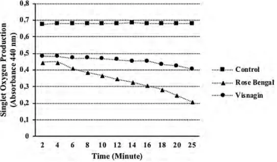
The decreasing in the absorbance indicates higher singlet oxygen production. Visnagin produced singlet oxygen by the RNO (p-Nitrosodimethylaniline) bleaching method. The singlet oxygen production of visnagin increased time dependently. After 24 min, the absorbance of visnagin was 0.404 ± 0.001 while the absorbance of Rose Bengal was almost 0.209 ± 0.001 (p < 0.05) (Fig. 1).
Doxorubicin has a broad-spectrum and widely used in the treatment of different types of cancer since the 1960s
Fig. 1 The singlet oxygen generation of visnagin and rose bengal by the RNO bleaching assay. Values are mean ± SEM,
n = 3, p < 0.05 0,8 ... . 0,7
··.::
:
·.
::
11
:
·.
:
:
:
:
•:
::::
·
:
•
·
·:::::•··::::
:
•:
:
:::
:·
·
:
:::
::
•
:
::::.
·
.::·.
:
:• ··
::
:
:·
.····
··
..
..
.
..
..
...
.
.
...
..
...
.
...
···
··
•
·
·
·
·
·
·
·
··
·
·
·•
·
···
·
··
..
..
..
...
....
....
.
.
...
..
:.:
,.
:
....
.
.
.
·--··:·
.
.
.
...
...
.
···
•
...
.
...
'...
.
..
•
·
··
··
·
·
·
.. .. . . ... ... , ... . , ,, , ,, , , , + . . . .. ••• .... •,·•U1•••...
•
·£•• ... u,t••••••••...
.
...
·
···-.
..
..
..
.
.
...
..
.
0,1 u••·--•••••t••••••••u .... • .. •••uu ... ••••• .. ••••u•t•••••u•
o---2 4 6 8 10 12 14 16 18 20 25 Time (Minute). ...
,
•
..
...
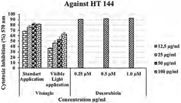
Control ... Rose Bengal ···--•··· Visnagin[22, 23]. We compared our results with doxorubicin, used as a positive control in this study. Visnagin showed cyto-toxic activity against HT 144 (human malign melanoma) cell lines in dose-dependent manner by the standard MTT assay, and by the illuminated MTT assay. In standard MTT assay, it showed 68.09 ± 1.06, 76.46 ± 2.39, 82.07 ± 1.94, and 80.93 ± 2.01% (p < 0.05) inhibitory activity against HT-144 cancer cell line at 12.5, 25, 50, and 100 µg/mL concentrations, respectively. Visnagin was more potent to inhibit the growth of melanoma cells, depending on the concentration by the standard MTT assay. When visible light was applied, the inhibition values reduced to 36.52 ± 19.3, 45.96 ± 4.10, 52.86 ± 5.08 and, 63.19 ± 2.94% (p < 0.05) at 12.5, 25, 50, and 100 µg/mL concentrations, respectively (Fig. 2). It has been observed that the light exposure had no desirable effect on the cytotoxicity by increasing concentra-tions. The inhibition value of visnagin was 8.06% at 100 µg/ mL against 3T3 cell lines; therefore, found nontoxic.
A few studies have been conducted on the cytotoxic and antitumor activity of visnagin, and its derivatives. Sayed et al. [24] have found that visnagin isolated from the aerial parts of Cyperus rotundus L., exhibited potent cytotoxic activity against the L5178y (mouse lymphoma) cell line. It induces 94% inhibition at 3 µg/mL concentration. The cytotoxic activity of visnagin, isolated from Cimicifugae
rhizoma (Ranunculaceae), against HL-60, MCF7, and
A549 cell lines were also evaluated. Visnagin was found to exhibit inhibitory activity against HL 60 cell line with the IC50 value of 18.1 µM. It has been observed that the cyto-toxic activity against other cell lines was weak [13]. The antitumor activity of some heteroaromatic benzofurans derived from visnagin against the HepG2 cancer cell line was compared with 5-fluorouracil and doxorubicin, known anticancer drugs. Some of the benzofuran derivatives exhibited a strong growth inhibition in a dose-dependent manner when compared to 5-fluorouracil, and doxorubicin [14]. Pakfetrat et al. [15] suggested that ethanolic extract
of A. visnaga has inhibitory effects on the cell growth of MCF-7 (human breast adenocarcinoma), and HeLa (human cervical cancer) cell lines. Visnagin isolated from Ammi
visnaga was evaluated against HeLa, HepG2, HCT 116,
and MCF-7 cell lines. The highest cytotoxic activity of visnagin exhibited activity against the HepG2 cell lines with the IC50 value of 10.9 + 0.68 µg/mL. Moreover, it
showed activity against HCT 116, MCF-7, and HeLa can-cer cell lines with IC50 values of 12.3 ± 0.94, 13.7 ± 0.942, and 35.5 ± 1.2 µg/mL, respectively [25].
In the present study, the ROS formed in HT 144 cells was detected by using DCFH-DA (2′,7′-dichloro-dihydro-fluorescein diacetate). The fluorescence of DCFH-DA, which was oxidized by ROS to DCF (dichlorofluores-cein), was measured at the excitation, and emission of 495/520 nm, respectively. Compared to H2O2, which was used as a standard, visnagin induced intracellular ROS production. Martelli et al. [11] reported that visnagin is a potent inducer of the intracellular reactive oxygen species (ROS) production, particularly for superoxide radicals and singlet oxygen.
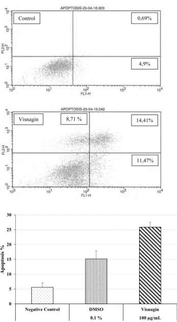
The dual staining with FITC Annexin V/PI identified the percentage of apoptosis by employing flow cytom-etry technique. The percentage of apoptosis in the HT 144 cell lines treated with 100 µg/mL of visnagin was 25.88%. The visnagin induced 11.47% early apoptosis (An+/PI-) and induced 14.41% late apoptosis (An+/PI+), and 8.71% of cells were necrotic (An-/PI+) (Fig. 3). Effect of visnagin on TNF-α production was determined by Human TNF-α ELISA kit. We found that visnagin has no effect on TNF-α secre-tion (Fig. 4). Our data are in line with the previous studies that apoptosis is induced due to the increased production of ROS in the apoptotic pathway, where death receptors play an essential role [26–29].
Our data demonstrated that visnagin has an antiprolif-erative effect on HT 144 cells in a dose-dependent man-ner. Visible light treatment inhibited more than 50% of cells at concentrations of 50, and 100 µg/mL. Visnagin induced apoptosis due to the intracellular generation of ROS. How-ever, it has no effect on TNF-α secretion. Thus, it can be said that other death receptors may induce the apoptosis.
Conclusions
Visnagin may be a potential lead for the treatment of mela-noma. Our data suggest that visnagin can inhibit the prolif-eration of human malignant melanoma cells by the induction of the intracellular ROS production, and the activation of the proapoptotic pathway. However, further molecular studies are required to investigate its effectiveness as an anti-cancer agent of visnagin.
Fig. 2 Cytotoxic activity of visnagin against HT 144 human
malig-nant melanoma cell line by the standard and illuminated MTT assays. Values are mean ± SEM, n = 3, p < 0.05
,.,
...
0 0 ~ Against HT 144 S1ondort Yisibl• 0.25 µM 0.5 µM Applicalion Lli,11 opp~Cl\liou Vlsnugin Ooiorubkiu C-Onrchtralion 11g/ml 1112,5 11glmL 1:175 µg/ml tlSO 11g/ml 01001111/hllAcknowledgements This study is a part of F.A.Ö’s Ph.D. thesis and was supported by the Research Fund of Istanbul University (Project Number: TP-19969). Prof. Dr. KerimanGünaydın would like to thank all staff of the Dr. Panjwani Center for Molecular Medicine and Drug Research (ICCBS), University of Karachi, Pakistan, for providing research facilities for her studies.
Compliance with ethical standards
Conflict of interest The authors declare that is no conflicts of interest associated with this publication.
References
1. Bamhill RL, Fandrey K, Levy MA, Mihm ML, Hyman B (1992) Angiogenesis and tumor progression of melanoma. Quantification of vascularity in melanocytic nevi and cutaneous malign mela-noma. Lab Invest 67:331–337
2. Watson M (2012) Drugs in clinical development for melanoma: summary and table. Pharm Med 26:171–183
3. Rogers HW, Weinstock MA, Hinckley MR, Feldman SR, Fleis-cher AB, Coldiron BM (2010) Incidence estimate of nonmela-noma skin cancer in the United States, 2006. Arch Dermatol 146:283–287
4. Matsuo Y, Kamitani T (2010) Parkinson’s disease-related protein, a-synuclein, in malignant melanoma. PLoS ONE 5:1–8
5. Disse M, Reich H, Lee PK, Schram SS (2016) A review of the association between parkinson disease and malignant melanoma. Dermatol Surg 42:141–146
6. Thompson JF, Scolyer RA, Kefford RF (2005) Cutaneous mela-noma. Lancet 365:687–701
7. Batistatou A, Cook MG, Massi D (2009) Histopathology report of cutaneous melanoma and sentinel lymph node in Europe: a web-based survey by the Dermatopathology Working Group of the European Society of Pathology. Virchows Arch 454:505–511 8. Soengas MS, Lowe SW (2003) Apoptosis and melanoma
chem-oresistance. Oncogene 22:3138–3151
9. Gunaydin K, Beyazit N (2004) The chemical investigations on the ripe fruits of Ammi visnaga (Lam.) Lamarck growing in Turkey. Nat Prod Res 18:169–175
10. Rauwald HW, Brehm O, Odenthal KP (1994) The Involvement of a Ca2+ channel blocking mode of action in the pharmacology of
Ammi visnaga fruits. Planta Med 60:101–105
11. Martelli P, Bovalini L, Fe S, Franchi GG, Bari M (1985) Active oxygen forms in photoreaction between DNA and furanochr-omones khellin and visnagin. FEBS Lett 189:255–257
12. Chen X, Kagan J (1993) Photosensitized cleavage and cross-linking of pBR322 DNA with khellin and visnagin. J Photochem Photobiol B 20:183–189
13. Cuong TD, Lim CJ, Kim SW, Park JE, Hung TM, Min BS (2011) Isolation of compounds from Cimicifugae Rhizoma and their cyto-toxic activity. Nat Prod Sci 17:80–84
14. El-Nakkady SS, Roaiah HF, El-Serwy WS, Soliman AM, El-Moez SIA, Abdel-Rahman AA-H (2012) Antitumor and antimicrobial activities of some hetero aromatic benzofurans derived from natu-rally occurring visnagin. Acta Pol Pharm 69:645–655
15. Pakfetrat H, Nemati N, Shiravi A (2015) Cytotoxicity effects of
Ammi visnaga extract on Hela and MCF-7 cancer cell line. Anim
Biol 7:25–33
16. Kraljic I, Mohsni S (1978) A new method for the detection of singlet oxygen in aqueous solutions. Photochem Photobiol 28:577–581 0 5 10 15 20 25 30
Negative Control DMSO Visnagin
0.1 % 100 µg/mL Apoptosis % 4,9% 11,47% 14,41% 0,69% Control Visnagin 8,71 %
Fig. 3 Flow cytometry detection of apoptosis in HT 144 cell lines treated with visnagin
Fig. 4 TNF-⍺ secretion (pg/ml) of visnagin. Values are mean ± SEM,
n = 3, p < 0.05 ~ g 2 +"'
2~
-g 0 g ,oo ~ g ., g :;~ 2-g 0 g ,oo !000 1800 ~ 1600 ~ 1400 ~1200.,
.~ 1000£
soo C !:: 600 Ca
400 200 0 -+- -APOPTOSIS-25-04-16.000 10 1 1o" Fl.,1-H APOPTOSIS-25-04· 16.042 101 ,o" Fl..l·H 'l'NF~a Secretion Cnnlrol 1o-' ,o" Visno~Jn 100 µGfmL 104 10'17. Aydoğmuş-Öztürk F, Günaydin K, Öztürk M, Jahan H, Duru ME, Choudhary MI (2018) Effect of Sideritis leptoclada against HT-144 human malignant melanoma. Melanoma Res 28:502–509 18. Wu D, Yotnda P (2011) Production and detection of reactive
oxy-gen species (ROS) in cancer. J Vis Exp 57:1–4
19. Jahan H, Choudhary MI, Shah Z, Khan KM (2017) Derivatives of 6-nitrobenzimidazole inhibit fructose-mediated protein glycation and intracellular reactive oxygen species production. Med Chem 13:577–584
20. Vermes I, Haanen C, Steffensnakken H, Reutelingsperger C (1995) A novel assay for apoptosis-flow cytometric detection of phosphatidylserine expression on early apoptotic cells using fluorescein-labeled annexin-V. J Immunol Methods 184:39–51 21. Iram N, Mildner M, Prior M, Petzelbauer P, Fiala C, Hacker S,
Schoppl A, Tschachler E, Elbe-Burger A (2012) Age-related changes in expression and function of Toll-like receptors in human skin. Development 139:4210–4219
22. Bristow MR, Mason JW, Billingham ME, Daniels JR (1978) Doxorubicin cardiomyopathy: evaluation by phonocardiography, endomyocardial biopsy, and cardiac catheterization. Ann Intern Med 88:168–175
23. Xi L (2016) Visnagin—a new protectant against doxorubicin car-diotoxicity? Inhibition of mitochondrial malate dehydrogenase 2 (MDH2) and beyond. Ann Transl Med 4:65–69
24. Sayed H, Mohamed MH, Farag SF, Mohamed GA, Proksch P (2007) A New Steroid Glycoside and Furochromones from
Cype-rus rotundus L. Nat Prod Res 21:343–350
25. Beltagy AM, Beltagy DM (2015) Chemical composition of Ammi
visnaga L. and new cytotoxic activity of its constituents khellin
and visnagin. J Pharm Sci Res 7:285–291
26. Circu ML, Aw TY (2010) Reactive oxygen species, cellular redox system and apoptosis. Free Radic Biol Med 48:749–762 27. Matthews N, Neale ML, Jackson SK, Stark JM (1987) Tumour
cell killing by tumour necrosis factor: inhibition by anaerobic conditions, free-radical scavengers and inhibitors of arachidonate metabolism. Immunology 62:153–155
28. Larrick JW, Wright SC (1990) Cytotoxic mechanism of tumor necrosis factor-alpha. FASEB J 4:3215–3223
29. Shakibaei M, Schulze-Tanzil G, Takada Y, Aggarwal BB (2005) Redox regulation of apoptosis by members of the TNF superfam-ily. Antioxid Redox Signal 7:482–496
Publisher’s Note Springer Nature remains neutral with regard to