IDENTIFICATION OF NOVEL GENETIC ELEMENTS
CONTROLLING TRANSCRIPTIONAL REGULATION OF
THE HUMAN Na
+/I
-SYMPORTER (NIS) GENE
A THESIS SUBMITTED TO
THE DEPARTMENT OF MOLECULAR BIOLOGY AND GENETICS AND THE INSTITUTE OF ENGINEERING AND SCIENCE OF
BILKENT UNIVERSITY
IN PARTIAL FULFILLMENT OF THE REQUIREMENTS FOR THE DEGREE OF DOCTOR OF PHILOSOPHY
BY
HANI ALOTAIBI AUGUST 2006
ii
I certify that I have read this thesis and that in my opinion it is fully adequate, in scope and in quality, as a thesis for the degree of Doctor of Philosophy.
Assist. Prof. Dr. Tamer Yağcı I certify that I have read this thesis and that in my opinion it is fully adequate, in scope and in quality, as a thesis for the degree of Doctor of Philosophy.
Assist. Prof. Dr. Elif Ayşe Erson
I certify that I have read this thesis and that in my opinion it is fully adequate, in scope and in quality, as a thesis for the degree of Doctor of Philosophy.
Prof. Dr. Ediz Demirpençe
I certify that I have read this thesis and that in my opinion it is fully adequate, in scope and in quality, as a thesis for the degree of Doctor of Philosophy.
Assist. Prof. Dr. Uygar H. Tazebay
I certify that I have read this thesis and that in my opinion it is fully adequate, in scope and in quality, as a thesis for the degree of Doctor of Philosophy.
Prof. Dr. Mehmet Öztürk
Approved for the Institute of Engineering and Science
Director of Institute of Engineering and Science
iii
ABSTRACT
IDENTIFICATION OF NOVEL GENETIC ELEMENTS CONTROLLING
TRANSCRIPTIONAL REGULATION OF THE HUMAN Na+/I
-SYMPORTER (NIS) GENE
Hani Alotaibi
Ph.D. in Molecular Biology and Genetics Supervisor: Assit Prof. Dr. Uygar H. Tazebay
August 2006, 91 Pages
The function of sodium iodide symporter (NIS) in mammary gland epithelial cells is essential for the accumulation of iodide in mother’s milk, which is the first source of iodide for the synthesis of thyroid hormones in the newborn. In addition to the lactating mammary gland, NIS expression has been also detected in breast tumors. Several hormones and ligands have been implicated in the functional expression of NIS in the mammary gland and breast cancer cell line models but the molecular determinants governing this expression are not yet identified. In this study we aimed to identify cis- and trans-acting elements regulating NIS expression in the breast cancer cell line MCF-7 in response to all-trans-retinoic acid (tRA), and to assess the possible role of 17-β-estradiol (E2) in regulating the expression of NIS. Using comparative bioinformatics, we have identified several regions that were conserved in human, mouse and rat in the sequences flanking and including the NIS gene. By using luciferase reporter assays, we have established that conserved clusters 3 and 4 respond to tRA in MCF-7. We have also shown that putative retinoic acid response elements controlling tRA-induced NIS expression in MCF-7 are located in the first intron of this gene. This tRA-responsive NIS expression was also correlated with the estrogen receptor status of mammary gland cell lines and we investigated roles of ERα in the regulation of NIS expression. We showed that the suppression of endogenous ERα by RNA interference resulted in down-regulation of both basal and tRA-induced NIS expression in MCF-7, furthermore, we have also shown that (E2) is capable of up-regulating NIS expression in MCF-7. In the ERα negative cell line MDA-MB-231, re-introduction of ERα resulted in NIS expression in a ligand independent manner. The role of ERα in the regulation of NIS expression was supported by the identification of an estrogen response element (ERE) in the promoter of NIS, this ERE was conserved in human, mouse and rat. We have also showed that this ERE could respond to E2 stimulation, and that ERα occupies the NIS promoter by binding to this novel element in vivo. These results indicate that E2 and ERα contribute to the regulation of NIS in the breast cancer cell line MCF-7.
iv
ÖZET
İNSAN Na+/I- SİMPORTIR GENİ TRANSKRİPSİYONUNUN DÜZENLENMESİNİ
KONTROL EDEN YENİ GENETİK ELEMANLARIN BELİRLENMESİ
Hani Alotaibi
Moleküler Biyoloji ve Genetik Doktorası Tez Yöneticisi: Yard. Doç. Dr. Uygar H. Tazebay
Ağustos 2006, 91 Sayfa
Na+/I- taşıyıcı protein (Na+/I- Symporter protein, NIS) aktivitesi anne sütüne iyot taşınması ve dolayısı ile yeni doğan bebeğin tiroid hormonları üretebilmesi için şarttır. Süt üreten meme dokusuna ek olarak, artış gösteren NIS ifadesi meme tümörlerinde de teşhis edilmiştir. Birkaç hormon ve ligandın meme hücrelerinde NIS geni işlevsel ifadesinde etkili olduğu tesbit edilmiş olsa da, bu ifadeye yol açan moleküler faktörler, veya genetik belirleyiciler henüz tamamen belirlenmemiştir. Bu çalışmada, MCF-7 insan meme kanseri hücre hattında all-trans-retinoik asit (tRA) muamelesi ile NIS geni ifadesine yol açan cis- ve trans- etkili faktörleri belirledik, ve 17-β-estradiol (E2)’ün NIS geni regülasyonuna etki mekanizmalarını inceledik. Çalışma kapsamında, önce karşılaştırmalı biyoenformatik yöntemleri kullanarak insan sıçan ve farede NIS geninde korunmuş ekzon dışı bölgeleri belirledik. Daha sonra bu bölgelerden cis-etkili faktör potansiyeli olanların lüsiferaz genini aktive edeceği deney düzenekleri ile korunmuş bölge 3 ve 4’ün MCF-7 hücrelerinde tRA ligandına yanıt verdiğini gösterdik. Ayrıca, tRA’ya yanıtı kontrol eden elemanların NIS geninin ilk intronu içerisinde olabileceğine dair veriler elde ettik. Bunlardan başka, NIS gen ifadesi tRA yanıtının hücrelerdeki östrojen reseptör alfa (ERα) varlığı ile korelasyon gösterdiğini ortaya koyarak, bu faktörün NIS gen ifadesini kontrol ederken girdiği moleküler etkileşimleri gen dizisi düzeyinde belirledik. Çalışmalarımızda, RNA interferans metodu kullanarak MCF-7 hücrelerinde ERα gen düzeyini düşürdüğümüzde, hem bazal NIS ifadesinin hem de tRA ile indüklenen NIS geni ifadesinin düştüğünü gördük. Aynı zamanda, E2 ligandının NIS gen ifadesini artırıcı etkisini gösterdik. Ayrıca, ERα ifadesi olmayan MDA-MB-231 meme kanseri hücre hatlarına sonradan ERα geni verdiğimizde NIS ifadesinin E2’den bağımsız olarak arttığını görerek, bu artışa yol açan kontrol mekanizmasını detaylı olarak inceledik. Bu analizlerde, NIS geni kontrol bölgesinde fare ve sıçanda da korunmuş bir ERα yanıt elemanı (Estrogen Response Element) olduğunu, bu ERE’nin E2’ye yanıt verdiğini, ve ERα faktörünün buraya hem in vitro hem de in vivo şartlarda bağlandığını gösterdik. Bu sonuçlar, ERα ve E2’nin meme kanseri hücrelerinde NIS geni transkripsiyonunu kontrol ettiğini ortaya koymuştur.
v
DEDICATION
To My Loving and Caring Wife
Neslihan
vi
ACKNOWLEDGEMENTS
First of all, I would like to express my gratitude to my supervisor, Assist. Prof. Uygar Tazebay, for his guidance, support and for his friendship.
I would like to thank Prof. Mehmet Öztürk for his interest and guidance throughout this project.
I would like to thank Prof. Roberto Di Lauro for providing the plasmid pGL3E1bLuc, the cell lines HeLa and FRTL-5 and for valuable discussions. Many thanks also to Prof. Domenico Salvatore, who provided the cell line MCF-7 and for interest in our work. The cell lines MDA-MB-231 and MDA-66 were kindly provided by Prof. Frank Gannon and we are grateful for that.
I would like also to thank all the members (past and present) of the Department of Molecular Biology and Genetics at Bilkent University for wonderful atmosphere and great friendship, especially Elif Yaman for valuable technical support. Many thanks also to Prof. Ediz Demirpençe for continuous support.
Last but not least, I would like to thank my family for their moral support and for their patience.
This project was supported by an EMBO short-term fellowship, and TÜBİTAK grant 104T231.
vii
TABLE OF CONTENTS
ABSTRACT... III ÖZET ...IV DEDICATION ... V ACKNOWLEDGEMENTS ...VI TABLE OF CONTENTS...VII LIST OF TABLES ... X LIST OF FIGURES ...XI1. INTRODUCTION ... 1
1.1. Biological Significance of Iodide Transport ... 1
1.1.1. Structure of the Na+/I- symporter ... 3
1.1.2. Expression of the Na+/I- symporter ... 4
1.1.3. Function of the Na+/I- symporter... 5
1.1.3.1. Iodide requirement in hormone biosynthesis and development... 6
1.1.4. Significance of iodide transport in the clinic ... 7
1.2. Regulation of Na+/I- Symporter Gene Expression ... 9
1.2.1. Regulation in thyroid gland... 9
1.2.1.1. NIS upstream enhancer ... 12
1.2.1.2. Regulation of NIS by retinoids in the thyroid... 13
1.2.2. Regulation of NIS in mammary gland ... 15
1.2.3. Regulation of NIS in human mammary carcinoma cell line MCF-7... 16
1.3. Aim of This Study... 18
1.3.1. Rationale ... 19
viii
2.1. Sequence Information and Databases ... 20
2.1.1. PCR amplification of conserved regions ... 20
2.2. Plasmids ... 22
2.2.1. Reporter constructs ... 22
2.2.1.1. Site directed mutagenesis... 23
2.2.2. Expression vectors ... 24 2.3. Cell Culture ... 24 2.3.1. Cell lines ... 24 2.3.2. Growth media... 25 2.3.3. Hormone induction ... 25 2.3.3.1. tRA induction... 25 2.3.3.2. E2 induction ... 25 2.3.4. Transfection ... 26
2.3.4.1. Transient transfection with ERα ... 26
2.4. Luciferase Reporter Assay ... 26
2.5. RNA Isolation ... 27
2.5.1. cDNA synthesis, RT-PCR... 27
2.6. Protein Isolation ... 28
2.6.1. Western blot analysis ... 28
2.7. Suppression of ERα by shRNA... 29
2.8. Electrophoretic Mobility Shift Assay (EMSA)... 29
2.8.1. Nuclear extract preparation... 31
2.8.2. Oligo-nucleotides ... 32
2.8.2.1. Biotin labeling... 32
2.9. Chromatin Immunoprecipitation (ChIP)... 33
3. RESULTS ... 35
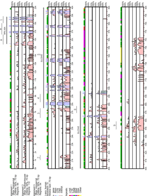
3.1. Comparative Genomics Identifies Potential cis-acting Elements Controlling NIS transcription ... 35
ix
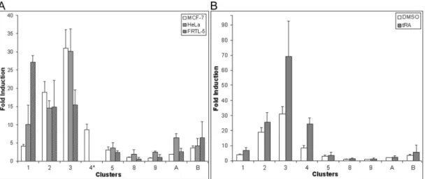
3.1.2. Conserved clusters 3 and 4 respond to tRA in MCF-7 ... 38
3.1.3. tRA Response in conserved cluster 3 is not attributable to a single retinoic acid responsive element ... 40
3.1.3.1. Conserved region 3-4 in NIS promoter accounts for most of NIS transcription ... 41
3.1.4. 5’UTR sequence has an up-regulatory effect on NIS transcription but does not increase the tRA responsiveness... 42
3.1.5. Combining clusters 3 and 4 has a synergistic tRA response in the absence of UTR sequences ... 44
3.1.6. Nkx-2.5 reduces the transcriptional potential of Cl.3 in HeLa cells... 46
3.2. Estrogen Receptor-α Activates Transcription of the Mammary Gland Sodium Iodide Symporter... 47
3.2.1. tRA-responsive NIS expression is correlated with the presence of a functional ERα ... 47
3.2.2. E2 up-regulates tRA-induced NIS expression in MCF-7 cells ... 49
3.2.3. Suppression of ERα by shRNA down-regulates NIS expression ... 51
3.2.4. Ectopic ERα expression in MDA-MB-231 up-regulates NIS expression 52 3.2.5. Identification of a novel non-canonical ERE in NIS promoter... 56
3.2.6. Physical interaction of ERα with NIS promoter in vivo ... 58
3.2.7. Physical interaction of ERα with NIS ERE in vitro in response to E2 .... 59
3.2.8. ERE mutations in the context of cluster 3 did not affect tRA responsiveness... 60
3.2.9. E2 up-regulates NIS expression in MCF-7 in a dose dependant manner 63 4. DISCUSSION ... 65
4.1. tRA Response Elements in Thyroid Cell Lines and in MCF-7 are Distinct ... 65
4.2. Estradiol and ERα are Involved in the Transcription of NIS... 70
4.3. Conclusion ... 78
x
LIST OF TABLES
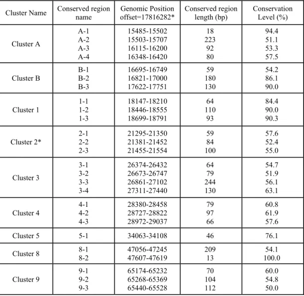
Table 2.1. Conserved region clusters used for PCR amplification. ... 21
Table 2.2. EMSA Binding reaction preparation chart... 30
Table 2.3. Oligo-nucleotide sequences used in EMSA reactions. ... 32
xi
LIST OF FIGURES
Figure 1.1. The current model for the secondary structure of NIS ... 4 Figure 1.2. An illustration of thyroid hormone bio-synthesis in thyroid follicular
cells. ... 7 Figure 1.3. A representation of the factors involved in the regulation of NIS
expression in the thyroid gland. ... 11 Figure 1.4. Regulatory elements involved in Nkx-2.5 dependant transcription of NIS
in MCF-7... 18 Figure 3.1. VISTA plot of conservation levels in a 90 kbp genomic DNA in human,
mouse and rat. ... 36 Figure 3.2. tRA dependant expression of NIS in MCF-7. ... 38 Figure 3.3. Conserved clusters 3 and 4 respond to tRA in MCF-7... 39 Figure 3.4. The retinoic acid responsive element in Cl.3 is not the major site for tRA
response in MCF-7... 41 Figure 3.5. Conserved region 3-4 in cluster 3 is essential for proper transcriptional
activation. ... 43 Figure 3.6. Combining clusters 3 and 4 has a synergistic tRA response in the absence of the UTR sequence... 45 Figure 3.7. Nkx-2.5 has a negative effect on luciferase gene expression in Cl.3 in
HeLa cells... 47 Figure 3.8. ERα positivity in mammary gland cell lines is correlated with tRA
responsive NIS expression. ... 48 Figure 3.9. Estradiol has an up-regulatory effect on the tRA-induced NIS expression
in MCF-7... 50 Figure 3.10. Suppression of ERα by shRNA down-regulates NIS expression in
xii
Figure 3.11. Transient expression of ERα in MDA-MB-231 cells leads to increased NIS expression in a ligand independent manner... 53 Figure 3.12. Stable expression of ERα in MDA-MB-231 cells leads to a higher basal
expression of NIS in a ligand independent manner. ... 54 Figure 3.13. Growth media composition affects basal transcription of NIS both in
MDA-66 and in MCF-7 cells. ... 55 Figure 3.14. A putative novel ERE sequence is located in close proximity of NIS
TATA box element in the promoter region... 57 Figure 3.15. The novel ERE element in NIS promoter activates transcription in
functional assays in response to E2... 58 Figure 3.16. ERα occupies the promoter of NIS in vivo. ... 59 Figure 3.17. E2 stimulates the interaction of ERα with the novel NIS ERE in gel
retardation assays. ... 61 Figure 3.18. Mutants of the novel ERE sequence in the context of Cl.3 did not affect
luciferase gene expression in response to tRA stimulation... 62 Figure 3.19. E2 up-regulates NIS expression in MCF-7 in a dose-dependant manner.
... 64 Figure 4.1. Proposed model of interactions of cis- and trans-acting factors involved
1
1. INTRODUCTION
1.1. Biological Significance of Iodide Transport
Iodine is the heaviest element normally metabolized in biological material (Wolff, 1964), and iodide is a limiting element for the synthesis of hormones, which are essential for proper growth and development of many organs (Stubbe et al., 1986). Because of the scarcity of this element (Wolff, 1964), several organisms evolved with a remarkable mechanism of collecting this rare element. One of the first organs to be known for its iodide concentrating mechanism is the thyroid gland (Carrasco, 1993). Moreover, indications to the presence of iodide concentrating mechanisms in other organs in human and animals date back to as early as 1856 when Claude Bernard described the presence of iodine in the salivary gland, and in 1859 as reports describing the presence of iodine in the milk of nursing mothers were available, as well as in hair, skin, ovaries, placenta, kidney, stomach and intestine [reviewed in (Brown-Grant, 1961)]. The availability of radio isotopes by 1945 improved the techniques by which lower amounts of iodide could be measured, and thus provided a better understanding regarding organs in which the iodide transporter was functional (Brown-Grant, 1961).
The expression of an iodide transporter in the thyroid has long been known, based on the fact that thyroid hormone synthesis requires the presence of an iodide concentrating mechanism in this organ (Wolff, 1964). The existence of mRNA capable of encoding a functional iodide transporter was illustrated by the expression of poly A RNA isolated from the thyroid cell line FRTL-5 in the oocytes of X. laevis, which led eventually to the cloning of the cDNA of the rat sodium iodide symporter (Vilijn and Carrasco, 1989; Dai et al., 1996). The cloning of the sodium iodide symporter (see below) provided an invaluable tool for the analysis of NIS mRNA (and later, protein) expression in different tissues or organs. Since then, NIS mRNA
2
expression was demonstrated in several other organs of higher vertebrates. Using RT-PCR, hNIS mRNA was detected in the thyroid, salivary gland, parotid gland, submandibular gland, pituitary gland, pancreas, testis, mammary gland, gastric mucosa, prostate, ovaries, kidney, and placenta (Smanik et al., 1997; Ajjan et al., 1998a; Spitzweg et al., 1998; Mitchell et al., 2001; Spitzweg et al., 2001). By Northern blot analysis, the expression of the human NIS mRNA was observed in thyroid, and in the parotid glands (Spitzweg et al., 1998).
The sodium iodide (Na+/I-) symporter (NIS, the official nomenclature given by HUGO gene nomenclature committee is SLC5A5) is the fifth member of the sodium/solute carrier family 5 (SLC5A), a family of proteins that mediate the active transport of a variety of molecules including iodide. Twelve members of this family have been described so far, including 7 members involved in the transport of glucose (members 1, 2, 4 and 9-12). This family belongs to the solute carrier super-family which includes 45 solute carrier families and one solute carrier organic anion transporter family (Wain et al., 2002; Wain et al., 2004).
The rat iodide transporter was the first to be cloned as a result of a functional screening of a human cDNA library from FRTL-5, a rat thyroid cell line, in Xenopus laevis oocytes (Dai et al., 1996). In the same year another report described the cloning of the human iodide transporter using cDNA prepared from human papillary carcinoma tissue; they amplified the hNIS cDNA fragment using primers derived from the nucleotide sequence of the rat mRNA of rNIS (Smanik et al., 1996). In the year 2000, Tazebay et al., showed that active transport of iodide in the lactating mammary gland in mice is mediated by the transporter encoded by the same gene (Tazebay et al., 2000). Later, the mouse sodium iodide symporter was cloned from thyroid and lactating mammary gland tissues (Perron et al., 2001; Pinke et al., 2001). The hNIS cDNA encodes a 643-amino acid protein with 84% homology to the rat and the mouse genes. This gene encoding the human iodide transporter was mapped to chromosomal location 19p13.2-p12 using fluorescence in situ hybridization (Smanik et al., 1997). The coding sequence of the hNIS gene is encoded by 15 exons, and the exon-intron junctions were also described (Smanik et al., 1997).
3
1.1.1. Structure of the Na+/I- symporter
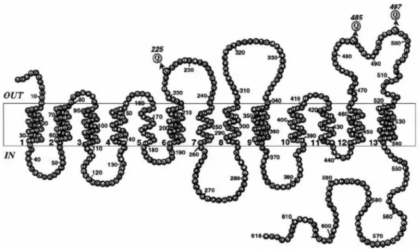
The first clues regarding the secondary structure of NIS came from the first report, in which the authors described the mRNA sequence of this gene (Dai et al., 1996). In that report, analysis of the predicted protein sequence based on hydropathy analysis and secondary structure algorithms suggested that the cloned cDNA encodes a protein with 12 putative trans-membrane domains, and due to its hydrophilic composition, the carboxy-terminus was placed at the cytoplasmic side of the plasma membrane (Dai et al., 1996).
The generation of an anti-NIS antibody allowed researchers to further analyze the structure and the post-translational modifications that lead to the mature functional protein. The cytoplasmic location of the carboxy terminus of NIS was confirmed using indirect immunofluorescence in permeabilized FRTL-5 cells (Levy et al., 1997). Moreover, using this antibody, membrane fractions from FRTL-5 cells or COS cells transfected with NIS cDNA revealed a prominent immunoreactive polypeptide with a molecular weight of about 87 kDa, different from the predicted mass (65 kDa). This difference was attributed to post-translational modifications at 3 putative Asparagine (Asn) residues at positions 225, 485 and 497 by N-linked glycosylation (Levy et al., 1997; Paire et al., 1997), two of which (residues 485 and 497) were located in the predicted sixth extra-cellular loop. Site directed mutagenesis of these putative glycosylation sites demonstrated that NIS is processed at three Asn sites instead of two; thus placing the third glycosylation site (previously predicted in the third intracellular loop at position 225) in the cytoplasmic side of the membrane, because N-linked glycosylation occur at exposed extracellular facing sites in the endoplasmic reticulum during protein processing (Levy et al., 1998).
Moreover, by using an amino-terminus FLAG-tagged NIS, engineered by site directed mutagenesis, Levy et al. (1998) showed that non-permeabilized as well as permeabilized cells were stained by anti-FLAG antibodies, an observation suggesting that the amino terminus of NIS is located at the extra-cellular side of the plasma membrane (Levy et al., 1998). Based on that, the authors suggested a revised model for the secondary structure of NIS, in which the amino terminus faces extracellularly.
4
According to this current model (Figure 1.1), NIS is an intrinsic membrane protein, with 13 transmembrane helices (Levy et al., 1998).
Figure 1.1. The current model for the secondary structure of NIS. In this revised model, NIS contains 13 transmembrane helices. According to this model, the hydrophilic loop containing Asn225 and the NH2 terminus faces extracellularly. All three N-linked glycosylation consensus sequences are indicated with Qs at positions 225, 485, and 497. Picture adapted from Levy et al. (1998).
1.1.2. Expression of the Na+/I- symporter
Ajjan et al. (1998) analyzed the expression of NIS in rat extra-thyroidal tissues by quantitative RT-PCR and they showed NIS transcripts in the stomach and in the lactating mammary gland and to a less extent in the small intestine (Ajjan et al., 1998b). Expression of rNIS in the stomach was also demonstrated by another group (Kotani et al., 1998). They compared the expression level of rNIS mRNA and protein in the rat stomach and the rat thyroid cell line FRTL-5, results of which revealed lower NIS protein in stomach in contrast to the level in FRTL-5, although
5
the mRNA expression level in the stomach was much higher than that of the thyroid cell line, this observation was reasoned to the degradation of the immature gastric NIS protein (Kotani et al., 1998).
Tazebay et al. (2000) analyzed the expression of NIS protein in the mammary gland of mice in various physiological stages. A functional expression of mNIS protein starts at mid pregnancy (day 11 of a 19 day gestation period in mice), and reaches highest levels towards the end of pregnancy (day 18), which continues during lactation, such expression of mNIS protein was absent from the mammary gland of nubile mice (Tazebay et al., 2000). They also provided evidence of an increased NIS expression in over 80% of human breast cancer samples compared to no expression in normal tissues from reductive mammoplasties, as well as in experimental mammary tumors induced by an activated Ras and Neu oncogenes under the control of the Murine Mammary Tumor Virus (MMTV) promoter in transgenic mice (Tazebay et al., 2000).
In a more recent study (Wapnir et al., 2003), NIS expression in a wide variety of normal and cancerous tissues was analyzed by immunohistochemical staining using an anti-NIS antibody. From 371 breast specimens (conventional whole tissue sections) analyzed, NIS expression was confirmed in 76% of invasive breast carcinoma and 88% of ductal carcinoma in situ samples, whereas 87% of normal counterparts were reported negative. NIS expression was also observed in a wide range of tumors of other origins, including prostate (74%), ovary (73%), lung (65%), colon (62.6%), andendometrium (56%). NIS protein was present in 75% of benign thyroidlesions and 73% of thyroid cancers (Wapnir et al., 2003).
Expression of NIS mRNA in mouse tissues has been described by RT-PCR as well; high levels of mNIS expression were reported in the thyroid, stomach and in the lactating mammary gland, but with lower levels in small intestine, skin, brain, testis, mammary gland, ovary, spleen and in prostate gland (Perron et al., 2001).
1.1.3. Function of the Na+/I- symporter
As the name implies, sodium/iodide symporter is the transporter responsible for the active transport of iodide from the blood stream into cells. This intra-cellular iodide
6
is then used in different physiological processes. This uptake process is sodium dependant (Bagchi and Fawcett, 1973), and NIS actively co-transports Na+ and I -with a stoichiometry of 2 Na+:1 I- (Eskandari et al., 1997). The sodium-driven transport of iodide is maintained by an ouabain sensitive sodium-potassium ATPase, which provides the energy required for this process [(Wolff and Halmi, 1963), and for a review see (Baker and Morris, 2004)]. NIS is also capable of transporting other ions with less affinity, including ClO3-, SCN-, SeCN-, NO3-, Br-, BF4-, IO4-, BrO3-,
but not perchlorate (ClO4-) (Eskandari et al., 1997). In fact, ClO4- is a well known
competitive inhibitor of iodide transport as well as NO3-, BF4-, SCN-,
2,4-dinitrophenol, and cardiac glycosides (Wolff, 1964; Carrasco, 1993; Eskandari et al., 1997).
1.1.3.1. Iodide requirement in hormone biosynthesis and development
It has been known for decades that thyroid hormone biosynthesis requires the presence of inorganic iodide, and the presence of an iodide trapping system is the first limiting step in this process (Wolff, 1964; Carrasco, 1993; Baker and Morris, 2004). Iodide is an essential constituent of the thyroid hormones triiodothyronine (T3) and thyroxin (T4). NIS located at the basolateral membrane of thyrocytes (Chambard et al., 1983), transports iodide into the cells, which is then transported across the apical membrane into the follicular lumen (or colloid) by different anion transporters, such as pendrin and apical iodide transporter (Bidart et al., 2000; Rodriguez et al., 2002). In the colloid, thyroid peroxidase covalently incorporates transported iodide into tyrosine residues of the thyroid hormone precursor, thyroglobulin, in a process known as organification (Igo et al., 1964; Carrasco, 1993). Iodinated thyroglobulin is then endocytosed, followed by phagolysosomal hydrolysis of the iodinated thyroglobulin releasing the thyroid hormones, which are then released into the blood stream, this process in mainly controlled by the thyroid stimulating hormone (Carrasco, 1993). A summary of these events is illustrated in Figure 1.2.
Thyroid hormones are essential for proper growth and maturation of skeletal muscles, nervous system and lungs of the fetus and the developing newborn (Stubbe et al., 1986).
7
As mentioned before, NIS is also expressed in the lactating mammary gland, and functions to secrete iodide into mother’s milk (Tazebay et al., 2000), thus providing the first source of thyroid hormones to the newborn. To date, the biological relevance of NIS expression and iodide transport in organs other than the thyroid and the lactating mammary gland is not clear and further research is required to reveal the significance of iodide in the physiological processes in these organ systems.
Figure 1.2. An illustration of thyroid hormone bio-synthesis in thyroid follicular cells. TSH stimulates the sodium dependent iodide transport into thyroid follicular cells, this process is maintained by the sodium-potassium ATPase, providing the energy required for this process. Iodide uptaken into follicular cells is transported to colloid by pendrin. Placed at the basolateral membrane, thyroperoxidase (TPO) catalyzes the incorporation of iodide into tyrosine residues of thyroglobulin (Tg) to produce thyroid hormones T3 and T4. Iodinated Tg will be endophagocytosed into the cells and the hormones will be released into blood stream. Figure from Baker and Morris (2004).
1.1.4. Significance of iodide transport in the clinic
The ability of the thyroid gland to transport iodide is an absolute requirement for the synthesis of thyroid hormones. This characteristic, mediated by functional NIS
8
expression is also observed in abnormalities of the thyroid such as thyroid nodules and thyroid cancer (Wollman and Reed, 1960). This function of NIS – as the key transporter of iodide – has emerged as a valuable tool for the diagnosis and treatment of thyroid cancer. For decades, radioactive iodide played a major therapeutic role in the postoperative management of differentiated thyroid carcinoma (DTC) because of its effectiveness to ablate remnant thyroid tissue and metastases. Moreover, the degree and pattern of iodide accumulation in the thyroid, as revealed by scintigraphic imaging, is used as an aid in the differential diagnosis of thyroid nodules.
This characteristic of the thyroid gland, i.e. expression of NIS that is capable of iodide uptake, has been reported in the mammary gland during lactation and in breast cancer (Cho et al., 2000; Tazebay et al., 2000). NIS expression was also observed in a high percentage of human breast cancer specimens (with various pathologies) in contrast to no expression in normal tissues obtained from reductive mammoplasties (Tazebay et al., 2000; Wapnir et al., 2003). These results suggest that radioiodide administration may be effective as an adjuvant to surgical treatment of primary breast cancer, and/or as a tool in the diagnosis and treatment of metastatic disease. A major characteristic of the healthy thyroid gland is that it exhibits NIS activity for life, within boundaries set by thyroid regulatory factors such as thyroid stimulating hormone and iodide itself (Eng et al., 1999; Riedel et al., 2001). In contrast, the potential effectiveness of radioiodide therapy in breast cancer depends on whether NIS becomes functionally expressed in malignant mammary cells, given that it is not functionally expressed in healthy cells, except during pregnancy and lactation. It is notable that a single transport protein – NIS – catalyzes the same fundamental process – active Na+-dependent I- transport – in both tissues, but is regulated differently in each of them. These differences affect not only how NIS functions under normal conditions, but also how it can play a role in cancer management in both tissues.
Unlike the thyroid gland (being the only known organ to incorporate iodide into thyroid proteins), in mammary gland cells iodide is secreted into the milk, and this difference creates a challenge for the application of an effective dose of radioiodide for the treatment of malignant cells of the breast (Zuckier et al., 2001).
9
Clearly, alternative strategies for detection of micrometastatic disease and for more effective and targeted systemic therapies are needed to improve survival in breast cancer, which remains the leading cause of cancer deaths in women (ages of 20-59) in developed countries (Greenlee et al., 2000).
Recently, several researchers have reported the possible use of radioiodide for the treatment of cancers by forced expression of NIS in tumors of several origins, such as in prostate cancer (Spitzweg et al., 2000; La Perle et al., 2002), hepatoma (Haberkorn et al., 2001), glioma (Cho et al., 2002), neuroendocrine tumor cells (Schipper et al., 2003), head and neck squamous cell carcinoma (Gaut et al., 2004), colon cancer (Mitrofanova et al., 2005; Scholz et al., 2005), pancreatic tumors (Dwyer et al., 2006a), and in ovarian tumor xenografts (Dwyer et al., 2006b).
Insofar, as NIS is functionally expressed to a sufficient degree in cancerous cells, whether of thyroid, breast, or any other origin, radioiodide emerges as a potential diagnostic and therapeutic tool. A considerable amount of work has already been carried out concerning transcriptional regulation of NIS in thyroid gland [see (Pasca di Magliano et al., 2000) and references within]. On the other hand, the molecular determinants of mammary gland NIS transcription are still relatively unknown. Therefore, an extensive study of cis- and trans-acting factors regulating the NIS gene in mammary gland might prove extremely valuable and informative for the efforts of establishing a novel diagnostic and/or therapeutic protocol against the breast disease.
1.2. Regulation of Na+/I- Symporter Gene Expression
1.2.1. Regulation in thyroid gland
In thyroid gland, thyroid stimulating hormone’s (TSH) action elevates the intracellular level of cyclic AMP (cAMP), and this elevation is an important modulator of gene expression in thyrocytes (Ikuyama et al., 1992; Armstrong et al., 1995). Such effect of TSH on iodide uptake in the thyroid was first reported in the 1960s when researchers described increased iodide uptake in thyroid gland of rats treated with TSH in a cycloheximide dependent manner. This suggested that TSH actually is responsible for the synthesis of an enzyme that mediates iodide uptake
10
(Halmi et al., 1960). This observation was later confirmed by Knopp and co-workers (1970) using bovine thyroid cells, and the inhibitory effect of actinomycin D when added together with TSH was also observed. In contrast, in cells treated with actinomycin D after 2 hours of TSH treatment iodide uptake was stimulated normally, suggesting that TSH treatment resulted in the synthesis of a specific RNA molecule. They also found that cyclohexamide blocked iodide uptake when added with TSH, however, if TSH and cyclohexamide were washed out after two hours, iodide uptake developed normally (Knopp et al., 1970). In the same study, they also observed a similar effect of TSH when they incubated the cells with cAMP, and that the cellular levels of cAMP responded to increasing or decreasing concentrations of TSH, concluding that TSH in thyroid cells activates adenyl cyclase so that cAMP production is augmented, and then the production of a specific RNA molecule, which in turn induces the formation of specific stimulatory protein (Knopp et al., 1970).
These early findings actually suggested a regulatory action of TSH on the expression of NIS in the thyroid gland. Regulation of NIS expression at the transcriptional level was more evident in research carried out after the cloning of the NIS gene (Dai et al., 1996; Smanik et al., 1996; Perron et al., 2001; Pinke et al., 2001), thus supporting results of earlier reports concerning regulation of NIS expression in thyrocytes. It has been shown that TSH activates the transcription of NIS via cAMP in a cyclohexamide-dependent manner (Kogai et al., 1997). Later on, several reports characterized this TSH stimulated NIS transcription, clarifying this regulatory mechanism and confirming that, in thyroid gland, TSH regulates NIS dependent iodide transport both at post-translational and at transcriptional level (Levy et al., 1997; Ohno et al., 1999; Riedel et al., 2001). Clues for TSH mediated post-translational regulation of NIS came from studies, in which membrane vesicles (prepared from FRTL-5 cells which lost iodide uptake as a result of prolonged deprivation of TSH) retained iodide uptake after stimulation by TSH, suggesting that NIS protein is present in the vesicles and a mechanism other than transcription might be required for proper NIS activity (Kaminsky et al., 1994).
The regulatory effect of TSH on NIS protein was illustrated later on; researchers found out that the half life of NIS protein increases from 3 days to 5 days
11
in the presence of TSH, and that NIS is a phosphoprotein, whose phosphorylation is mediated by TSH (Riedel et al., 2001). Moreover, TSH was found to modulate the intra-cellular distribution of NIS; in the presence of TSH, NIS is mainly located at the plasma membrane, whereas in TSH deprived cells NIS was translocated to intra-cellular compartments (Riedel et al., 2001).
At transcriptional level, TSH dependent expression of NIS is mediated by an adenylate cyclase-cAMP pathway (Carrasco, 1993; Ohno et al., 1999). Several groups isolated the 5’ regulatory region of rat and human NIS genes in order to study cis- and trans-acting elements that regulate NIS transcription in thyrocytes (Endo et al., 1997; Tong et al., 1997; Behr et al., 1998; Ryu et al., 1998; Ohno et al., 1999). It was reported that, a novel transcription factor, named “NIS TSH-responsive factor-1” or NTF-1, mediates the transcriptional regulatory effect of TSH, mediated by cAMP, on NIS promoter through a TSH responsive element (TRE) located between positions -420 and -385 of the rat NIS promoter in a thyroid specific manner (Ohmori et al., 1998). Figure 1.3 illustrates the positions of cis-acting elements involved in the regulation of NIS transcription in the thyroid gland.
Figure 1.3. A representation of the factors involved in the regulation of NIS expression in the thyroid gland. Small boxes on the horizontal line represent the position of the known binding sites of the factors regulating the thyroid specific transcription of NIS. The position of TATA element and the first exon is indicated with horizontal boxes. The position of transcription start site is indicated by the right angle arrow, numbering is relative to the first base of the start codon.
12
Thyroid transcription factor-1 (TTF-1), which belongs to the Nk2 family of homeobox-containing genes in Drosophila (Guazzi et al., 1990) was implicated in the regulation of several thyroid specific genes such as thyroid peroxidase (TPO), thyroglobulin (Tg) and thyroid stimulating hormone receptor (TSH-R) (Damante and Di Lauro, 1994). It was also found to activate the transcription of NIS in thyroid cells, and functional TTF1 binding sites were found between nucleotide positions -245 to –230 bp of the rNIS promoter (Endo et al., 1997). Mutations in the NTF-1 binding site (TRE) causing loss of the TSH response also resulted in a decrease in the 1-induced promoter activity in transfection experiments. Suggesting that TTF-1-mediated thyroid specific expression of NIS is controlled by the TSH/cAMP-pathway (Endo et al., 1997).
1.2.1.1. NIS upstream enhancer
The isolation and the cloning of the promoter and upstream regulatory region of the rat NIS gene facilitated the search for cis- and trans-acting genetic elements mediating thyroid-specific and TSH-regulated transcriptional activation (Endo et al., 1997; Tong et al., 1997; Ohno et al., 1999). These studies resulted in the identification of a thyroid-specific transcriptional regulatory cis-acting element at the 5'-flanking region of the rat NIS gene (Ohno et al., 1999). This enhancer region (NIS upstream enhancer, or NUE), is located between nucleotides -2495 to -2264 and contains binding sites for Pax8, a paired domain factor which is present both in thyroid and in kidney, and TTF-1, a homeodomain containing protein present in the developing thyroid, lung and diencephalon. In DNase I footprinting studies carried by Ohno et al. (1999), it has been shown that Pax8 actually binds to two sites in this newly identified enhancer. Mutational analysis of these binding sites has shown that Pax8 binding as well as NIS transcription is reduced when these sequences are modified, suggesting a functional role for Pax8 in NUE transcriptional activity.
TTF-1 also binds at two different sequences in NUE element, and one of these two binding sites overlaps with Pax8 binding site, while the other is closely located (~20 nucleotides) but distinct from the second site. It was demonstrated that, when located at the 5' of the thymidine kinase (TK) promoter controlling a marker gene, the NUE element can activate the transcription of the marker in nonthyroid cell
13
lines in a cAMP-dependent manner, only if this construct is cotransfected with a Pax8 expression vector, and not with a TTF-1 expression vector. This suggests that Pax8 (and not TTF-1) is required for the transcriptional activation and cAMP stimulation of the NUE. Moreover, a mutation at another site containing a degenerate cAMP responsive element (CRE-like) sequence (5'-TGACGCA-3') does not interfere with Pax8 binding to NUE, but almost abolishes NUE activation by protein kinase A (PKA). This indicates that an unidentified element that binds to this degenerate CRE-like sequence acts synergistically with Pax8 in order to establish TSH-controlled transcription of NIS (Ohno et al., 1999).
Further work revealed that this CRE-like element in the rNUE (named NUC) is a bona fide CRE and that it can be recognized by various members of the AP-1 and CREB family of transcription factors that modulate the transcriptional activity of NUE. Further more, using tethered dimers of b-Zip molecules, it has been shown that specific homo- or hetero-dimers of AP-1 can synergistically stimulate NUE activity in concert with Pax-8 (Chun et al., 2004).
Thyroid specific transcription factor-1 (TTF-1), NIS TSH-responsive factor-1 (NTF-1) and Pax8 are the major transcription factors that have been implicated in thyroid specific transcription of rNIS (Endo et al., 1997; Ohmori et al., 1998; Ohno et al., 1999). In accord with that, the Human NIS upstream enhancer (hNUE) was also identified; it was found to be localized at –9847 to –8968 bp relative to the hNIS gene start codon. It contains functional Pax8 and TTF-1 binding sites and a CRE-like sequence (Taki et al., 2002). The enhancer was shown to be cell specific; it activates NIS transcription only in thyroid cell lines, and not in MCF-7 breast cancer or JEG-3 choriocarcinoma cells (Taki et al., 2002).
1.2.1.2. Regulation of NIS by retinoids in the thyroid
As presented earlier, the ability of thyroid tumors to retain iodide uptake was used for decades for the treatment and diagnosis of thyroid cancer. However, in cases with advanced tumorigenesis, thyroid cells suspend this characteristic of iodide uptake due to progressive tumor associated de-differentiation of thyrocytes, thus leading to ineffective radioiodide therapy. It has been shown recently that in patients with
14
radioiodide resistant tumors, treatment with retinoic acid (RA: a well known agent with differentiation-inducing properties) reactivates the iodide uptake mechanism, and thus restoring the possibility of radioiodide based therapy (Simon et al., 1996). Further characterization of this stimulatory effect of RA in thyroid cell models revealed that, RA treatment of normal non-transformed thyrocytes resulted in decreased iodide uptake and reduced NIS expression. On the other hand, both NIS mRNA and iodide uptake were elevated in human follicular thyroid carcinoma cell lines, suggesting that RA treatment could be used to up-regulate NIS expression and thus iodide uptake in tumor cells to be targeted differentially by radioiodide treatment (Schmutzler et al., 1997).
Retinoic acids are derivatives of vitamin A, which play an important role in several physiological processes during embryonic development and in adult life (Pfahl and Chytil, 1996). They are also known for their potent proliferation-inhibiting and differentiation-inducing properties. Retinoic acid signals are mediated by nuclear receptors (Retinoic acid receptors, RAR and Retinoic X receptors, RXR), action of which can be seen as receptor-receptor interactions, or receptor-DNA interactions, as well as interactions with other regulatory proteins (Pfahl and Chytil, 1996). Transcription activation function of RARs is mediated by binding to DNA sequences called retinoic acid response elements (RARE) in the promoter of target genes (Giguere, 1994). The binding site of RARs may vary, depending on the target gene, and the consensus sequence is a hexamer (PuGG/TTCA). The classical RARE is composed of two direct repeats of this core motif, which are usually separated by 5 nucleotides, although functional direct repeats separated by 1, 2 or 10 nucleotides have been also reported (Giguere, 1994; Kato et al., 1995).
The molecular determinants controlling this RA induced NIS expression in thyroid cells were investigated; it has been shown that RA exerts its up-regulatory effect on hNIS promoter through a RARE located at -1375 relative to the ATG codon (Schmutzler et al., 2002). It has been shown that RAR binds to this element (DR10: 5’-AGGTCAn10GGGTCC) and activates NIS transcription in response to RA
stimulation, and that the RA stimulation and RAR binding were abolished due to mutations in either half site of this element (Schmutzler et al., 2002). This evidence
15
of a direct stimulatory action of RA on NIS expression in thyroid cell lines, as well as the success in RA redifferentiation prior to radioiodide therapy, encouraged investigators to study the feasibility of radioiodide therapy after RA treatment in cancer patients with tumors of other origins (Spitzweg et al., 2003; Abu et al., 2005).
1.2.2. Regulation of NIS in mammary gland
In addition to the thyroid gland where iodide is used for thyroid hormone biosynthesis, iodide is also concentrated in lactating mammary gland, and sufficient supply of iodide in milk to the nursing newborn is essential for proper development of the nervous system, skeletal muscles, and lungs (Carrasco, 1993). Tazebay and colleagues previously carried out a study to assess the functionality as well as the regulation of NIS protein expression in various extrathyroidal tissues by western blots, and scintigraphic imaging in live animals after radioiodide or technetium-pertechnetate (99mTcO
4-, a substrate of NIS with shorter radioactive half-life as
compared to radioiodide, 131I-) injections (Tazebay et al., 2000). They have provided immunological and biochemical evidence that iodide uptake in the lactating mammary gland is mediated by the same protein as in the thyroid, but with significant differences in the hormonal regulation of its expression in these two tissues.
in vivo experiments in mice demonstrated that in normal physiology, NIS expression is strictly linked to mammary development in gestation, and to lactation (Tazebay et al., 2000). After delivery, NIS accumulation in mammary epithelial cells depends on suckling in a reversible manner. Non-lactating mammary gland tissue in female mice does not express NIS (and does not accumulate iodide) unless animals receive subcutaneous oxytocin treatments for three consecutive days. On the other hand, a similar treatment in ovariectomized mice is not sufficient for NIS up-regulation, and in such surgically treated animals administration of estradiol (E2) together with oxytocin is essential for functional expression of NIS. In these animals, a hormone combination including oxytocin and prolactin in addition to E2 leads to a robust NIS expression comparable to that in lactating mammary glands (Cho et al., 2000; Tazebay et al., 2000). The fact that E2 treatment was only essential in ovariectomized animals, whereas lactogenic hormones were sufficient for functional
16
NIS expression in normal (surgically untreated) virgin mice suggested that ovary functions and endogenous estrogens are essential in up-regulating NIS expression (Tazebay et al., 2000).
The effect of prolactin was also described in studies performed using mouse mammary gland explants (Rillema and Rowady, 1997), it has been shown that physiological concentrations of prolactin were sufficient to increase the expression of NIS protein accompanied by an increased iodide uptake. This increase in NIS protein levels was inhibited by cyclohexamide and actinomycin D, suggestive of a prolactin role at both translational and transcriptional levels (Rillema et al., 2000). In another study, the effect of prolactin was also manifested as it increased the iodide uptake when added to mouse mammary gland explants isolated from mid pregnancy mice and incubated with insulin and cortisol (Rillema et al., 2002). In the same study, an effect of insulin on the iodide uptake was also observed, by itself or in combination with any of the other hormones applied. It was interesting to observe a significant increase in iodide uptake mediated by insulin in explants isolated from virgin mice too, in contrast to prolactin, which did not affect iodide uptake when applied alone. The increase in iodide uptake in response to the triple hormone treatment of explants from pregnant mice compared to no effect on virgin animal explants suggested that lactogenic hormone stimulation of iodide uptake required differentiated mammary gland cells (Rillema et al., 2002). However, the molecular determinants contributing to this regulation by lactogenic and steroidal hormones affecting NIS expression or iodide uptake in the mammary gland during lactation were not described in those reports.
1.2.3. Regulation of NIS in human mammary carcinoma cell line MCF-7
In order to identify cis-acting elements responsible for hormone dependant regulation of NIS expression, an appropriate cell line model where functional analyses could be performed should be found. Despite a wide search by many groups including ourselves, a mammary gland cell line where NIS expression is regulated in response to estradiol, oxytocin or prolactin could not yet be identified. Nevertheless, RA which has a role in development, differentiation, and cell growth has been shown to up-regulate functional NIS expression in estrogen receptor (ER) positive, RA
17
receptor positive, MCF-7 human breast carcinoma cell line (Kogai et al., 2000). In this cell line, NIS was shown to be inducible in response to 9-cis-retinoic acid (9cRA) and all-trans-retinoic acid (tRA), ligands that were previously known to induce iodide transport activity in dedifferentiated thyroid tumor metastatic tissues in humans (Kogai et al., 2000; Schmutzler and Kohrle, 2000; Tanosaki et al., 2003). Kogai et al. (2000) have shown that tRA have up-regulated both NIS expression and iodide transport in MCF-7 cells in a dose dependent manner. The absence of a similar increase in NIS mRNA levels in the ERα- MDA-MB-231 cell line after tRA treatment has led the authors to consider that ERα positivity of MCF-7 may have led to increased levels of RAR in the presence of E2, which may provide cellular conditions favorable for NIS expression (Kogai et al., 2000). A correlation between the ERα status of mammary cell lines and 9-cis-retinoic acid (9cRA, a ligand for both RAR/RXR heterodimers and RXR/RXR homodimers) inducibility of NIS gene was also previously indicated (Tanosaki et al., 2003).
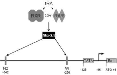
In a separate study, Nkx-2.5, a homeobox transcription factor, was indicated as the mediator of tRA responsive NIS expression in MCF-7 cells (Dentice et al., 2004). In MCF-7, RA was shown to first up-regulate the expression of Nkx-2.5 mRNA (reaching maximum levels after 6 hours), followed by NIS mRNA with maximum level at 12 hours, the iodide uptake increase was linear and followed the increase in NIS expression. Nkx-2.5 was also shown to interact with two sites in the rat NIS promoter; the site W was at -250 and the site N2 was at -542 relative to the ATG codon (Figure 1.4). They have also reported a correlation between Nkx-2.5 expression and lactation in mice (Dentice et al., 2004).
Systemic tRA treatment in immunodeficient mice with MCF-7 xenograft tumors resulted in a significant increase in NIS expression and in iodide uptake. Similar treatment was also evident in transgenic mice carrying the oncogene polyoma virus middle T antigen (Kogai et al., 2004) and the iodide accumulation in other organs was not affected by tRA treatment. The RA induced-NIS expression in MCF-7 was also found to be stimulated by dexamethasone. In the presence of tRA, dexamethasone increased NIS mRNA levels and iodide uptake significantly, in
18
addition to an inhibition of radioiodide efflux, resulting in a better drug combination to increase radioiodide toxicity in breast cancer cells (Unterholzner et al., 2006).
Figure 1.4. Regulatory elements involved in Nkx-2.5 dependant transcription of NIS in MCF-7. It was proposed that upon tRA stimulation, RA receptors will activate Nkx-2.5, which in turn will bind to two sites (N2 and W) in the promoter of rNIS leading to transcription. The position of these elements is indicated, and the numbering is relative to the start codon.
Recently, it was reported that in MCF-7 cells, treatment with prolactin, insulin, insulin-like growth factor-I (IGF-I), and IGF-II resulted in an increased expression of NIS both at transcriptional and at the protein levels. These ligands also resulted in an increased iodide uptake in MCF-7 cells in culture (Arturi et al., 2005).
1.3. Aim of This Study
As presented above, the transcriptional regulation of the mammary gland NIS gene is not yet fully understood. Several hormones and ligands have been implicated in the increased functionality of this gene aiming to develop strategies for novel methods for the diagnosis and treatment of breast cancer. Since NIS is expressed in the normal mammary gland only during lactation, and in breast cancer, understanding the
19
molecular determinants regulating its expression and function will ultimately provide a broader vision on possible therapeutic applications.
Our aim is to identify cis- and trans-acting genetic elements involved in the transcriptional regulation of NIS in the mammary gland, and in breast cancer models.
1.3.1. Rationale
It has been reported recently that conserved patterns of gene expression could reflect conserved patterns of regulatory mechanisms and potentially conserved cis-acting elements (Negre et al., 2005). Thus conserved regions in the sequences around and including NIS gene could harbor regulatory cis-acting elements involved in the mammary gland specific expression. We carried out a comparative bioinformatics analysis of the sequences flanking and including the NIS gene in human, mouse and rat searching for conserved regions, which possibly contain distal control regions regulating the expression of NIS in response to tRA in MCF-7 breast cancer cells. Such putative control elements were cloned in front of a luciferase reporter gene and they were used in functional assay experiments. This has led to the identification of several putative control regions.
It has been also shown that E2 is one of the hormones regulating functional NIS expression in the mammary gland, which when administered alone was sufficient to up-regulate its expression (Tazebay et al., 2000). Moreover, the well known tRA-induced expression in MCF-7 was correlated with presence of estrogen receptor (ER), in contrast to MDA-MB-231, in which ER negativity and lack of RA response correlated. We studied the effect of individual or combined nuclear receptor ligands, such as 17-β-estradiol and tRA, on NIS transcription in the breast cancer cell line model MCF-7. We found out that tRA induces NIS expression in cell lines that were both ERα+ and RARα+. We also showed that ectopic expression of ERα in MDA-MB-231 restored basal NIS expression in a ligand independent manner. We show evidence to the direct involvement of ERα in NIS expression by the a functional ERE in NIS promoter.
20
2. MATERIALS AND METHODS
2.1. Sequence Information and Databases
Genomic DNA sequences to be analyzed for DNA conservation in non-coding sequences were obtained from the Genome Browser database at the University of California Santa Cruz (Kent et al., 2002). The initial analysis was performed on a 310 kbp sequence from Homo sapiens (Release July 2003; Chr.19:17622612-17932611), Rattus norvegicus (Release June 2003; Chr. 16: 18813016-19123015), and Mus musculus (Release October 2003; Chr. 8: 71126739-71436738). Annotations for the human sequence were obtained from the Genome Vista database (Couronne et al., 2003), and the sequences were aligned with the mVista tool (Frazer et al., 2004) using a conservation level of 60% at a window length of 75 bp with the human sequence at the x axis.
The second analysis was performed on a shorter stretch of DNA of 90 kbp from H. sapiens (Release July 2003; Chr. 19: 17816282-17906905), R. norvegicus (release June 2003; Chr. 16: 19018698-19107000), and from M. musculus (Release October 2003; Chr. 8: 71152446-71242000). Annotations for the human sequence were obtained from the Genome Vista database, and sequences were aligned with the mVista tool using the human sequence at the x axis and at 50% conservation level and a window length of 75 bp.
2.1.1. PCR amplification of conserved regions
Common conserved regions among human, mouse and rat were selected for PCR amplification. Conserved regions clustered within 1 kbp were amplified as one larger fragment and subsequently cloned into the reporter vector (see Results section 3.1). PCR amplification was performed in 25 µl reaction volumes containing 0.8X PCR buffer, 3 mM MgCl2, 200 µM dNTP, 10 pmoles of each primer, 5% DMSO, 1 U of
21
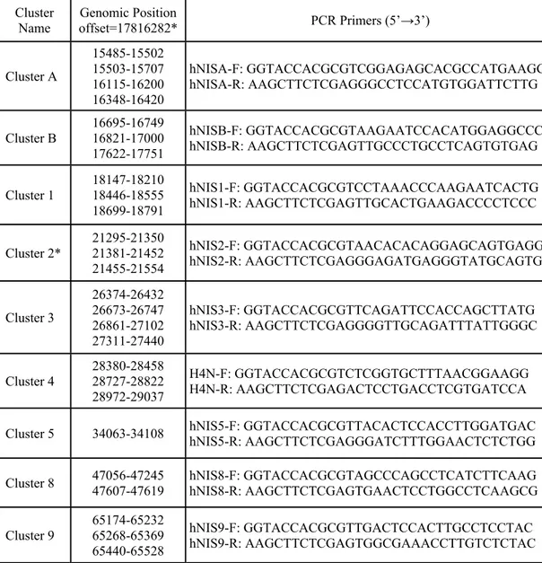
Taq DNA polymerase (Roche) and 100 ng of human genomic DNA. Thermal cycler conditions were an initial denaturation step at 94°C for 4 min; a loop cycle of 94°C, 30sec / 62°C, 30sec / 72°C, 40sec; and a final extension at 72°C for 7 minutes. Table 2.1 illustrates the position of each conserved region cluster and primers used for PCR amplification.
Table 2.1. Conserved region clusters used for PCR amplification. Genomic position of each cluster is indicated according to the human sequence of chromosome 19 Release July 2003.
Cluster
Name offset=17816282*Genomic Position PCR Primers (5’→3’) Cluster A 15485-15502 15503-15707 16115-16200 16348-16420 hNISA-F: GGTACCACGCGTCGGAGAGCACGCCATGAAGG hNISA-R: AAGCTTCTCGAGGGCCTCCATGTGGATTCTTG Cluster B 16695-16749 16821-17000 17622-17751 hNISB-F: GGTACCACGCGTAAGAATCCACATGGAGGCCC hNISB-R: AAGCTTCTCGAGTTGCCCTGCCTCAGTGTGAG Cluster 1 18147-18210 18446-18555 18699-18791 hNIS1-F: GGTACCACGCGTCCTAAACCCAAGAATCACTG hNIS1-R: AAGCTTCTCGAGTTGCACTGAAGACCCCTCCC Cluster 2* 21295-21350 21381-21452 21455-21554 hNIS2-F: GGTACCACGCGTAACACACAGGAGCAGTGAGG hNIS2-R: AAGCTTCTCGAGGGAGATGAGGGTATGCAGTG Cluster 3 26374-26432 26673-26747 26861-27102 27311-27440 hNIS3-F: GGTACCACGCGTTCAGATTCCACCAGCTTATG hNIS3-R: AAGCTTCTCGAGGGGTTGCAGATTTATTGGGC Cluster 4 28380-28458 28727-28822 28972-29037 H4N-F: GGTACCACGCGTCTCGGTGCTTTAACGGAAGG H4N-R: AAGCTTCTCGAGACTCCTGACCTCGTGATCCA Cluster 5 34063-34108 hNIS5-F: GGTACCACGCGTTACACTCCACCTTGGATGAC hNIS5-R: AAGCTTCTCGAGGGATCTTTGGAACTCTCTGG Cluster 8 47056-47245 47607-47619 hNIS8-F: GGTACCACGCGTAGCCCAGCCTCATCTTCAAG hNIS8-R: AAGCTTCTCGAGTGAACTCCTGGCCTCAAGCG
Cluster 9 65174-65232 65268-65369 65440-65528 hNIS9-F: GGTACCACGCGTTGACTCCACTTGCCTCCTAC hNIS9-R: AAGCTTCTCGAGTGGCGAAACCTTGTCTCTAC * Offset value for cluster 2 regions was (17816282) based on Human-mouse comparison, each interval represents the individual conserved regions.
22
2.2. Plasmids
2.2.1. Reporter constructs
The luciferase reporter vectors, pGL3E1bLuc and phRL-TK were kindly provided by Roberto Di Lauro, Naples, Italy. The reporter pGL3E1bLuc is a modified version of the original plasmid pGL3-Basic (Promega), which contains the E1b TATA element (5’-TCG AGT CTA GAG GGT ATA TAA TGG ATC-3’) between XhoI/BglII sites of pGL3-Basic; destroying the BglII site while keeping the XhoI site intact. PCR products containing conserved regions were cloned into MluI/XhoI sites of pGL3E1bLuc to produce the reporter plasmids named cluster A, cluster B and clusters 1, 2, 3, 4, 5, 8 and 9.
Cluster 3 derivatives containing individual or combined conserved regions were prepared by restriction endonuclease digestion of internal sequences, or by PCR. The plasmid Cl.3-ex containing the conserved region 3-1 was prepared by removing EspI/XhoI fragment from cluster 3, likewise plasmids Cl.3 -pm, Cl.3 ∆3-4, Cl.3 -xp, Cl.3 -SacI and Cl.3 -EspI were prepared by removing the PvuII/MluI, the PstI/SmaI, the XhoI/PvuII, the SacI and the EspI fragments respectively.
The plasmid Cl.3-2 was prepared by cloning a PCR product containing the conserved region 3-2 using primers 3-2F: 5'-CTG CAG ACG CGT AGG CTG AGC TGA GAC TTG AA-3' and 3-2R: 5'-AAG CTT CTC GAG GAG GAA TAA ATG GGA CGT GG-3' into MluI/XhoI sites of pGL3E1bLuc, likewise Cl.3-3 was prepared by inserting a PCR fragment containing the conserved region 3-3 (PCR primers were 3-3F: 5'-GGT ACC ACG CGT CCA TTT ATT CCT CTG AGG CA-3' and 3-3R: 5'-ACT AGT CTC GAG TGT CTG CTG TTG ACA GGT GG-3') into MluI/XhoI sites of pGL3E1bLuc.
The plasmid Cl.3/4 was prepared by removing the insert containing the conserved cluster 4 from Cl.4 (first, digesting with MluI followed by klenow filling of the cohesive ends and then digesting with XhoI) and inserting this fragment into the BamHI (klenow filled to produce blunt ends) and SalI sites of Cl.3 following the luciferase reporter gene.
23
Constructs containing the human NIS 5’UTR were prepared by inserting a PCR product (PCR primers were hNIS5UTR-F 5’-TGG CCT GTC TGT CCC AGT CCA GGG CTG A-3’ and hNIS5UTR-R 5’-TCT CCA CGG CCT CCA TGG AGG GCG GGT GCG GA-3’) amplified from human genomic DNA into the SmaI/NcoI sites of the reporter constructs.
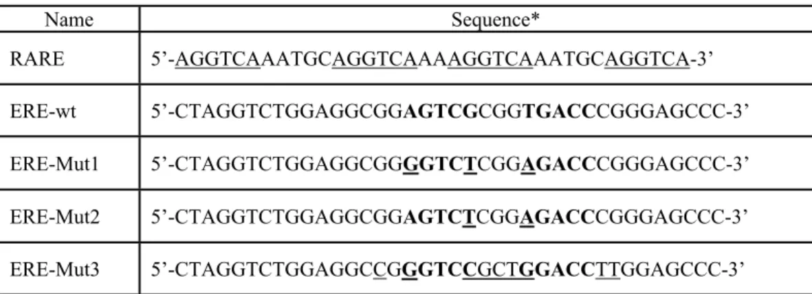
The reporter pPS2XERE was prepared by ligating a synthetic double strand (ds) oligo-nucleotide containing two tandem copies of the pS2 ERE into MluI/XhoI sites of pGL3E1bLuc. The DNA sequence of this oligo-nucleotide was 5’-CGC GTA AGG TCA CGG TGG CCA CAC GCG TAA GGT CAC GGT GGC CAC CCC GTC-3’. Likewise, pNIS2XERE was created by inserting a synthetic ds oligo-nucleotide containing two tandem copies of the putative NIS ERE (5’-CGC GTA GGC GGA GTC GCG GTG ACC CGG CGG AGT CGC GGT GAC CCG GGA GC-3’) into the MluI/XhoI sites of pGL3E1bLuc. The reporter plasmid pRARE-Luc was prepared by inserting a ds oligo-nucleotide containing 3 DR5 elements separated by 2 nucleotides in the MluI/XhoI sites of pGL3E1bLuc, the sequence of this oligo was (only the upper strand is shown and the direct repeat elements are underlined) 5’-CGC GTA GGT CAA ATG CAG GTC AAA AGG TCA AAT GCA GGT CAA AAG GTC AAA TGC AGG TCA C-3’. The reporter pC5-Luc was prepared by inserting a ds oligo nucleotide containing 5 tandem repeats of the C element harboring TTF-1/Nkx-2.5 binding site (Dentice et al., 2004), the sequence of which was 5-CCC AGT CAA GTG TTC TT-3’ in the MluI/XhoI sites of pGL3E1bLuc. 2.2.1.1. Site directed mutagenesis
Constructs harboring the retinoic acid response element (5’-AGG TCA AAG TCC TCC TGG GTC C-3’; bases 125-146 in cluster 3), were subjected to site directed mutagenesis based on data from Schmutzler et al. (2002). PCR based mutagenesis was performed in 50 µl reaction volumes containing 40 ng of target plasmid DNA, 1X Pfu buffer, 200 µM dNTP mix, 100 ng of each primer and 2.5 U of Pfu-Turbo DNA polymerase (Stratagene). The primer pair (the mutant nucleotides are underlined) sense a125t/g127a: 5'-GAC CAG AAC CTC CAG TGA TCA AAG TCC TCC TGG G-3' and antisense a125t/g127a: 5'-CCC AGG AGG ACT TTG
24
ATC ACT GGA GGT TCT GGT C-3'. Reaction conditions were an initial denaturation step at 95ºC for 30 seconds followed by 15 cycles of 95ºC for 30 seconds / 55ºC for 1 minute / 68ºC for 6 minutes and 20 seconds. Following the PCR reaction the tubes were cooled to 37ºC on ice and then 1 µl of DpnI (10U/µl; Fermentas) was added and incubated at 37ºC for 1 hour. Following the DpnI digestion of the parental (methylated) strand, 5 µl were used for transformation of super-competent E. coli (DH5α) cells.
Plasmids were rescued from single colonies and checked for the presence of the mutation by automated DNA sequencing. Similar protocol was used to create the ERE mutants in plasmids harboring the estrogen responsive element. The oligo nucleotide sequences of primers used for the mutagenesis are shown in table 2.3.
2.2.2. Expression vectors
The expression plasmid for Nkx-2.5 was kindly provided by Domenico Salvatore, Naples, Italy. The expression vector for ERα (pCMV-ERα) was prepared by inserting the EcoRI fragment (containing ERα coding sequence) form the plasmid pSG5ERpuro (kindly provided by Patrick Balaguer, Montpellier, France) into pcDNA3.1C (Invitrogen). Oligo-nucleotides for the knockdown of ERα were designed and supplied by Oligoengine, WA (N-19 targets 458 and 499 on NM_000125 were ER458: 5’-TTC AGA TAA TCG ACG CCA G-3’, and for sh-ER499: 5’-GTA CCA ATG ACA AGG GAA G-3’). These shRNA oligos were then cloned in the BglII/XhoI sites of pSuper-GFP/Neo (pSR, Oligoengine, WA) to generate the two knockdown constructs, pSR-ER-458 and pSR-ER-499.
2.3. Cell Culture
2.3.1. Cell lines
Human breast cancer cell lines BT-474, T-47D, BT-20, 453, MDA-MB-468, hTERT-HME1, MCF-7, MDA-MB-231 and MDA-66; Human cervical cancer cell line HeLa; rat thyroid cell line FRTL-5 and the monkey kidney cell line COS-7 were used in this study.
25
2.3.2. Growth media
Mammary gland, HeLa and COS-7 cell lines were maintained in high glucose Dulbecco’s modified Eagle’s medium (Gibco) [supplemented with 10% fetal bovine serum (FBS), 1% penicillin/streptomycin (P/S) and 1% L-glutamine (Biochrom)], abbreviated in the text as reg-DMEM, at 37°C in a 5% CO2 incubator. MDA-66 was
maintained in the above medium with the addition of 0.4 mg/ml Hygromycin (Roche). FRTL-5 cells were maintained in Coon's modified Ham's F12 medium supplemented with 2 mM glutamine, 5% FBS, 1% P/S and the 6 hormone mix (10 µg Insulin, 10 nM Hydrocortisone, 5 µg/ml Transferrin, 10 ng gly-his-lys acetate, 10 ng/ml somatostatin and 10 mU/ml Thyroid Stimulating Hormone (TSH)) at 37ºC in a 5% CO2 incubator. These cells display a sensitivity to TSH-induced increase in
cAMP levels, thus the presence of TSH is an absolute growth requirement.
2.3.3. Hormone induction
All-trans-retinoic acid (tRA) and 17-β-estradiol (E2) were purchased from Sigma. tRA was dissolved in DMSO, E2 was dissolved in ethanol as 10 mM stock solutions, and stored protected from light at -20°C.
2.3.3.1. tRA induction
In general tRA induction experiments were performed in reg-DMEM, unless otherwise indicated. tRA was applied to a final concentration of 1 µM for 12 hours when analyzing the expression of NIS by RT-PCR (24 hours induction was applied when performing luciferase reporter assay with tRA stimulation). After hormone induction, cells were rinsed with cold PBS, and harvested by trypsinization, cell pellets were divided into two tubes; RNA and protein extracts were prepared from the same sample.
2.3.3.2. E2 induction
Induction with E2 was performed in sf-DMEM (a phenol-red free DMEM (Sigma) supplemented with 10% dextran-coated-charcoal stripped FBS, 1% P/S and 1% L-glutamine). Two days before the addition of hormones, cells were fed with sf-DMEM in order to deplete the culture media from endogenous steroids and retinoids.
26
E2 was applied to a final concentration of 10 nM for 3 hours. After hormone induction cells were collected as mentioned above and divided into two tubes.
2.3.4. Transfection
Cell lines were transfected with plasmid DNAs using FuGENE-6 reagent (Roche). FuGENE:DNA ratios were determined experimentally to be 3:1 for MCF-7, HeLa, FRTL-5 and COS-7 and 6:1 for both MDA-MB-231 and for MDA-66. In general, cell lines were seeded in the appropriate culture container (depending on the assay to be performed) at an experimentally optimized density (depending on the cell line) so that they reach confluence at the time of the assay. For MCF-7 and FRTL-5, cells (90%-100% confluence in 100 mm dishes) are harvested by trypsinization and washed once with reg-DMEM, then resuspended in 10 ml complete medium, and diluted 1:7 in culture medium (3 ml cells in 20 ml medium), diluted cells are then seeded in 24-well plates for transfection prior to luciferase reporter assay; 400 µl cells per well. For MDA-MB-231 and MDA-66 dilution was 1:3 (6 ml cells in 20 ml medium), and for HeLa cells dilution ratio was 1:2.
2.3.4.1. Transient transfection with ERα
MDA-MB-231 cells were transfected with pCMV-ERα in 100 mm dishes, using FuGENE-6 (as described above) and 5 µg of the expression vector. Two days after transfection, media were replaced with fresh sf-DMEM containing 10 nM E2 or 1 µM tRA, or a combination of both hormones. After hormone induction cells were collected as mentioned above.
COS-7 cells grown in 150 mm dishes were transfected with the ERα expressing plasmid as described above. Two days after transfection, cells were treated with 10 nM E2 for 3 hours or with vehicle (EtOH) prior to harvesting. ERα transfected COS-7 cell were used for nuclear extract preparation.
2.4. Luciferase Reporter Assay
Cells were seeded in 24-well plates in reg-DMEM; so that they reach confluence at the time of the assay. Two days later, and 1 hour prior to transfection, cells were
27
washed twice with PBS, and the medium was replaced with culture medium (reg-DMEM or sf-(reg-DMEM depending on the hormone to be used) lacking antibiotics. Transfection was carried out with 200 ng of reporter vector plus 3 ng of phRL-TK to normalize for transfection efficiency. For E2 induction, two days post transfection, medium was changed with fresh sf-DMEM containing 10 nM E2 (or EtOH as vehicle control) and continued incubation for 6 hours, while in the case of tRA induction, 12 hours after transfection, culture medium was replaced with reg-DMEM containing 1 µM tRA (or DMSO as vehicle control) and continued incubation for 24 hours. Then the cells were harvested and luciferase reporter assay was performed using the Dual-Glo Luciferase Assay system (Promega). Luciferase values for all samples were normalized by first subtracting the background of no-transfection control, and then dividing firefly luciferase values over those of Renilla luciferase. Fold induction is relative to the value of the empty vector pGL3E1bLuc.
2.5. RNA Isolation
The expression level of NIS, pS2, RIP140 and GAPDH was monitored by semi-quantitative RT-PCR. RNA was prepared from cell pellets using the Nucleospin RNA II kit (Macherey-Nagel) as recommended by the manufacturer. The RNA preparation protocol included on-column DNase treatment step, minimizing the presence of genomic DNA in the RNA samples. RNA concentrations were determined spectrophotometrically.
2.5.1. cDNA synthesis, RT-PCR
In general 2 µg of total RNA were used for cDNA synthesis using the Revert-Aid First Strand cDNA Synthesis Kit (Fermentas). Primers for semi-quantitative RT-PCR amplified corresponding transcripts from positions spanning two or more exonic sequences, except for the RIP140, for which, a -RT (where cDNA is prepared using the same amount of RNA but without adding reverse transcriptase to the tube) control was included to ensure the amplification from cDNA only. PCR primers were RT-NIS-F: 5’-CTC ATC CTG AAC CAA GTG AC-3’, RT-NIS-R2: 5’-TAC ATG GAG AGC CAC ACC A-3’, RT-pS2-F: 5’-CCA TGG AGA ACA AGG TGA TCT GC-3’, RT-pS2-R2: GTC AAT CTG TGT TGT GAG CCG AG-3’, GAPDH-F: