ISSN 1991-3494 (Print) ҚАЗАҚСТАН РЕСПУБЛИКАСЫ ҰЛТТЫҚ ҒЫЛЫМ АКАДЕМИЯСЫНЫҢ
Х А Б А Р Ш Ы С Ы
ВЕСТНИК
НАЦИОНАЛЬНОЙ АКАДЕМИИ НАУК РЕСПУБЛИКИ КАЗАХСТАНTHE BULLETIN
THE NATIONAL ACADEMY OF SCIENCES OF THE REPUBLIC OF KAZAKHSTANPUBLISHED SINCE 1944
JANUARY – FEBRUARY 2020
2
NAS RK is pleased to announce that Bulletin of NAS RK scientific journal has been accepted for indexing in the Emerging Sources Citation Index, a new edition of Web of Science. Content in this index is under consideration by Clarivate Analytics to be accepted in the Science Citation Index Expanded, the Social Sciences Citation Index, and the Arts & Humanities Citation Index. The quality and depth of content Web of Science offers to researchers, authors, publishers, and institutions sets it apart from other research databases. The inclusion of Bulletin of NAS RK in the Emerging Sources Citation Index demonstrates our dedication to providing the most relevant and influential multidiscipline content to our community.
Қазақстан Республикасы Ұлттық ғылым академиясы "ҚР ҰҒА Хабаршысы" ғылыми журна-лының Web of Science-тің жаңаланған нұсқасы Emerging Sources Citation Index-те индекстелуге қабылданғанын хабарлайды. Бұл индекстелу барысында Clarivate Analytics компаниясы журналды одан әрі the Science Citation Index Expanded, the Social Sciences Citation Index және the Arts & Humanities Citation Index-ке қабылдау мәселесін қарастыруда. Web of Science зерттеушілер, авторлар, баспашылар мен мекемелерге контент тереңдігі мен сапасын ұсынады. ҚР ҰҒА Хабаршысының Emerging Sources Citation Index-ке енуі біздің қоғамдастық үшін ең өзекті және беделді мультидисциплинарлы контентке адалдығымызды білдіреді.
НАН РК сообщает, что научный журнал «Вестник НАН РК» был принят для индексирования в Emerging Sources CitationIndex, обновленной версии Web of Science. Содержание в этом индек-сировании находится в стадии рассмотрения компанией Clarivate Analytics для дальнейшего принятия журнала в the Science Citation Index Expanded, the Social Sciences Citation Index и the Arts & Humanities Citation Index. Web of Science предлагает качество и глубину контента для исследователей, авторов, издателей и учреждений. Включение Вестника НАН РК в Emerging Sources Citation Index демонстрирует нашу приверженность к наиболее актуальному и влиятельному мультидисциплинарному контенту для нашего сообщества.
3 Б а с р е д а к т о р ы х.ғ.д., проф., ҚР ҰҒА академигі М.Ж. Жұрынов Р е д а к ц и я а л қ а с ы: Абиев Р.Ш. проф. (Ресей) Абишев М.Е. проф., корр.-мүшесі (Қазақстан) Аврамов К.В. проф. (Украина) Аппель Юрген проф. (Германия) Баймуқанов Д.А. проф., корр.-мүшесі (Қазақстан) Байтулин И.О. проф., академик (Қазақстан) Банас Иозеф проф. (Польша) Берсимбаев Р.И. проф., академик (Қазақстан) Велесько С. проф. (Германия) Велихов Е.П. проф., РҒА академигі (Ресей) Гашимзаде Ф. проф., академик (Әзірбайжан) Гончарук В.В. проф., академик (Украина) Давлетов А.Е. проф., корр.-мүшесі (Қазақстан) Джрбашян Р.Т. проф., академик (Армения) Қалимолдаев М.Н. проф., академик (Қазақстан), бас ред. орынбасары Лаверов Н.П. проф., академик РАН (Россия) Лупашку Ф. проф., корр.-мүшесі (Молдова) Мохд Хасан Селамат проф. (Малайзия) Мырхалықов Ж.У. проф., академик (Қазақстан) Новак Изабелла проф. (Польша) Огарь Н.П. проф., корр.-мүшесі (Қазақстан) Полещук О.Х. проф. (Ресей) Поняев А.И. проф. (Ресей) Сагиян А.С. проф., академик (Армения) Сатубалдин С.С. проф., академик (Қазақстан) Таткеева Г.Г. проф., корр.-мүшесі (Қазақстан) Умбетаев И. проф., академик (Қазақстан) Хрипунов Г.С. проф. (Украина) Юлдашбаев Ю.А. проф., РҒАакадемигі (Ресей) Якубова М.М. проф., академик (Тәжікстан) «Қазақстан Республикасы Ұлттық ғылым академиясының Хабаршысы». ISSN 2518-1467 (Online), ISSN 1991-3494 (Print) Меншіктенуші: «Қазақстан Республикасының Ұлттық ғылым академиясы»РҚБ (Алматы қ.). Қазақстан республикасының Мәдениет пен ақпарат министрлігінің Ақпарат және мұрағат комитетінде 01.06.2006 ж. берілген №5551-Ж мерзімдік басылым тіркеуіне қойылу туралы куәлік. Мерзімділігі: жылына 6 рет. Тиражы: 2000 дана. Редакцияның мекенжайы: 050010, Алматы қ., Шевченко көш., 28, 219 бөл., 220, тел.: 272-13-19, 272-13-18, http://www.bulletin-science.kz/index.php/en/ © Қазақстан Республикасының Ұлттық ғылым академиясы, 2020 Типографияның мекенжайы: «NurNaz GRACE», Алматы қ., Рысқұлов көш., 103.
4 Г л а в н ы й р е д а к т о р д.х.н., проф. академик НАН РК М.Ж. Журинов Р е д а к ц и о н н а я к о л л е г и я: Абиев Р.Ш. проф. (Россия) Абишев М.Е. проф., член-корр. (Казахстан) Аврамов К.В. проф. (Украина) Аппель Юрген проф. (Германия) Баймуканов Д.А. проф., чл.-корр. (Казахстан) Байтулин И.О. проф., академик (Казахстан) Банас Иозеф проф.(Польша) Берсимбаев Р.И.проф., академик (Казахстан) Велесько С. проф. (Германия) Велихов Е.П. проф., академик РАН (Россия) Гашимзаде Ф. проф., академик (Азербайджан) Гончарук В.В. проф., академик (Украина) Давлетов А.Е. проф., чл.-корр. (Казахстан) Джрбашян Р.Т. проф., академик (Армения) Калимолдаев М.Н. академик (Казахстан), зам. гл. ред. Лаверов Н.П. проф., академик РАН (Россия) Лупашку Ф. проф., чл.-корр. (Молдова) Мохд Хасан Селамат проф. (Малайзия) Мырхалыков Ж.У. проф., академик (Казахстан) Новак Изабелла проф. (Польша) Огарь Н.П. проф., чл.-корр. (Казахстан) Полещук О.Х. проф. (Россия) ПоняевА.И. проф. (Россия) Сагиян А.С. проф., академик (Армения) Сатубалдин С.С. проф., академик (Казахстан) Таткеева Г.Г. проф., чл.-корр. (Казахстан) Умбетаев И. проф., академик (Казахстан) Хрипунов Г.С. проф. (Украина) Юлдашбаев Ю.А. проф., академик РАН (Россия) Якубова М.М. проф., академик (Таджикистан) «Вестник Национальной академии наук Республики Казахстан». ISSN 2518-1467 (Online), ISSN 1991-3494 (Print) Собственник: РОО «Национальная академия наук Республики Казахстан» (г. Алматы). Свидетельство о постановке на учет периодического печатного издания в Комитете информации и архивов Министерства культуры и информации Республики Казахстан №5551-Ж, выданное 01.06.2006 г. Периодичность: 6 раз в год. Тираж: 2000 экземпляров. Адрес редакции: 050010, г. Алматы, ул. Шевченко, 28, ком. 219, 220, тел. 272-13-19, 272-13-18. www: nauka-nanrk.kz, bulletin-science.kz © Национальная академия наук Республики Казахстан, 2020 Адрес типографии: «NurNazGRACE», г. Алматы, ул. Рыскулова, 103.
5
E d i t o r i n c h i e f
doctor of chemistry, professor, academician of NAS RK
М.Zh. Zhurinov
E d i t o r i a l b o a r d:
Abiyev R.Sh. prof. (Russia)
Abishev М.Ye. prof., corr. member. (Kazakhstan) Avramov K.V. prof. (Ukraine)
Appel Jurgen, prof. (Germany)
Baimukanov D.А. prof., corr. member. (Kazakhstan) Baitullin I.О. prof., academician (Kazakhstan) Joseph Banas, prof. (Poland)
Bersimbayev R.I. prof., academician (Kazakhstan) Velesco S., prof. (Germany)
Velikhov Ye.P. prof., academician of RAS (Russia) Gashimzade F. prof., academician (Azerbaijan) Goncharuk V.V. prof., academician (Ukraine) Davletov А.Ye. prof., corr. member. (Kazakhstan) Dzhrbashian R.Т. prof., academician (Armenia)
Kalimoldayev М.N. prof., academician (Kazakhstan), deputy editor in chief Laverov N.P. prof., academicianof RAS (Russia)
Lupashku F. prof., corr. member. (Moldova) Mohd Hassan Selamat, prof. (Malaysia)
Myrkhalykov Zh.U. prof., academician (Kazakhstan) Nowak Isabella, prof. (Poland)
Ogar N.P. prof., corr. member. (Kazakhstan) Poleshchuk О.Kh. prof. (Russia)
Ponyaev А.I. prof. (Russia)
Sagiyan А.S. prof., academician (Armenia) Satubaldin S.S. prof., academician (Kazakhstan) Tatkeyeva G.G. prof., corr. member. (Kazakhstan) Umbetayev I. prof., academician (Kazakhstan) Khripunov G.S. prof. (Ukraine)
Yuldashbayev Y.A., prof., academician of RAS (Russia) Yakubova М.М. prof., academician (Tadjikistan)
Bulletin of the National Academy of Sciences of the Republic of Kazakhstan.
ISSN 2518-1467 (Online), ISSN 1991-3494 (Print)
Owner: RPA "National Academy of Sciences of the Republic of Kazakhstan" (Almaty).
The certificate of registration of a periodic printed publication in the Committee of Information and Archives of the Ministry of Culture and Information of the Republic of Kazakhstan N 5551-Ж, issued 01.06.2006.
Periodicity: 6 times a year. Circulation: 2000 copies.
Editorial address: 28, Shevchenko str., of. 219, 220, Almaty, 050010, tel. 272-13-19, 272-13-18, http://nauka-nanrk.kz /, http://bulletin-science.kz
© National Academy of Sciences of the Republic of Kazakhstan, 2020 Address of printing house: «NurNaz GRACE», 103, Ryskulov str, Almaty.
64
BULLETIN OF NATIONAL ACADEMY OF SCIENCES
OF THE REPUBLIC OF KAZAKHSTAN ISSN 1991-3494
Volume 1, Number 383 (2020), 64 – 71 https://doi.org/10.32014/2020.2518-1467.8
UDC 616-036.22
A. Balgimbayeva1, G. Shabdarbaeva1, L. Zhanteliyeva1,
A. Ibazhanova1, Uğur Uslu2, D. Khussainov1
1Kazakh National Agrarian University, Almaty, Kazakhstan; 2University of Selcuk, Medicine Faculty, Konya, Turkey.
E-mail: [email protected], [email protected], [email protected], [email protected]
DIAGNOSTICS AND TREATMENT OF DIOCTOPHYMOSIS IN DOGS
Abstract. This article includes materials on the rare disease in domestic and wild carnivores, dioctophimosis,
caused by a helminth from the group of aphasmidia nematodes, Dioctophyme renale, which is parasitic in the kidneys, recently registered in veterinary clinics in Almaty. The disease has important social significance - a person is susceptible to it, the infection of which occurs when raw fish is eaten. In Almaty, according to the statistics of veterinary clinics in recent years (2018-2019), 17 cases of dioctophimosis in dogs have been registered, that is up to 0.3% of the number of dogs examined for helminthiases. The appearance of this disease, which is relatively new for our region, can have a significant impact on the epizootiological and epidemiological situation. The results of in vivo laboratory diagnosis of dioctophimosis in dogs by ovoscopic methods are presented, with the provision of microphotos of the parasite eggs; methods of instrumental diagnostics - ultrasound examination (ultrasound) of the kidneys with the provision of photos proving the presence of a parasite in the renal pelvis. Since the only treatment for dioctophimosis is surgery and extraction of dioctophyma from the affected organ (kidney), our own original materials are presented for a step by step surgical intervention to extract helminth from the kidneys of dogs and treat animals. The condition of the operated animals is satisfactory, the prognosis for surgery is favorable.
Key words: dogs, carnivores, helminths, helminthiases, monitoring, nematodes, epizootiological and
epidemio-logical situation, hematuria, dioctophimosis, ultrasound, OIE, WHO, invasive material, urine, feces, kidneys, surgical treatment.
Relevance of research. Dioctophimosis (code according to ICD-10 - B83.9) is a helminthiasis disease
of silver-black foxes, dogs, jackals, marten and other carnivores, as well as humans when eating raw fish. The disease is characterized by damage to the kidneys, renal pelvis, ureters, bladder, abdominal cavity, sometimes the liver, blood vessels and heart; manifested by intoxication of the body, uremic phenomena, bloody urine. The literature describes cases of human infection with dioctophyma and rarely cattle. The disease relates to the group of helminthiases caused by aphasmidia nematodes [1,2].
The systematic position of the dioctophimosis pathogen [1]:
Kinqdon – Animalia; Type – Nemathelminthes; Class – Nematoda; Subclass – Adenophorea; Order – Dioctophymida; Suborder – Dioctophymata; Superfamily – Dioctophymoidea; Family – Dioctophymidae; Genus – Dioctophyme; Specie – Dioctophyme renale.
The Dioctophyme renale pathogen, is a nematode referring to the Dioctophymidae family, the only specie of the Dioctophyme. D. renale genus, represents sexual dimorphism and is considered the largest nematode, which infects the domestic animals, males can reach a length of 40 cm, and females can reach up to 100 cm [3]. The female has the rounded tail end, its vulva opens at the level of the initial part of the esophagus. The male has the tail end, which ends with a bell-shaped bursa, from which a spicule protrudes. The papillary tubercles in the form of two circles are around the mouth of the helminth [2].
Reproduction occurs by discharging eggs of 0.077 - 0.083x0.046 - 0.047 mm, brown, with lids at the poles. The egg shell consists of three layers: the outer, inner and vitelline membrane. The surface of the egg is dotted with small impressions like pockets. Eggs are secreted into the environment with the urine of a sick animal [2].
Dioctophyma are biohelminths. The pathogen development takes place with the participation of two hosts: definitive and intermediate hosts.
65
Definitive hosts: The main definitive hosts are ermines and canids. However, this nematode has been reported in pigs, cats, horses, cattle and other mammals such as wild cat, cheetah, coati, focid densification, mongoose rat [4]; and about 24 cases have been reported in humans [5], therefore, it is considered a zoonotic disease. Intermediate hosts: Branchiobdella or Lumbriculus oligochaetes.
Additional and reservoir hosts: fish (sabrefish, perch, barbel, mackerel, spike, shovel, pike, catfish, gambusia and some other cyprinids), in the body of which helminth larvae are encapsulated, remaining viable for a long time [2].
Sexually mature females, localized in a sick animal in the renal pelvis, ureters, and bladder, lay eggs, which are excreted with the urine into the external environment. Eggs, which fall into the water, are developed within 25 to 30 days. They form larvae, which reach 0.28 - 0.31 mm in length.
The eggs with developed larvae are swallowed together with detritus by the first intermediate hosts – oligochaetes of the Branchiobdella or Lumbriculus genus. In the intestine of the oligochaetes, larva emerges from the egg and migrates into the abdominal blood vessel, where it grows and develops. After 45-60 days, depending on the water temperature, the first molt occurs and the larva turns into the second stage, reaching 0.885-1.181 mm in length. After 3.5 - 4 months the larva molts for a second time and turns into a third-stage larva, reaching 6.905 - 8.018 mm in length. At this stage of larval development, young males and females are formed.
Oligochaetes infected by invasive larvae are eaten by fish, the second intermediate hosts (sicklefish, perch, barbel, shemaya, ship, shovelnose sturgeon, pike, catfish, gambusia and some other cyprinids) [2].
The further development of the third stage larva occurs only in the organism of the definitive host, where it can enter together with the oligochaetes or with infected fish. Larvae of dioctofimide, once entered in the intestines of the final host (carnivores or humans), penetrate its wall, enter the body cavity and migrate through the body, reaching the renal pelvis. At this time, the larvae molt twice. The life cycle of dioctofimide is completed in 8.5 - 9 months.
The lifespan of a nematode in the organism of a definitive host is up to 3–5 years [2].
Epizootological data. Locally, dioctophimosis of carnivores is found in areas of Transcaucasia, Central Asia, Kazakhstan, the Far East, Karelia and Siberia in large river basins. Dioctophimosis is registered in far abroad countries: Italy, Austria, Germany, Holland, France, England, Japan, North and South America [2]. For example, infection of dogs in some areas of North America reaches up to 37%, in Kazakhstan it ranges from 1 to 9% [6]. This parasite has a worldwide distribution, however, most reports have been received from South-East Asia [7.8].
Epidemiology. Human dioctophimosis is registered in many countries of the world: Argentina, Para-guay, Brazil, the USA, a number of European countries, Iran, South Vietnam, China, Japan. In the CIS countries, it is more often found in Tajikistan, Uzbekistan, Kazakhstan, isolated cases are registered in Leningrad and Arkhangelsk regions. Since annelids (intermediate hosts of Dioctophyme renale) live on the wet coasts of water bodies, endemic foci are confined to river valleys and lakes [6].
Symptoms and pathogenesis. Mostly sexually mature helminths are localized in animals in the kidneys and less often in the abdominal cavity. But, before getting there, nematodes migrate from the stomach of a mammal, where they get with fish or oligochaetes. They invade the muscle layer of the stomach wall, causing a hematoma. Then they migrate to the body cavity and are closer to the liver. Penetrate into its parenchyma, and then into the renal pelvis. As a result, its wall is greatly stretched and thinned, the kidney atrophies. Sometimes the nematode enters the urethra. All this leads to significant violations of these organs and painful phenomena of the whole organism. An animal or a human does not have appetite, vomiting, general exhaustion, and an oppressed state appear. A muddy, bloody liquid accumulates inside the renal pelvis, its smell resembles that of urine. The mucous membrane of the renal pelvis becomes grayish-white or yellowish in color. In places of degenerated areas, the lime salt are deposited in the form of multiple lumps [2,6].
Pathological changes in the human body at the onset of the disease are associated with the migration of larvae into the abdominal cavity and into the liver, which is accompanied by hemorrhages, serous-fibrinous inflammation of the mesentery, venous congestion in the liver, and the formation of granulomas and scars at the sites of damage. During parasitization of adult helminths in the human renal pelvis, the kidney parenchyma is destroyed step by step, and the capsule is preserved only. The right cavity, directly adjacent to the duodenum, from which the parasite larvae migrate, is more often affected [6,9].
66
The most characteristic symptoms of the disease are renal colic, pyuria, hematuria, oliguria and anuria. Often there are complaints of pain in the lumbar region, which spread throughout the abdomen.
Complications in humans are renal coma [6,10].
In fish, Dioctophyme renale larvae reach 6.9 - 8.2 mm in length and 0.19 - 0.2 mm in width. They are localized in the internal organs: in the intestinal wall, on the peritoneum, mesentery, in the gonads, a connective tissue capsule is formed around them.
Diagnosis. To make a diagnosis during life, urine is examined for the parasite eggs and an ultrasound examination of the kidneys is performed. Posthumously, disease is diagnosed on the basis of an autopsy of animals and finding a nematode in the kidney, less commonly in other organs. When examining the fish, encapsulated larvae are found in the intestine, mesentery, and gonads [2].
Treatment. The only treatment at this time is surgery and extraction of Dioctophyme renale from the affected organ. There is evidence of attempts to treat animals with ivomek, praziquantel, levomizole, etc. [2,6,11].
Prevention of the disease is to identify dysfunctional water bodies and prohibit to feed the animals with raw fish. In areas unfavorable due to dioctophimosis, dogs are not allowed to sites of catching and cutting fish. The diet of fur-bearing animals excludes the raw fresh fish caught in reservoirs unsuccessful due to this invasion. They explanatory work is conducted among fisheries workers and among the population on the prevention of dioctophimosis [12,13,14,15,16].
The epidemiological and epidemiological situation with many helminthiases of carnivores, especially in zoonotic helminthiases in Kazakhstan and in neighboring countries, should be improved. In the world, especially in tropical and subtropical countries, the epidemiological and epidemiological situation regarding zoonotic helminthiases is very problematic. For example, according to the classification of the Office International des Epizooties (OIE), some helminthiases, in particular echinococcosis, which is spread among population and productive animals by the carnivorous, are included in the list of the most common diseases and in many countries are included in national programs to eliminate the disease. The annual costs for treating patients and losses in animal husbandry are amounted to USD 3 bln. Kazakhstan is among the regions permanently unfavorable for echinococcosis. World experience shows that establishing the incidence of helminth infections in animals allows us to assess the degree of threat to humans. According to the World Health Organization (WHO), every year a quarter of the population (more than 1.4 billion people) is infected with parasites, the most significant group of which is helminths. Nematodoses are the most common of the helminthiases [17].
Kazakhstan also has a difficult epidemiological and epidemiological situation regarding zoonotic helminthiases. From the State report on the situation in Kazakhstan, it follows that the epidemiological situation with parasitosis in the country poses a threat to public health. About 20,000 cases of parasitosis are recorded annually in Kazakhstan, the intensive incidence rate is on average about 24.2 per 100 thousand people. A number of researchers (Kereyev Ya.M., 2010; Shalmenov M.Sh., 2005; Akshulakov S.K., 2002; Amireyev S.A., 2002; Shabdarbayeva G.S., Abdibekova A.M., Shapiyeva Zh.Zh., 2012; Lider L.A., 2009; Shabdarbayeva G.S. et al., 2016) note a large infection of carnivores and humans with zoonotic helminthiases [18-23].
In Kazakhstan, a number of scientists performed researches related to monitoring of helminthiases of carnivorous zoonotic helminthiases. 11 species of helminths from different systematic groups were registered in the studied carnivorous. From the class of trematodes, only 1 species was noted – Opistorhis felineus; from the cestode class 4 species: Multiceps multiceps, Dipilidium caninum, Echinococcus granulosus, Alveococcus multilocularis; from the class of nematodes of 6 species: Dioctophyme renale, Toxocara canis, Toxascaris leonina, Ancylostoma caninum, Trichocephalus vulpis, Dirofilaria immitis. The greatest invasion in carnivores was noted by various nematodes, in particular, the species Toxascaris leonina that amounted to 28.9% of the total number of infected animals. A rather high infection was observed in carnivorous with toxocariasis: 22.4%. Significant invasion was also noted by the species from the cestodes Dipilidium caninum, which is 13.8%. All the above types of helminths are dangerous in human infection and can cause significant damage to his health. The remaining helminths, including dioctophimosis (Dioctophyme renale) are presented in isolated cases – from 0.7% to 5.8%. The average helminth infection in the studied dogs was 79.6% [24]. Based on the monitoring results, recommendations were suggested on the control of zoonotic helminthiases and on methods for detecting larval stages of helminths in fish [25,26].
67
In connection with the increase in the number of livestock, the types of their diseases are also increasing today. Since invasive diseases occur in all types of domestic animals, they cause enormous damage to the national economy. Many invasive diseases, in particular parasitic diseases lead to mass death of animals [27].
Results. The studies were carried out in Almaty clinics. 17 cases of a rare helminthiasis nematode
disease – dioctophimosis in dogs were registered during 2018-2019.
During the anamnesis, the nature of nutrition of the dogs was ascertained whether the animals were fed with raw fish or not. If so, where was the fish come or purchased from?
Clinical examination was carried out using methods of animal thermometry, focusing on the nature of urination: difficult or not; urination frequency; pain during urination; color and texture of urine.
Then, several portions of urine were obtained from dogs, and urine was centrifuged at 1000 rpm within 1 minute. The supernatant was carefully drained, the suspension remaining at the bottom of the centrifuge tube was pipetted by portions on a glass slide and viewed under a microscope at 10×40 magnification. Characteristic Dioctophyme renale eggs were found (figure 1). The preliminary clinical diagnosis in all cases was confirmed by intravital urine examination.
During ultrasound examination of the kidneys (ultrasound) contours of 1 or 2 helminths were found in the renal pelvis in dogs. The phenomena of kidney atrophy from squeezing of tissues by a parasite and coagulated helminth in the renal pelvis were observed (figures 2–4).
Figure 1 – Dioctophyme renale eggs at 10x40 magnification Figure 2 – Dog’s kidney ultrasound Results
Figure 3 – Dioctophyme renale in the dog’s kidney pelvis Figure 4 – Dog’s kidneys ultrasound examination results
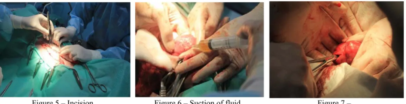
Having established a positive diagnosis and location of helminths, surgical treatment of dioctophimosis was performed. The progress of operation to extract Dioctophyme renale from the kidney is shown in figu-res 5–16.
Figure 5 – Incision of the abdominal wall
Figure 6 – Suction of fluid from the tissues around the kidney
Figure 7 – Incision of the kidney wall
68
At first, the type of anesthesia for the animal was determined. The need and dosage of anesthesia were calculated and given according to the weight of the animal. Next, the access points to the kidney were determined, the incision site was planned, the incision site was treated with aseptic antimicrobial agents, and the abdominal wall was incised. Then, reaching the kidney, suction of fluid collected around the kidney as a result of the inflammatory process was performed.
A small incision was made in the kidney wall, the forceps were carefully inserted into the incision, the helminth was fixed, and removed carefully through the incision using the twisting method (figures 5–9).
The extracted helminths were identified by genus and species, measured, museum preparations were prepared from them, which replenished the parasitological museum of the department.
Figure 8 – Capturing helminth
with forceps Figure 9 – Removing helminth from the kidney Figure 10 – Helminth extracted from the dog’s kidney
Figure 11 – Dioctophyme renale from the dog’s kidney
Figure 12 – Female and male of Dioctophyme renale
from the dog’s kidney
Figure 13 – Measurement of Dioctophyme renale
Figure 14 – Head end of Dioctophyme renale
Figure 15 – Suture on the wall of the abdominal cavity after surgery
Figure 16 – Postoperative anesthesia in a dog
The condition of the patients after the surgery was satisfactory in all cases, there was no temperature reaction, the food was taken with pleasure, the wound healed by primary intention. Urination is not difficult, the kidneys regained their function.
Conclusion. Taking into account the geographical location of Almaty, the lack of large rivers around
the metropolis with the above species composition of fish, it becomes clear why there is a very small infection of carnivorous with dioctophimosis. Cases of infection of the dog with dioctophimosis in our clinical practice are apparently imported cases. But infection of carnivores and humans from invasive fish imported from regions unsuccessful for this disease is not excluded.
69 А. И. Балгимбаева1, Г. С. Шабдарбаева1, Л. О. Жантелиева1, А. С. Ибажанова1, Угур Услу2, Д. М. Хусаинов1 1Қазақ ұлттық аграрлық университеті, Алматы, Қазақстан; 2Сельчук университеті, медицина факультеті, Конья, Туркия ИТТЕРДІҢ ДИОКТОФИМОЗЫН БАЛАУ ЖӘНЕ ЕМДЕУ Аннотация. Мақалада соңғы уақытта Алматы қаласының ветеринариялық клиникаларында тіркелген, үй және жабайы етқоректі жануарлардың сирек кездесетін ауруы – диоктофимоз, бүйректерде паразиттелетін афазмидиялық нематод тобының гельминті тудыратын Dioctophyme renale бойынша материалдар ұсынылған. Аурудың маңызды әлеуметтік мәні бар - адам оған сезімтал, инфекция шикі балық жеген кезде пайда болады. Ет қоректі гельминтоздардың мониторингі бойынша зерттеулер Алматы қаласының клиникаларында жүргізілді. Зоонозды гельминтоздардың мониторингіне көп көңіл бөлінді. Алматы қаласында әртүрлі жүйелі топтар гельминттерінің 11 түрі тіркелген. Трематод класынан тек 1 түр ғана белгіленген – Opistorhis felineus; цестод класынан 4 түр: Multiceps multiceps, Dipilidium caninum, Echinococcus granulosus, Alveococcus
multilo-cularis; нематод класынан 6 түр: Dioctophyme renale, Toxocara canis, Toxascaris leonina, Ancylostoma caninum, Trichocephalus vulpis, Dirofilaria immitis.
Ет қоректілердің ең жоғарғы инвазиялануы әртүрлі нематодтармен, атап айтқанда, Toxascaris leonina түрімен белгіленген және жұқтырылған жануарлардың жалпы санының 28,9% құрайды. Өте жоғары залал ет қоректік токсокарозда байқалады, ол 22,4% құрады. Сондай-ақ, зақымадалу таспа құрттарға жататын Dipili-dium caninum-да көрінді, ол - 13,8% құрады. Гельминттердің жоғарыда аталған барлық түрлері адам инфекция-сы кезінде қауіпті және оның денсаулығына айтарлықтай зиян келтіруі мүмкін. Қалған гельминттер, оның ішінде диоктофимоз (Dioctophyme renale) оқшауланған жағдайларда - 0,7% -дан 5,8%-ға дейін көрінді. Зерттелген иттердегі гельминттердің орташа деңгейі 79,6% құрады. Біздің зерттеулер бойынша 2018-2019 жж. сирек кездесетін гельминтоздардың нематодозды зооноздық ауруы - иттердің диоктофимозының 17 жағдайы тіркелді, бұл гельминтозға тексерілгендердің 0,3% құрайды. Анамнез кезінде иттердің тамақтану сипаты анықталды, яғни, жануарларды шикі балықпен азықтан-дырды ма? Егер иә болса, балық қайдан әкелінген немесе сатып алынған. Клиникалық тексеру жануарларды термометрия әдісімен жүргізілді, зәр шығару сипатына: қиындау неме-се жоқ; зәр шығару жиілігі; зәр шығару кезіндегі ауырсыну; зәр түсі мен консистенциясына назар аударды. Содан кейін иттерден бірнеше рет зәрді алып, зәрді 1000 айн/мин.1 минут ішінде центрифугадан өткіздік. Центрифугалық пробирканың түбінде қалған тұнба үстіндегі сұйықтықты абайлап төгіп, тамшылатқышпен заттық шыныға тамызып және микроскоптың 10х40 ұлғайған көрсеткішімен қарады. Диоктофимидке тән жұмыртқа табылды (1-сурет). Несепті алдын ала клиникалық балау барлық жағдайларда жануар тірі кезінде зерттелген. Бүйрекке ультрадыбыстық зерттеу жүргізу кезінде (УДЗ) бүйрек таяқшасында иттерде 1 немесе 2 гельминт контуры табылды. Бүйрек түтікшелерінде бүктелген гельминт және паразит тіндерінің қысылуынан бүйректің атрофиясы байқалады. Біздің өңірімізге қатысты жаңа аурудың пайда болуы зоонозды гельминтоздардың эпизоотологиялық-эпидемиологиялық жағдайына айтарлықтай әсер етуі мүмкін. Иттердегі диоктофимозды тірі кезіндегі зерт-ханалық диагностикасының нәтижелері, паразит жұмыртқаларының микрофотографиясын ұсына отырып, овоскопиялық әдістермен; аспаптық диагностика әдістерімен – бүйректі ультрадыбыстық зерттеу (УДЗ), бүй-ректің лоханкасында паразиттің болуын дәлелдейтін фотосуреттерді қолдана отырып ұсынылған. Диокто-фимозды емдеудің жалғыз әдісі хирургиялық араласу және зақымданған мүшелерден (бүйректен) диокто-фимдерді алу болып табылатындықтан, иттерді бүйректен гельминтті алу және жануарларды емдеу мақ-сатында кезең-кезеңмен оперативтік араласу бойынша өзіндік бірегей материалдар келтірілген. Операциядан өткен жануарлардың жағдайы қанағаттанарлық, операциядан өткен кездегі болжам қолайлы. Түйін сөздер: иттер, ет қоректілер, гельминттер, гельминтоздар, мониторинг, нематодтар, эпизоотоло-гиялық-эпидемиологиялық жағдай, гематурия, диоктофимоз, УДЗ, ХЭБ, ДДҰ, инвазирленген материал, несеп, нәжіс, бүйрек, операциялық емдеу. А. И.Балгимбаева1, Г. С.Шабдарбаева1, Л. О.Жантелиева1, А. С.Ибажанова1, Угур Услу2, Д. М.Хусаинов1 1Казахский национальный аграрный университет, Алматы, Казахстан; 2Сельчукский университет, медицинский факультет, Конья, Турция ДИАГНОСТИКА И ЛЕЧЕНИЕ ДИОКТОФИМОЗА СОБАК Аннотация. В статье представлены материалы по зарегистрированным в последнее время в ветеринар-ных клиниках г. Алматы редком заболевании домашних и диких плотоядветеринар-ных – диоктофимозе, вызываемом гельминтом из группы афазмидиевых нематод – Dioctophyme renale, паразитирующего в почках. Заболевание
70 имеет важное социальное значение – к нему восприимчив человек, заражение которого происходит при употреблении в пищу сырой рыбы. Исследования по мониторингу гельминтозов плотоядных проведены в клиниках г. Алматы. Большое внимание было уделено мониторингу зоонозных гельминтозов. Зарегистрировано у исследованных плото-ядных г. Алматы 11 видов гельминтов из разных систематических групп. Из класса трематод отмечен только 1 вид – Opistorhis felineus; из класса цестод 4 вида: Multiceps multiceps, Dipilidium caninum, Echinococcus
granulosus, Alveococcus multilocularis; из класса нематод 6 видов: Dioctophyme renale, Toxocara canis, Toxascaris leonina, Ancylostoma caninum, Trichocephalus vulpis, Dirofilaria immitis.
Наибольшая инвазированность у плотоядных отмечена различными нематодами, в частности, видом Toxascaris leoninа и составляет 28,9% от общего числа зараженных животных. Довольно высокая зараженность отмечена у плотоядных токсокарозом, которая составила 22,4%. Значительная инвазированность отмечена также видом из цестод Dipilidium caninum, составила 13,8%. Все указанные выше виды гельминтов представляют опасность в заражении человека и могут наносить значительный ущерб его здоровью. Остальные гельминты, в том числе и диоктофимоз (Dioctophyme renale), представлены в единичных случаях – от 0,7% до 5,8%. Средняя зараженность гельминтами исследованных собак составила 79,6%. Нами в течение 2018-2019 гг. зарегистрировано 17 случаев редкого гельминтозного нематодозного зоонозного заболевания – диоктофимоза собак. При анамнезе выясняли характер питания собак, кормили ли животных сырой рыбой. Если да, то откуда была привезена или закуплена рыба. Клиническое обследование проводили методами термометрии животных, акцентировали внимание на характер мочеиспускания: затрудненное или нет; частоту мочеиспускания; болезненность при мочеиспус-кании; на цвет и консистенцию мочи. Далее получали несколько порций мочи от собак, центрифугировали мочу при 1000 об/мин. в течение 1 минуты. Осторожно сливали надосадочную жидкость, оставшуюся на дне центрифужной пробирки взвесь, пипеткой порциями переносили на предметное стекло и просматривали под микроскопом при увеличении 10х40. Обнаружены характерные яйца диоктофимид (рисунок 1). Предварительный клинический диагноз во всех случаях был подтвержден прижизненным исследованием мочи. При проведении ультразвукового исследования почек (УЗИ) в почечной лоханке у собак были обнару-жены контуры 1 или 2 гельминта. Наблюдались явления атрофии почки от сдавливания тканей паразитом и свернувшийся гельминт в почечной лоханке. Появление данного, относительно нового для нашего региона заболевания, может оказать значительное влияние на эпизоотолого-эпидемиологическую обстановку зоонозных гельминтозов. Представлены резуль-таты прижизненной лабораторной диагностики диоктофимоза у собак овоскопическими методами, с предос-тавлением микрофотографий яиц паразита; методами инструментальной диагностики – ультразвуковым ис-следованием (УЗИ) почек с предоставлением фотографий, доказывающих наличие паразита в лоханке почки. Так как единственным методом лечения диоктофимоза является хирургическое вмешательство и извлечение диоктофим из пораженного органа (почки), приведены собственные оригинальные материалы по поэтапному оперативному вмешательству с целью извлечения гельминта из почек собак и лечения животных. Состояние прооперированных животных удовлетворительное, прогноз при проведении оперативного вмеша-тельства благоприятный. Ключевые слова: собаки, плотоядные, гельминты, гельминтозы, мониторинг, нематоды, эпизоотолого-эпидемиологическая обстановка, гематурия, диоктофимоз, УЗИ, МЭБ, ВОЗ, инвазированный материал, моча, фекалии, почки, оперативное лечение.
Information about authors:
Balgimbayeva A. Kazakh National Agrarian University, Almaty, Kazakhstan; https://orcid.org/0000-0002-4187-6733 Shabdarbaeva G. Kazakh National Agrarian University, Almaty, Kazakhstan; [email protected]; https://orcid.org/0000-0001-5708-5162
Zhanteliyeva Laura, Kazakh National Agrarian University, Almaty, Kazakhstan; [email protected]; https://orcid.org/0000-0002-7564-2089
Ibazhanova A., Kazakh National Agrarian University, Almaty, Kazakhstan; [email protected]; https://orcid.org/0000-0002-2833-1413
Uğur USLU, University of Selcuk, Medicine Faculty. Konya, Turkey; [email protected]; [email protected]; https://orcid.org/0000-0003-3456-312X
Khussainov Damir, Kazakh National Agrarian University, Almaty, Kazakhstan; [email protected]; https://orcid.org/0000-0002-0244-8381
71
REFERENCES
[1] Fedorov K.P. i dr. Osnovy obshhej i prikladnoj veterinarnoj parazitologii. Novosibirsk, 2004. P. 650-655.
[2] Akbaev M.Sh., Vasilevich F.I., Akbaev R.M., Vodjanov A.A., Kosminkov N.E., Pashkin P.I., Jatusevich A.I. Parazitologija i invazionnye bolezni zhivotnyh//M., «KolosS», 2008. P. 347-349.
[3] Ferreira, V.L., Medeiros, F.P., July, J.R., Raso, T.F., 2010. Dioctophyma renale in a dog: clinical diagnosis and surgical treatment. Vet. Parasitol. 168, 151–155 doi:10.1016 /j.vetpar.2009.10.013
[4] Measures, L., 2001. Dioctophymatosis. In: Samuel, W., Pybus, M., Kocan, A. (Eds.), Parasitic Diseases of Wild Mammals. Iowa State University Press/Ames, Iowa, P. 357–364. ISBN 0-8138-2978-X
[5] Chauhan, S., Kaval, S., Tewari, S., 2016. Dioctophymiasis: a rare case report. J. Clin. Diagn. Res. 10, DD01–DD02. doi: 10.7860 / JCDR / 2016 / 17394.7305
[6] Sergiev V.P., Lobzin Ju.V., Kozlov S.S. Parazitarnye bolezni cheloveka (protozoozy i gel'mintozy). Rukovodstvo dlja vrachej. Sankt-Peterburg, «Foliant», 2006, P. 461-463.
[7] Hajialilo, E., Mobedi, I., Masoud, J., Hasanpour, H., Mowlavi, G., 2015. Dioctophyme renale in Vulpes vulpes from the Caspian Sea littoral of Iran. Iran. J. Public. Health. 44, 698–700. PMID:26284212 PMCID: PMC4537628
[8] Zolhavarieh, S.M., Norian, A., Yavari, M., 2016. Dioctophyma renale (Goeze, 1782) infection in a domestic dog from Hamedan, Western Iran. Iran. J. Parasitol. 11, 131–135 Available at: http://ijpa.tums.ac.ir
[9] A. S. Ibazhanova, G. S. Shabdarbyeva, N. P. Ivanov, A. M. Namet, M. A. Aliyev, D. M. Bekenov//Кetosis of cattle in the farm "Вayserke-agro» News of the national academy of sciences of the republic of Kazakhstan series of agricultural sciences. ISSN 2224-526х Vol. 2, N 50 (2019), 45–50 https://doi.org/10.32014/2019 2224-526Х.15
[10] Amireev S.A. Jepidemiologija. Chastnaja jepidemiologija. 2 tom. Almaty, 2002. 687 p. [11] Urkhart G. i dr. Veterinarnaja parazitologija. M., «Akvarium», 2000. P. 124-125.
[12] Zhavoronok S.V., MiCura V.M., Krasavcev E.L., Mihajlov M.I., Karpov I.A., Semenov V.M. – Tropicheskie i parazitarnye bolezni//Minsk, «Vyshjejnaja shkola», 2014. 349 p.
[13] Belov, A.D. Bolezni sobak /A.D. Belov, E.P. Danilov, I.I. Dikur i dr. M. Kolos, 1995. 2-e izd. 205 p. [14] Berkinbaj O., Ahmetsadykov N. Bolezni zverej i ptic. Uchebnik. «Prints», Almaty, 2009, 432 p. [15] Lider L.A. Invazionnye bolezni plotojadnyh i pushnyh zverej. Tipografija KATU, Astana, 2009, 180 p.
[16] Romanenko N.A. i dr. Sanitarnaja parazitologija. M., «Medicina», 2000. 1 Mezhdunarodnye rukovodjashhie principy tehniki bezopasnosti JuNEP v oblasti biotehnologii // JuNEP. 1995. 39 p.
[17] Informacionnyj bjulleten' № 99 Vsemirnoj organizacii zdravoohranenija. Ijul' 2013 g. [Jelektronnyj resurs]. – Rezhim dostupa: http:// www. who. int/ mediacentre/ factsheets/fs099/ru/
[18] Kereev Ja.M. Jehinokokkoz zhivotnyh. Monografija. Ural'sk, 2010. 197 p.
[19] Shalmenov M.Sh. Rekomendacii po profilaktike jehinokokkoza s/h zhivotnyh v Zapadno-Kazahstanskoj oblasti. – Ural'sk, 2005. 18 p.
[20] Akshulakov S.K. Jehinokokkoz cheloveka //Almaty, 2002. 86 p.
[21] Shabdarbaeva G.S., Abdibekova A.M., Shapieva Zh.Zh. Antropozoonozy i mery ih profilaktiki v Respublike Kazahstan//Monografija. Almaty, «S-Print», 2012, 104 p.
[22] Shabdarbaeva G.S., Abdybekova A.M., Shalmenov M.Sh., Shapieva Zh.Zh. Jepizootologo-jepidemiologicheskij analiz zoonoznyh gel'mintozov v zapadnyh regionah Kazahstana//Materialy Mezhdunarodnoj nauchno-prakticheskoj konferencii «Veterinarija v HHІ veke: problemy, metody, reshenija», KazATU, Astana. 2016. P. 216-220.
[23] «Rekomendacii po kontrolju zoonoznyh gel'mintozov v Kazahstane»//Rekomendacii. Almaty, Izd-vo «Print-Master», 2017. 50 p. (Avtory: G.S.Shabdarbaeva; D.M.Husainov, S.S.Amirgalieva, H.B.Abeuov; G.E.Turganbaeva, A.S.Ibazhanova, Zh.Zh.Kenzhebekova; A.I.Balgimbaeva.
[24] Mandrykina Ul'jana, Shabdarbaeva G.S. Sluchaj dioktofimoza u sobaki v g.Almaty//Materialy X Mezhdunarodnoj studencheskoj jelektronnoj nauchnoj konferencii «Studencheskij nauchnyj forum-2018», Moskva, 2018. Sajt: http://scienceforum.ru/2018/3/7953.
[25] Metodicheskie rekomendacii «Metody issledovanija ryb»//Metodicheskie rekomendacii utverzhdeny na NTS NII problem animalogii pri KazNAU. Izd-vo «PRINT-MASTER», Almaty, 2017. 35 p. 1,5 p.l. (Shabdarbaeva G.S., Isbekov K.B., Asylbekova S.Zh., Kojshybaeva K.K., Ibazhanova A.S., Kenzhebekova Zh.Zh., Turganbaeva G.E.).
[26] «Rekomendacii po patologicheskoj morfologii parazitarnyh boleznej ryb»//Rekomendacii utverzhdeny na NTS NII problem animalogii pri KazNAU. Izd-vo «PRINT-MASTER», Almaty, 2017. 20 p. 1,5 p.l. (Shabdarbaeva G.S., Isbekov K.B., Asylbekova S.Zh., Kojshybaeva K.K., Ibazhanova A.S., Kenzhebekova Zh.Zh., Turganbaeva G.E.).
[27] Shabdarbaeva G., Ibazhanova A., Kenzhebekova Zh., Balgimbayeva A., Zhanteliyeva L. Clinical-morphology of
moneyosiosis in sheep // журнал «Известия НАН РК. Серия аграрных наук», №3(45) 2018. ISSN 2224-526X. Май-июнь. P. 67-72.
312
Publication Ethics and Publication Malpractice
in the journals of the National Academy of Sciences of the Republic of Kazakhstan
For information on Ethics in publishing and Ethical guidelines for journal publication see http://www.elsevier.com/publishingethics and http://www.elsevier.com/journal-authors/ethics.
Submission of an article to the National Academy of Sciences of the Republic of Kazakhstan implies that the described work has not been published previously (except in the form of an abstract or as part of a
published lecture or academic thesis or as an electronic preprint,
see http://www.elsevier.com/postingpolicy), that it is not under consideration for publication elsewhere, that its publication is approved by all authors and tacitly or explicitly by the responsible authorities where the work was carried out, and that, if accepted, it will not be published elsewhere in the same form, in English or in any other language, including electronically without the written consent of the copyright-holder. In particular, translations into English of papers already published in another language are not accepted.
No other forms of scientific misconduct are allowed, such as plagiarism, falsification, fraudulent data, incorrect interpretation of other works, incorrect citations, etc. The National Academy of Sciences of the Republic of Kazakhstan follows the Code of Conduct of the Committee on Publication Ethics (COPE), and
follows the COPE Flowcharts for Resolving Cases of Suspected Misconduct
(http://publicationethics.org/files/u2/New_Code.pdf). To verify originality, your article may be checked by the Cross Check originality detection service http://www.elsevier.com/editors/plagdetect.
The authors are obliged to participate in peer review process and be ready to provide corrections, clarifications, retractions and apologies when needed. All authors of a paper should have significantly contributed to the research.
The reviewers should provide objective judgments and should point out relevant published works which are not yet cited. Reviewed articles should be treated confidentially. The reviewers will be chosen in such a way that there is no conflict of interests with respect to the research, the authors and/or the research funders. The editors have complete responsibility and authority to reject or accept a paper, and they will only accept a paper when reasonably certain. They will preserve anonymity of reviewers and promote publication of corrections, clarifications, retractions and apologies when needed. The acceptance of a paper automatically implies the copyright transfer to the National Academy of Sciences of the Republic of Kazakhstan.
The Editorial Board of the National Academy of Sciences of the Republic of Kazakhstan will monitor and safeguard publishing ethics.
Правила оформления статьи для публикации в журнале смотреть на сайте:
www:nauka-nanrk.kz
ISSN 2518-1467 (Online), ISSN 1991-3494 (Print)