108
©Copyright 2020 by the Turkish Spine Society / The Journal of Turkish Spinal Surgery published by Galenos Publishing House.
CASE REPORT
J Turk Spinal Surg 2020;31(2):108-10ABSTRACT
INTRODUCTION
Spontaneous spinal epidural hematoma (SSEH) is a rare condition that causes spinal cord compression. In many patients, the cause of SSEH is anticoagulant therapy. Interestingly, the anticoagulation status of most patients with SSEH is in the therapeutic international normalised ratio (INR) range. If a patient receiving anticoagulant therapy has local or reflected pain and loss of strength and sensation, SSEH should be considered. Spinal magnetic resonance imaging (MRI) is important for early diagnosis and treatment. Surgical intervention may be required in the management of SSEH. However, spontaneous recovery can also be observed in patients who are followed up conservatively(1,2).
In most patients, SSEH is located in the cervicothoracic or thoracolumbar junction. It is usually detected in the posterior or posterolateral areas of the spinal canal. The reason for the common occurrence of the hematoma in these areas may be the laxity of the epidural venous plexus. The incidence of SSEH
is approximately one in one million individuals per year. It is more common in the age groups of 15-20 years and 47-75 years and occurs predominantly in men. The clinical picture is usually acute. Bladder and intestinal dysfunctions are common. The rate of spinal epidural hematomas due to anticoagulation therapy is 17% of the reported cases(3,4).
In this article, we present the case of a 53-year-old female patient with SSEH who received warfarin treatment following a coronary bypass.
CASE REPORT
A 53-year-old female patient presented to the emergency department with severe back pain and paraparesis that lasted for 24 h. She was brought to the examination room in a wheelchair. The muscle strength of both lower extremities was 1/5, and sensory loss was noted below T11. She had bladder and bowel dysfunctions. The patient had no history of trauma. She had a medical history of hepatitis C and hypertension and had previously undergone coronary bypass, for which she was Spontaneous spinal epidural hematoma (SSEH) is a rare but clinically important disease, and delayed diagnosis may have serious consequences. When a patient presents to the emergency department with sudden, unexplained neck, back or low back pain during anticoagulant therapy, the possibility of SSEH should be considered. Magnetic resonance imaging (MRI) is important for early diagnosis of SSEH. In this article, we present a rare case of SSEH in the thoracolumbar region due to warfarin treatment in a 53-year-old woman and compare her clinical features and treatment approach with similar patients in the literature.
The patient was admitted to the emergency department with severe back pain and paraparesis. She was using warfarin as a part of anticoagulant therapy following a coronary bypass. Her blood test results showed that the international normalised ratio was 4.13. Spinal MRI revealed an SSEH extending between T11 and L1 levels and compressing the spinal cord. The hematoma was evacuated by surgery 1 day after the preoperative preparations. The patient was mobilised on the fifth postoperative day. On the 40th postoperative day, she visited the outpatient clinic with a full
recovery.
In the literature, SSEH is usually presented in case reports and warfarin-induced SSEH is a very rare condition.
Keywords: Anticoagulant treatment, spontaneous spinal epidural hematoma, Warfarin
Address for Correspondence: Özkan Özger, İstinye University Faculty of Medicine, Department of Neurosurgery, İstanbul, Turkey E-mail: [email protected] Received: 27.01.2020 Accepted: 20.04.2020
ORCID ID: orcid.org/0000-0001-7257-8379
1İstinye University Faculty of Medicine, Department of Neurosurgery, İstanbul, Turkey 2Rumeli University, Reyap Hospital, Clinic of Neurosurgery, İstanbul, Turkey
Özkan ÖZGER
1, Necati KAPLAN
2SPONTANEOUS SPINAL EPIDURAL HEMATOMA FOLLOWING
WARFARIN TREATMENT: CASE REPORT
DOI: 10.4274/jtss.galenos.2020.93
Presented in: This article was accepted as a poster presentation at the Aging Spine Symposium organized by the Turkish Neurosurgical Society Spinal and Peripheral Nerve Surgery
Özger and Kaplan. Spontaneous Spinal Epidural Hematoma Following Warfarin Treatment J Turk Spinal Surg 2020;31(2):108-10
109
on uncontrolled warfarin therapy. Her prothrombin time was 49.6 s (9.5-13.8), activated partial thromboplastin time was 116 s (22.7-31.8) and INR was 4.13. These values were higher than the therapeutic levels.
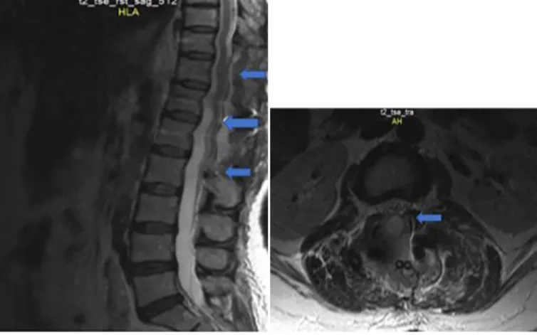
Her lumbar MRI revealed an SSEH, which extended along with the corpus of the T11–L1 vertebrae, mostly to the left of the spinal canal, causing 70% stenosis of the spinal canal and severely compressing it (Figure 1).
Fresh frozen plasma (FFP) and vitamin K were administered to the patient to correct the preoperative INR value. The INR value was 1.5 s 1 day after admission. The patient’s general condition did not improve after one day of admission. Therefore, we decided to perform surgery.
Under general anaesthesia, the patient underwent a laminectomy along the SSEH and where the spinal cord was decompressed. Thoracolumbar instrumentation was performed following three distant laminectomies because of the risk of kyphosis development in the thoracolumbar junction in the future. On the fifth postoperative day, the muscle strength of both lower extremities was 3/5. She was mobilised with assistance. On the 40th postoperative day, she visited the
outpatient clinic with a full recovery. The muscle strength of both lower extremities was 5/5. The control MRI showed relief of the spinal cord (Figure 2).
Six years postoperatively, the patient had no signs or symptoms, except for mild kyphosis.
DISCUSSION
In 1869, Jackson described the first case of a spinal epidural hematoma. Thereafter, the number of patients with SSEH who underwent surgical intervention exceeded 600. In recent years, case reports of patients with SSEH who have shown improvement with conservative treatment have also been published. With the invention of MRI, the number of cases diagnosed with SSEH increased. The internal vertebral venous plexus is the most likely source of SSEH. Most hematomas are
located in the posterolateral or posterior part of the spinal canal(4).
Epidural haemorrhages can be classified as idiopathic, spontaneous and secondary. SSEH accounts for 0.3-0.9% of spinal epidural hematomas. In 2010, the case of a 42-year-old male patient with SSEH was reported. He used aspirin and warfarin for deep vein thrombosis, and his INR value was 2.2. His spinal MRI revealed an SSEH at the right posterolateral area between the T12 and L1 levels. He was started on FFP, vitamin K and dexamethasone treatment after the discontinuation of the anticoagulant therapy. The patient who presented with myeloradiculopathy showed complete neurological improvement on the sixth day and radiological improvement in the eighth week(3). This case is interesting because the patient
had a complete recovery, both neurologically and radiologically, with conservative follow-up and medical treatment.
In 2017, the case of a 76-year-old female patient with an SSEH between C2 and S5 vertebrae levels was published. The patient had a history of pulmonary embolism. She had developed acute quadriplegia and was intubated while receiving warfarin treatment. Her INR value was 14.04. After normalisation of her INR, neurological recovery began. Although no surgical treatment was performed, complete neurological and radiological improvement was seen at the 3-month follow-up(5). The spontaneous resolution of SSEH, which extends
along almost the entire spinal canal, and both neurological and radiological recovery in a short time raises the question of whether surgical treatment is actually needed.
Another 86-year-old male patient receiving warfarin treatment for chronic atrial fibrillation developed bladder dysfunction and painful paraparesis. An SSEH was detected between C7 and T6 vertebrae levels and showed improvement with conservative treatment(6). In an article published in 2007, a 70-year-old female
patient receiving warfarin treatment developed severe low back pain and paraplegia. Despite surgical treatment, her clinical
Figure 1. a) Preoperative T2 MRI sagittal section of the
thoraco-lumbar spine shows hyperintense T11-L1 SSEH and cord compres-sion (blue arrows); b) Axial view of SSEH in the thoracolumbar
spine in a preoperative T2 MRI section (blue arrow)
MRI: Magnetic resonance imaging, SSEH: Spontaneous spinal epidural hematoma
Figure 2. a) Postoperative thoracolumbar T2 MRI sagittal section
showed complete evacuation of SSHE and anterior subarachnoid space of the spinal cord was enlarged (blue arrows); b) No SSEH
is apparent in a postoperative T2 MRI axial section (blue arrow) MRI: Magnetic resonance imaging, SSEH: Spontaneous spinal epidural haematoma
Özger and Kaplan. Spontaneous Spinal Epidural Hematoma Following Warfarin Treatment J Turk Spinal Surg 2020;31(2):108-10
110
signs did not improve even after 14 months postoperatively(7).
As illustrated, there have been case reports indicating that the non-surgical approach might be more moderate because of the unsuccessful cases after surgical treatment.
Between 2009 and 2016, a case series of 10 patients with a mean age of 70 years who underwent surgery for SSEH was published. It was reported that 50% of these patients recovered postoperatively. In addition, the time delay from the onset of symptoms to surgery could not be correlated with the outcome(8).
In our case, the time between the onset of symptoms and the surgical procedure was 1 day. However, this article emphasizes that the effect of delayed time on the outcome is not significant. In a 2004 article, a 60-year-old female patient presented to the emergency department with acute paraplegia. An SSEH extending between T1 and T12 vertebra levels was detected. This was the first report of an SSEH that had an extent spanning the entire thoracic level. The patient recovered completely, both neurologically and radiologically(9). The authors reported that
although the surgical procedure was successful, conservative follow-up may also be an option in neurologically stable cases with SSEH.
In 2018, the authors reported a tumour-like spontaneous lumbar epidural hematoma in a 50-year-old female patient receiving warfarin therapy. The patient underwent surgical treatment. Lumbar MRI performed 1 year later showed that the SSEH was completely absorbed(10). In this case, the authors
argued that surgical intervention should generally be preferred in patients with progressive spinal compression symptoms. This issue in patients with SSEH who were receiving warfarin treatment is essentially similar to that in our patient. In our case, we achieved successful results with surgical intervention. However, there are several opinions in the literature. Although the clinical evidence is weak, some cases show complete recovery with conservative follow-up. Therefore, although it is said that SSEH treatment requires urgent surgery, a complete consensus has not been formed because of the rarity of cases. In conclusion; surgical treatment should be given priority in patients with SSEH receiving anticoagulant therapy, such as warfarin, if progressive neurological deficit is noted. However, patients who do not have neurological deficits or those who recover neurologically while being prepped for surgery can be followed up conservatively. In both cases, vitamin K and FFP
supplementation should be administered to patients with an INR value above the therapeutic levels.
Ethics
Informed Consent: Informed consent was obtained from the
patient and patient’s daughter.
Authorship Contributions
Surgical and Medical Practices: Ö.Ö., N.K., Concept: Ö.Ö., N.K., Design: Ö.Ö., N.K., Data Collection or Processing: Ö.Ö., N.K., Analysis or Interpretation: Ö.Ö., N.K., Literature Search: Ö.Ö., N.K., Writing: Ö.Ö., N.K.
Conflict of Interest: No conflict of interest was declared by the
authors.
Financial Disclosure: The authors declared that this study
received no financial support.
REFERENCES
1. Kirazli Y, Akkoc Y, Kanyilmaz S. Spinal epidural hematoma associated with oral anticoagulation therapy. Am J Phys Med Rehabil. 2004;83:220-23.
2. Yabe H, Ishii A, Niikawa N, Matsubayashi H, Kakei M, Kawakami M, et al. An elderly patient who developed spontaneous spinal epidural hematoma during warfarin therapy. Intern Med. 2012;51:1429-32. 3. Sagar A, Hassan K. Drug interaction as cause of spontaneously
resolving epidural spinal hematoma on warfarin therapy. J Neurosci Rural Pract. 2010;1:39-42.
4. Sandvig A, Jonsson H. Spontaneous chronic epidural hematoma in the lumbar spine associated with Warfarin intake: a case report. Springerplus. 2016;5:1832.
5. Raasck K, Khoury J, Aoude A, Abduljabbar F, Jarzem P. Nonsurgical management of an extensive spontaneous spinal epidural hematoma causing quadriplegia and respiratory distress in a choledocholithiasis patient: A case report. Medicine (Baltimore). 2017;96:e9368. 6. Zuliani G, Guerra G, Dalla Nora E, Fainardi E. Spinal anterior epidural
hematoma in an elderly man with unrecognized lupic anticoagulant taking warfarin. Aging Clin Exp Res. 2011;3:498-500.
7. Adam M, Leblebici B, Akman MN. Spontaneous spinal epidural hematoma related to warfarin therapy: A case report. J Back Musculoskelet Rehabil. 2007;20:11-14.
8. Raj R, Seppälä M, Siironen J. Spontaneous spinal epidural hematoma: a surgical case series of ten patients. World Neurosurgery. 2016;93:55-9. 9. Kotil K, Akcetin MA, Kuscuoglu U, Eras M, Acar C, Bilge T. Spontaneous
Thoracic Spinal Epidural Hematoma Causing Paraplegia. Turk Neurosurg. 2004;14:45-8.
10. Kayalar AE, Gel G, Elbir Ç, Hastürk AE. Tumor-Like Spontaneous Lumbar Epidural Hematoma in a Patient Under Anticoagulant Treatment. J Turk Spinal Surg. 2018;29:45-8.