our patients could be the disappearance of marked MR jet agitated blood stasis in LA cavity and the reduced mitral valve area due to clip. Another possibility is that the acute increase in LV afterload induced by removing the low-impedance regurgitant flow may have contributed to LASEC formation.
The occurrence of a heart thrombus associated Mitraclip proce-dure is rare and, to our knowledge, has been previously reported only in the LA and LV with routine pre-discharge echocardiography few days after implantation (2-5). In our second case, thrombus formation was observed during the Mitraclip procedure. However, there is no data in the literature on LASEC formation after or during the Mitraclip proce-dure.
In our second case, the mechanism of interatrial thrombus forma-tion after mitraclip implantaforma-tion might be the disappearance of severe MR jet agitated blood stasis in LA cavity. However, the reduced mitral valve area due to clip, endocardial damage during septal puncture, an inflammatory response to foreign body (guide catheter) contact with the atrial septum and the duration of the Mitraclip procedure may have contributed to a prothrombotic or hypercoagulable state, which could be responsible for thrombus formation. The thrombus could also origi-nate from the inside of the guide catheter while moving the guide catheter out. In addition, immediately after the Mitraclip procedure, a thrombus was observed despite having an ACT of 260 s. This case may also illustrate the need to be cautious despite achieving ACTs of >250 s during the Mitraclip procedure especially in the presence of AF.
Conclusion
This report shows that thrombus and SEC formation in the LA may occur during percutaneous mitral valve repair with the MitraClip sys-tem of severe MR.
Video 1. Transesophageal echocardiography demonstrates a trace residual MR and marked LASEC in left atrium and left atrial append-age in case 1
Video 2. LASEC was clearly absent immediately before grasping the leaflets in case 1
Video 3. Transesophageal echocardiography demonstrates a mobile echogenic and fluctuating mass seemed to be attached to the inter-atrial septum at the septal puncture site and mild LASEC immedi-ately after the guide catheter removal from the interatrial septum in case 2
References
1. Movsowitz C, Movsowitz HD, Jacobs LE, Meyerowitz CB, Podolsky LA, Kotler MN. Significant mitral regurgitation is protective against left atrial spontaneous echo contrast and thrombus as assessed by transesophageal echocardiography. J Am Soc Echocardiogr 1993; 6: 107-14. [CrossRef]
2. Bekeredjian R, Mereles D, Pleger S, Krumsdorf U, Katus HA, Rottbauer W. Large atrial thrombus formation after MitraClip implantation: is anticoagulation mandatory? J Heart Valve Dis 2011; 20: 146-8.
3. Orban M, Lesevic H, Massberg S, Hausleiter J. Left ventricular thrombus formation after successful percutaneous edge-to-edge mitral valve repair. Eur Heart J 2013; 34: 942. [CrossRef]
4. Pleger ST, Schulz-Schönhagen M, Geis N, Mereles D, Chorianopoulos E, Antaredja M, et al. One year clinical efficacy and reverse cardiac remodelling in patients with severe mitral regurgitation and reduced ejection fraction after MitraClip implantation. Eur J Heart Fail 2013; 15: 919-27. [CrossRef]
5. Orban M, Braun D, Sonne C, Orban M, Thaler R, Grebmer C, et al. Dangerous liaison: successful percutaneous edge-to-edge mitral valve repair in patients with end-stage systolic heart failure can cause left ventricular thrombus formation. EuroIntervention 2013 Oct 30. pii: 20130326-01. Address for Correspondence: Dr. Ayşe Saatcı Yaşar,
Atatürk Eğitim ve Araştırma Hastanesi, Kardiyoloji Kliniği, Bilkent, 06530, Ankara-Türkiye
Phone: +90 312 291 25 25
E-mail: [email protected] Available Online Date: 25.06.2014
©Copyright 2014 by Turkish Society of Cardiology - Available online at www.anakarder.com DOI:10.5152/akd.2014.5355
Cryoablation of an anteroseptal
acces-sory pathway through the jugular and
subclavian veins in a patient with
interruption of the inferior vena cava
and azygos continuation
Basri Amasyalı, Taner Şen, Ayhan Kılıç1
Department of Cardiology, Faculty of Medicine, Dumlupınar University; Kütahya-Turkey
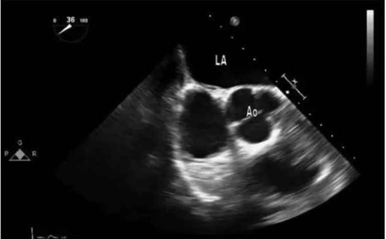
1Department of Cardiology, Gülhane Military Medical Academy; Ankara-Turkey Figure 3. Basal short axis view before procedure in case 2
Figure 4. Basal short axis view showing echogenic mass attached to the interatrial septum at the septal puncture site in case 2
Case Reports Anadolu Kardiyol Derg 2014; 14: 549-57
Introduction
Interruption of the inferior vena cava (IVC) is a rare entity, usually accompanied by azygos or hemiazygos continuation. Venous blood from the caudal part of the body reaches the heart via the azygos vein and superior vena cava. Interruption of the IVC with azygos continua-tion is seen in 0.6% of patients with congenital heart disease and less than 0.3% of individuals without any other variations or anomalies (1). The importance of this anomaly is that it may coexist with congenital heart defects and may interfere with catheter manipulation. On the other hand, ablation of anteroseptally located accessory pathways can be challenging because of the increased risk of atrioventricular (AV) block. This report presents successful cryoablation of an anteroseptal accessory pathway through the jugular and subclavian veins in a young adult with interrupted inferior vena cava and azygos continuation, in whom the femoral approach was not possible due to the helix-like structure of the azygos system.
Case Report
A 32-year-old male patient was admitted with symptoms of palpita-tion and dizziness in the last five years, occurring 2-3 times in a month and lasting 1-3 hours despite various antiarrhythmic drugs.
Physical examination and laboratory findings were normal. Heart rate was 80 beats/min and blood pressure was 120/65 mm Hg. A 12-lead ECG during sinus rhythm confirmed the presence of ventricular pre-excitation consistent with an anteroseptally located accessory
path-way. Echocardiographic examination was reported as normal and there was no sign of any structural heart disease.
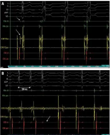
After informed consent, he was taken to the electrophysiology laboratory. Three venous sheaths were introduced to the right femoral vein but catheters could not be advanced to the heart. Contrast venog-raphy revealed an interrupted inferior vena cava and dilated, helix-like azygos and hemiazygos veins (Fig. 1). Then, upper torso was prepared and one right jugular and two left subclavian sheaths were introduced. A quadripolar steerable catheter (Marinr CS, Medtronic, MN, USA) was placed into the coronary sinus and another diagnostic quadripolar catheter (Soloist, Medtronic, MN, USA) was placed at the His bundle region. Intracardiac mapping with a 6 mm-tipped cryoablation catheter (Freezor Xtra, Medtronic CryoCath LP, Canada) revealed an anterosep-tally located accessory pathway (Fig. 2A). The anterograde and retro-grade effective refractory period of the accessory pathway were 500-300 msec and 500-320 msec, respectively. The Wenckebach point was at 290 msn. Programmed atrial stimulation induced an orthodromic tachycardia with a tachycardia cycle length of 360 msec (Fig. 2B). We preferred to ablate the accessory pathway during sinus rhythm because of the risk of catheter dislodgement and less effective energy delivery to the targeted point during high heart rates, although less common during cryoablation. Besides, presence of intermittent pre-excitation made it possible to follow the His potential during ablation under sinus Figure 1. Interruption of inferior vena cava and azygos continuation.
(A) Ablation catheter (arrow) cannot be advanced through tortuous azygos veins. (B) Interruption of the inferior vena cava. (C) Dilated and accordion-like azygos and hemiazygos veins. (D) Azygos vein emptying into the superior vena cava
A
C
B
D
Figure 2. (A) Anteroseptally located accessory pathway (arrows point the His deflection) showing intermittent pre-excitation. (B) Successful cryoablation of the anteroseptal accessory pathway during orthodromic tachycardia (tachycardia cycle length: 360 ms). Note the tachycardia terminates with ventriculoatrial block (arrow) followed normally conducted sinus beats without any sign of pre-excitation CRYO - cryoablation catheter; CS - coronary sinus; d - distal; px - proximal
A
B
Case Reports
rhythm. A target site with continuous atrioventricular conduction and absence of His deflection was identified for cryoablation; however, the accessory pathway conduction persisted with cryomapping at -30°C under sinus rhythm. Thus, a decision was made to proceed with cry-omapping under induced tachycardia. As the tachycardia was seen to terminate at the ventriculoatrial limb and the following beats were free of pre-excitation, the catheter tip was further cooled for cryoablation
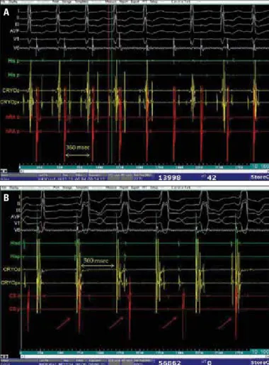
(Fig. 2B). The same target site was cooled to -60°C twice for 4 minutes (Fig. 3). After the ablation procedure, there was no sign of anterograde and retrograde accessory pathway conduction during burst pacing from atrium and ventricle, respectively (Fig. 4A and B). Accessory path-way conduction did not recur after 30 minutes and the procedure was concluded. Total procedure time was 95 minutes and total fluoroscopy time was 22 minutes. After 9 months, the patient is still asymptomatic and there is no preexcitation on the surface ECG.
Discussion
In this paper, we report accomplishment of an already risky antero-septal accessory pathway ablation further complicated by coexisting interrupted IVC and azygos continuation, necessitating alternative routes to reach the right heart.
Interruption of the IVC with azygos continuation is a rare congenital anomaly, in which the IVC is interrupted below the hepatic vein and venous return beyond this point is restored by the dilated azygos and hemiazygos veins draining into the superior vena cava (2).
Currently, cryoablation is effective and safe for patients with acces-sory pathway, with success rates approaching nearly 100% with acceptable complication rates (3). However, ablation of anteroseptally located accessory pathways carries relatively increased risk of AV block and is more complicated as compared to other locations. The most commonly used method for introduction of the ablation catheters to the heart is the femoral approach. However, the femoral approach is usually rendered impractical in the presence of IVC interruption and azygos continuation. There are few reports of successful ablation of accessory pathways in different locations in the presence of IVC inter-ruption and azygos continuation (4-6). To the best of our knowledge, this case report is the first to describe successful cryoablation of an antero-septal accessory pathway using the superior approach via the jugular and subclavian vein in an adult patient in the presence of IVC interrup-tion with azygos continuainterrup-tion.
In the presence of IVC interruption with azygos continuation, positioning and manipulation of the recording and mapping catheters are more difficult due to the longer course and sharp angulation of the azygos vein draining into the superior vena cava. In the literature, some authors have used the femoral approach, coursing through the femoral vein, azygos vein and the superior vena cava into the right atrium while others have used a superior venous approach for abla-tion of different tachyarrhythmias (4-7). The superior approach (via the jugular or subclavian vein) has the advantage of improved cath-eter stability and higher cathcath-eter tip temperatures. On the other hand, the known risk of AV block associated with anteroseptal path-way ablation adds to the importance of catheter stability in these cases. Even if we were able to advance the catheters with the infe-rior approach, stability of the ablation catheter might not be as good as that of the superior approach.
Conclusion
In this case, we preferred jugular and subclavian venous access routes to overcome the inability to advance the catheters through the accordion-like azygos venous system and also to improve catheter stability.
Figure 3. Cryoablation catheter at the site of successful ablation. (A) right anterior oblique view. (B) left anterior oblique view
Abl - ablation; CS - coronary sinus
A B
Figure 4. (A) After the ablation procedure, the Wenckebach cycle length was 360 msec and there was no sign of anterograde accessory pathway conduction. Note that the HV interval is 42 msec. (B) Ventriculoatrial dissociation (arrows) is seen during burst pacing from ventricle with a cycle length of 500 msec
A
B
Case Reports Anadolu Kardiyol Derg 2014; 14: 549-57
References
1. Martinez Garcia MA, Pastor A, Ferrando D, Nieto ML. Casual recognition of an azygous continuation of the inferior vena cava in a patient with lung cancer. Respiration 1999; 66: 66-8. [CrossRef]
2. Chuang VP, Mena CE, Hoskins PA. Congenital anomalies of inferior vena cava. Review of embryogenesis and presentation of a simplified classifi-cation. Br J Radiol 1974; 47: 206-13. [CrossRef]
3. Drago F, Righi D, Placidi S, Russo MS, Di Mambro C, Silvetti MS, et al. Cryoablation of right-sided accessory pathways in children: report of efficacy and safety after 10-year experience and follow-up. Europace 2013; 15: 1651-6. [CrossRef]
4. Guerra Ramos JM, Font ER, Moya I Mitjans A. Radiofrequency catheter ablation of an accessory pathway through an anomalous inferior vena cava with azygos continuation. Europace 2004; 6: 134-7. [CrossRef]
5. Inama G, Vergara G, Gramegna L, Rillo M, Fuochi C, Furlanello F. Catheter ablation of Wolff-Parkinson-White syndrome associated with congenital absence of inferior vena cava. J Interv Card Electrophysiol 1998; 2: 301-4. [CrossRef]
6. Liu QM, Zhou SH, Ouyang FF. Successful radiofrequency ablation of a right posteroseptal accessory pathway through an anomalous inferior vena cava and azygos continuation in a patient with incomplete situs inversus. Cardiol J 2009; 16: 164-7.
7. Pérez-Silva A, Merino JL, Peinado R, Lopez-Sendon J. Atrial flutter abla-tion through the azygous continuaabla-tion in a patient with inferior vena cava interruption. Europace 2011; 13: 442-3. [CrossRef]
Address for Correspondence: Dr. Basri Amasyalı,
Dumlupınar Üniversitesi Tıp Fakültesi, Kardiyoloji Anabilim Dalı, 43000, Kütahya-Türkiye
Phone: +90 274 265 20 31 Fax: +90 274 265 20 16 E-mail: [email protected] Available Online Date: 25.06.2014
©Copyright 2014 by Turkish Society of Cardiology - Available online at www.anakarder.com DOI:10.5152/akd.2014.5512
A rare cause of circulatory shock
Cihan Altın, Arzu İzmir*, Sevda Osmanoğlu**, Esin Gezmiş***, Afşin Sağduyu****
Departments of Cardiology, *Chest Diseases, **Internal Medicine, ***Radiology and ****Psychiatry, Faculty of Medicine, Başkent University; İzmir-Turkey
Introduction
Circulatory shock is a life-threatening clinical syndrome character-ized by hypotension, tachycardia and symptoms of end-organ damage/ failure. Hypovolemic, cardiogenic, distributive, obstructive and neuro-genic shocks are the main types of circulatory shock. To find/diagnose the cause and to give the accurate treatment according to the underly-ing event are crucial in the management of circulatory shock. Herein we present an interesting shock case with unknown etiology which underlines the importance of detailed anamnesis and systemic exami-nation (1).
Case Report
A 43-year-old male patient who works as a stockbroker, was admit-ted to our emergency unit with complaints of nausea, vomiting and dizziness preceding 4-6 hours. On physical examination; he was
con-scious and oriented, his skin was pale, cold and clammy. He had hypo-tension (70/40 mm Hg) and sinus tachycardia. Other physical and neu-rological examinations were normal. On his first anamnesis; there was no history of systemic disease or medication. Only he had had a viral upper respiratory infection two weeks ago. There was no suspected toxin exposure except eating cultivated mushroom 8 hours ago. Multi-systemic examination and multiple consultations were done in order to find out the predisposing factor of this circulatory shock. Which type of shock is this? What is responsible for this clinical syndrome?
His hemogram and biochemical parameters including troponine-I were unremarkable except elevated renal function tests (Creatinine: 2.11 mg/dL). Arterial blood gases revealed hypoxia and hypocapnia. Except sinus tachycardia his all electrocardiographic and echocardi-graphic findings were normal. Our patient did not have any infection symptoms and his all sepsis parameters were unremarkable. Therefore; hypovolemic, cardiogenic and septic shocks were ruled out. Computerize tomography (CT) was performed to rule out/find if the reason was pul-monary embolism. However pulpul-monary embolism was not detected and thoracoabdominal CT was also reported as normal. Mushroom intoxi-cation was not considered because there was no elevation in patients liver function tests. Diagnosis could have been adrenal insufficiency induced by viral infection. Nevertheless patients’ morning cortisol level was normal and there was no significant response on hemodynamic parameters after intravenous steroid therapy. We were not able to rule out adrenal insufficiency with these findings so that Synacthen test was planned.
Despite appropriate and sufficient hydrations (crystalloid and col-loid), patient’s hypotension, hypoxia and oliguria did not improve. Renal impairment progressed. Additionally the patient became complicated by pulmonary edema which might be the result of prolonged hypoten-sion and fluid resuscitation (Fig. 1). To evaluate intravascular volume, central venous catheterization was planned. Before this invasive pro-cedure the patient confessed that he got 20 pills of amlodine (100 mg) as a suicide attempt because he had lost a high amount of money in stock market. During all these non-invasive diagnostic tests he did not confess this suicide attempt. There was no doubt for suicide attempt because he had no depressive mood or behaviors during his hospital-ization. Because we could not provide further treatments, the patient was referred to another clinic.
Discussion
Amlodipine is one of the longest half-life (30-50 hours) dihydropyri-dine calcium channel blocker (CCB) with a large volume of distribution (2). Toxicity may be seen in doses up to 5-10 times the therapeutic dose and occurs within 30-60 minutes following ingestion (3). In severe cases; it can result in prolonged hypotension (up to10 days), dysrhyth-mias and cardiac arrest (2). Our patient developed prolonged hypoten-sion and hypoxia without significant effect on systolic functions and cardiac pacemaker activity. He was complicated with acute renal fail-ure and pulmonary edema. Non-cardiogenic pulmonary edema associ-ated with CCB overdose is previously described in the literature (3-5). The pulmonary capillary transudations related to pre-capillary vasodila-tation was reported as the possible mechanism of pulmonary edema (5). CCB overdose is frequently complicated by renal failure, related to the severe hypoperfusion and end-organ ischemia (2).
There is no specific efficacious antidote for CCB intoxication. Hyperinsulinemic euglycemia using dextrose and insulin infusion,
cal-Case Reports