Yazışma Adresi/Address for Correspondence: Dr. Mehmet Öztürk, Selcuk University Medical Faculty, Department of Radiology, Konya, Turkey. E-mail: [email protected]
Geliş tarihi/Received: 08.08.2017 Kabul tarihi/Accepted: 08.12.2017
ARAŞTIRMA / RESEARCH
Measurement of the distance and angle between the aorta and superior
mesenteric artery and correlation to body-mass index in adolescents
Adölesanlarda süperior mezenterik arter ve aorta arasında mesafe ve açı ölçümü ve
vücut kitle indeksi ile ilişkisi
Mehmet Öztürk
1, Emine Çalışkan
2, Mehmet Sait Menzilcioğlu
3, Serdal Çitil
31Selçuk University, Medical Faculty, Department of Radiology, Division of Pediatric Radiology, Konya, Turkey 2Istanbul University Istanbul Faculty of Medicine Department of Radiology, Istanbul, Turkey
3Ministry of Health Kahramanmaraş Necip Fazıl City Hospital, Kahramanmaraş, Turkey
Cukurova Medical Journal 2018;43(3):568-573
Abstract Öz
Purpose: The aim of this study was to identify the variations in the angle and distance values between the superior mesenteric artery and abdominal aorta with body mass index in healthy adolescents.
Materials and Methods: This retrospective study included a total of 246 adolescent cases; 121 girls and 125 boys. The aorta-mesenteric angle and distance measurements were completed on contrast abdominal computed tomography axial and reformatted sagittal images. Body mass index (kg/m²) was calculated. Results: Mean age was 162.8 and 163.8 months for girls and boys, respectively, with no difference identified. Mean body mass index values for girls and boys were 17.8 and 18.6 respectively, with no significant difference identified. The superior mesenteric artery angle, duodenal and left renal vein distance values were 41.1 degrees, 11.2 mm, and 8.2 mm for girls and 42.7 degrees, 12.6 mm and 9.2 mm for boys with no variation identified according to gender. In both genders and in all cases there was a positive correlation between body mass index with superior mesenteric artery angle, duodenal and left renal vein distances.
Conclusion: In the healthy adolescent population, the aorta-mesenteric angle and distance measurements show positive correlation with body mass index. The values determined may be used as reference values for diagnosis of compression syndromes related to the mesenteric artery.
Amaç: Bu çalışmanın amacı sağlıklı adölesanlarda süperior mezenterik arter ile abdominal aorta arasındaki açı ve uzaklık değerlerinin vücut kitle indeksine göre değişimini saptamaktır.
Gereç ve Yöntem: Bu retrospektif çalışmaya 121’i kız ve 125’i erkek olmak üzere toplam 246 adölesan olgu dahil edildi. Kontrastlı batın bilgisayarlı tomografide aksiyal ve sagittal reformat görüntüler üzerinde aorta-mezenterik açı ve mesafe ölçümleri yapıldı. Vücut kitle indeksi (kg/m²) hesaplandı.
Bulgular: Yaş ortalaması kızlarda ve erkeklerde sırasıyla 162.8 ve 163.8 ay olup aralarında farklılık saptanmadı. Ortalama vücut kitle indeksi değerleri kızlarda ve erkeklerde sırasıyla 17.8 ve 18.6 olup aralarında anlamlı değişiklik saptanmadı. Süperior mezenterik arter açısı, doudenal ve sol renal ven mesafesi değerleri sırasıyla kızlarda 41.1 derece, 11.2 mm, 8.2 mm ve erkeklerde 42.7 derece, 12.6 mm, 9.2 mm olup cinsiyete göre değişiklik saptanmadı. Her iki cinsiyette ve tüm olgularda vücut kitle indeksi ile süperiyor mezenterik arter açısı, doudenal ve sol renal ven mesafeleri pozitif korelasyon göstermekteydi. Sonuç: Sağlıklı adölesan popülasyonunda aortamesenterik açı ve mesafe ölçümleri vücut kitle indeksi ile pozitif korelasyon göstermektedir. Ortaya çıkan değerler, süperior mezenterik arter ile ilgili sıkışma sendromlarının tanısında referans değerler olarak kullanılabilir.
Key words: Aortomesenteric angle, superior mesenteric
INTRODUCTION
The superior mesenteric artery (SMA) separates from the abdominal aorta (AA) at the level of the 1st lumbar vertebra behind the neck of the pancreas at an acute angle called the SMA angle or aorto-mesenteric angle. The left renal vein (LRV) and third section of the duodenum pass from right to left in the opening between these two arteries. Findings resulting from compression of these structures have been defined in the literature as Wilkie and nutcracker syndromes (NS).
Wilkie syndrome (WS), also known as SMA compression syndrome, was first described by von Rokitanski in 1861 while Wilkie described the pathophysiology and clinical findings in detail1,2. In WS there is external compression of the third section of the duodenum. In healthy individuals the mesenteric fat tissue and lymphatic tissue in the paraaortic field and around the SMA protect the duodenum from compression. However, in thin individuals with rapid weight loss fatty tissue reduces and the resistance to compression lessens. Symptoms of blockage of the upper gastrointestinal system occur without any intraluminal obstacle on endoscopic examination.
Compression of the LRV between the SMA and AA, known as NS, was first described by Grant3. In NS, intraluminal pressure increase occurs due to trapping of the LRV in the mesoaortic angle. The etiology includes reasons such as renal pitosis, abnormally high left renal vein, abnormally narrow angle of the SMA aortic branch, pancreas masses, lymphadenomegaly, and scarce retroperitoneal fat tissue4. The venous congestion may cause sensitivity of the left side of the vagina, dyspareunia, dysuria, dysmenorrhea, varicose vein formation in scrotal, gluteal and lower extremity regions and less frequently gastrointestinal symptoms like stomach pain5.
In the literature there are studies stating the importance of the angle and distance between the SMA and AA in the etiology of compression syndromes. Additionally, the amount of retroperitoneal fat tissue is known to affect the angle and distance between the SMA and AA 6,7. Body mass index (BMI) provides direct information about the fat mass in the body8. In children body morphology and fat distribution may display variability depending on gender, age, peri and post
pubertal periods. There are studies on the angle and distance in the childhood period, but there is insufficient data about the adolescent age group, especially9.
The aim of the study is to measure the aorta-mesenteric angle and distance with contrast abdominal computed tomography (CT) in healthy adolescents and research the correlation with BMI.
MATERIALS AND METHODS
This retrospective study, completed after receiving permission from the local clinical research ethics committee of Selcuk University Medical Faculty (Ethics committee year and number: 2017/ 359), included a total of 246 cases with ages varying from 10 to 17 years, with contrast abdominal CT in the Picture Archiving Communication Systems archive of Radiology department from March 2012 to March 2017. The ages, gender, weights, and height of the cases were obtained by scanning files from the hospital medical archive unit. The BMI was calculated as the weight (kg) divided by the square of the height [weight (kg)/height (m)2]. Cases with intraabdominal mass and fluid, paraaortic lymphadenopathy, scoliosis identified, insufficient contrast material opacification in the mesenteric artery and previous abdominal surgery history were excluded from the study.
CT technique and measurements
Images were obtained using a 16 slice multislice helical CT device (Somatom Sensation 16, Siemens, AG, Erlanger, Germany). All cases had gantry rotation time 0.5 s, tube voltage 140 kVp and tube flow 100-380 mA used. All patients were initially administered 800-1000 mL diatrizoate meglumine (Urovist-AngiograWn; Schering, Germany) at 2-3% dilute concentration by mouth over 45 minutes. Standard intravenous contrast material protocol included 2 mg/kg iopromide (Ultravist 300, Schering AG. Berlin, Germany) injected at 2 ml/s with an automatic injector (CT Injector; Ulrich Medical, Ulm-Jungingen, Germany). According to our standard abdominal CT protocol, with the patient in supine position 10 mm slices were obtained at 8 mm intervals. Later 2 mm slice thickness axial images were reconstructed with a 512x512 pixel image matrix using central sharp convolution kernel (B30f). All image data were sent to a work station (Leonardo, Siemens AG, Erlanger,
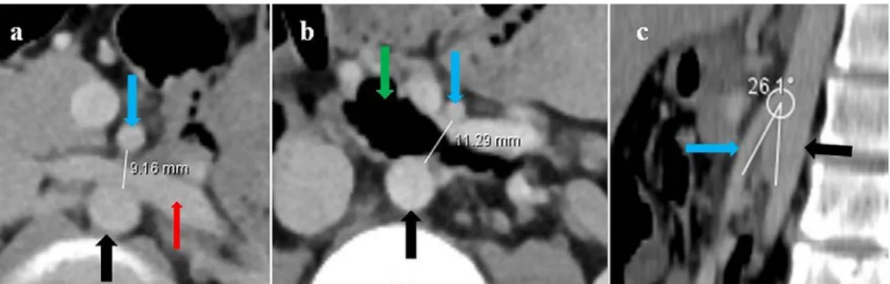
570 Germany) for electronic interpretation. To assess the branch configuration from AA to SMA, sagittal or oblique-sagittal multiplanar reconstruction images were obtained. The angle and distances between the SMA and aorta were measured by the same radiologist 3 times and then the mean was calculated. The distance between the SMA and AA was measured as the maximum distance between the anterior wall of the aorta and the posterior wall of the superior mesenteric artery at the level where the
left renal vein and duodenum intersect (Figure 1 a, b). The angle between these veins was measured on reformatted sagittal-oblique sagittal images at the same level. For angle measurements a line was drawn to an imaginary point between the SMA root and where the SMA began to descend parallel to the AA (Figure 1 c). Measurements were made with electronic calipers. Angles were drawn by hand and degrees were automatically calculated.
Figure 1 a, b, c. Contrast abdominal CT investigation: Measurement distances on axial slices at level of LRV (a) and duodenum (b). Aorta-mesenteric angle measurements on sagittal reformatted images (c).
Statistical analysis
For analysis of variables SPSS 22.0 (IBM Corporation, Armonk, New York, United States) program was used. Normal distribution of data was assessed with the Shapiro-Wilk test. Comparison of quantitative data from two independent groups used the Mann-Whitney U (exact) test with Monte Carlo results. To investigate the correlation between variables, Kendall’s tau-b test was used. Quantitative variables are given as mean ± sd (standard deviation) and median range (maximum-minimum) in tables. Variables were investigated at the 95% confidence interval with p values smaller than 0.05 accepted as significant.
RESULTS
Mean age was 162.8 and 163.8 months for girls and boys, respectively, with no difference identified (p=0.978). Mean BMI values for girls and boys were 17.8 and 18.6 respectively, with no significant difference identified (p=0.782). The SMA angle, duodenal and LRV distance values were 41.1 degrees, 11.2 mm, and 8.2 mm for girls and 42.7 degrees, 12.6 mm and 9.2 mm for boys with no
variation identified according to gender (p=0.932, 0.958 and 0.926, respectively). Descriptive data and SMA angle-distance values for the study population are summarized in Table 1. The correlation of variables in the study population is given in Table 2. In both genders and in all cases there was a positive correlation between BMI with SMA angle, duodenal and LRV distances (p<0.001, for each) (Figure 2, 3, 4).
DISCUSSION
This study aiming to assess the correlation of the distance and angle between the SMA and AA with age, gender and BMI in healthy adolescents completed measurements with contrast abdominal CT and determined important results. The first result is determination of normal values for adolescents according to gender and BMI. The second is the identification of a positive increasing correlation between BMI and the aorta-mesenteric distance and angle. The third result is that CT is an objective and reliable radiologic method to provide data for diagnosis of aorta-mesenteric compression syndromes.
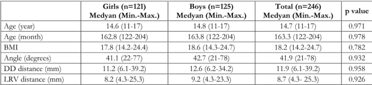
Table 1. Descriptive data and Superior mesenteric artery angle-distance values for the study population Girls (n=121)
Medyan (Min.-Max.) Medyan (Min.-Max.) Boys (n=125) Medyan (Min.-Max.) p value Total (n=246)
Age (year) 14.6 (11-17) 14.8 (11-17) 14.7 (11-17) 0.971 Age (month) 162.8 (122-204) 163.8 (122-204) 163.3 (122-204) 0.978 BMI 17.8 (14.2-24.4) 18.6 (14.3-24.7) 18.2 (14.2-24.7) 0.782 Angle (degrees) 41.1 (22-77) 42.7 (21-78) 41.9 (21-78) 0.932 DD distance (mm) 11.2 (6.1-39.2) 12.6 (6.2-34.2) 11.9 (6.1-39.2) 0.958 LRV distance (mm) 8.2 (4.3-25.3) 9.2 (4.3-23.3) 8.7 (4.3- 25.3) 0.926 Table 2. Correlations of variables in the study population
Angle (degrees) Duodenal distance (mm) Left renal vein distance (mm)
r p r p r p Girls Age (year) -0.017 0.782 -0.005 0.940 0.014 0.827 Age (month) -0.005 0.934 0.016 0.787 0.029 0.622 Body-mass index (kg/m²) 0.508 <0.001 0.465 <0.001 0.437 <0.001 Boys Age (year) 0.013 0.838 0.007 0.906 0.002 0.968 Age (month) 0.022 0.710 0.024 0.685 0.014 0.817 Body-mass index (kg/m²) 0.502 <0.001 0.436 <0.001 0.438 <0.001 Total Age (year) -0.002 0.967 0.001 0.986 0.008 0.853 Age (month) 0.009 0.835 0.019 0.648 0.021 0.613 Body-mass index(kg/m²) 0.499 <0.001 0.445 <0.001 0.430 <0.001
Figure 2. Distribution graphics of the SMA angle according to BMI in the study population.
Figure 3. Distribution graphics of the duodenal distances according to BMI in the study population.
Figure 4. Distribution graphics of the LRV distances according to BMI in the study population. The SMA separates from the anterior of the
abdominal aorta immediately inferior of the celiac truncus at the level of the L1-2 vertebra corpus with a sharp angle. The retroperitoneal and mesenteric fat and lymphatic tissue wrapping the SMA plays an important role in keeping this distance and angle large enough and protects the duodenum from compression between the SMA and aorta7. Studies
in healthy adults have reported the aorta-mesenteric angle is between 38-60 with a mean of 45 degrees10. At the level of the third section of the duodenum between the SMA and AA, the distance was identified as 10-28 mm11.
Ozkurt et al. in a study of healthy adults identified a positive increasing correlation between BMI and aorta-mesenteric angle and distance in both
572 genders12. Another study of normal adults in the Indian population completed measurements at three different levels from the branching of the SMA from the AA and assessed the correlation with BMI. They identified a positive increasing correlation with BMI at all three levels13.
Arthurs et al. in a study of 205 healthy children measured separate distances at the levels of the LRV and duodenum and the aorta-mesenteric angle to evaluate the correlation with visceral intraabdominal fat tissue volume. They found the aorta-mesenteric angle was 45.6±19.6 degrees with distance at the LRV level of 8.6±3.9 mm and at the duodenal level of 11.3±4.8 mm. They identified a positive correlation of intraabdominal fat tissue volume with aorta-mesenteric angle and distances9. This study only included the adolescent age group and produced similar results to the study by Arthurs et al. Additionally, BMI was used instead of intraabdominal fat tissue volume, with a positive correlation identified with aorta-mesenteric angle and distance values.
The mechanism of NS is not fully understood, with debates continuing on how to make definite diagnosis. Due to changing hemodynamics as a result of compression of the LRV with the SMA, intermittent hypertension may occur in the proximal LRV. Definite diagnosis of NS is made by measuring pressure gradients between the LRV and inferior vena cava with renal venography. However, the use of radiation during this procedure forms a risk for children and adolescents14,15. Measurements made with the non-invasive method of renal color Doppler ultrasound have measured high peak velocity in the LRV in symptomatic NS cases. The highest velocity rates before and after the LRV passes under the SMA are significantly high in children with NS; however the LRV rate interval is very wide in normal children16. Previous CT studies have reported an aorta-mesenteric distance less than 5 mm is significant between symptomatic and non-symptomatic children with NS17. However, it is not clear which cases should be treated. This study of healthy adolescents determined the normal angle and distance values with CT. These results may be used as reference values to differentiate normal and abnormal cases.
There are significant limitations to our study. The study did not include cases with clinical identification of aorta-mesenteric compression syndrome. As a result, the normal values for
aorta-mesenteric angle and distance were not compared with a patient group. However, our aim was to ease identification based on CT by determining the normal values in adolescents and variation according to BMI.
In conclusion, CT measurements of the healthy adolescent population determined the normal values according to age, gender and BMI. Additionally, a positive increasing correlation was identified between BMI and the aorta-mesenteric angle and distance. CT is a very useful radiologic method for diagnosis of aorta-mesenteric compression syndromes.
REFERENCES
1. Rokitansky C. Handbuch der Pathologischen Anatomie, ed 1. Wien, Braunmüller &Seidel, 1842. 2. Wilkie DP. Chronic duodenal ileus. Am J Med Sci.
1927;173:643-9.
3. Grant JCB. Method of Anatomy. Baltimore, Williams & Wilkins, 1937.
4. Ahmed K, Sampath R, Khan MS. Current trends in the diagnosis and management of renal nutcracker syndrome: a review. Eur J Vasc Endovasc Surg. 2006;31:410-6.
5. Tüzün KH, Arslan C, Deşer SB, Ersoy E, Junusbekov Y. Nutcracker sendromu: olgu sunumu. Damar Cerrahisi Dergisi. 2012;21:51-4.
6. Applegate GR, Cohen AJ. Dynamic CT in superior mesenteric artery syndrome. J Comput Assist Tomogr. 1988;12:976–80.
7. Ahmed AR, Taylor I. Superior mesenteric artery syndrome. Postgrad Med. 1997;73:776-8.
8. Bedogni G, Pietrobelli A, Heymsfield SB, Borghi A, Manzieri AM, Morini P et al. Is body mass index a measure of adiposity in elderly women? Obes Res. 2001;9:17-20.
9. Arthurs OJ, Mehta U, Set PA. Nutcracker and SMA syndromes: What is the normal SMA angle in children? Eur J Radiol. 2012;81:854-6.
10. Baltazar U, Dunn J, Floresguerra C, Schmidt L, Browder W. Superior mesenteric artery syndrome: an uncommon cause of intestinal obstruction. South Med J. 2000;93:606-8.
11. Payawal JH, Cohen AJ, Stamos MJ Superior mesenteric artery syndrome involving the duodenum and jejunum. Emerg Radiol. 2004;10:273-5.
12. Ozkurt H, Cenker MM, Bas N, Erturk SM, Basak M. Measurement of the distance and angle between the aorta and superior mesenteric artery: normal values in different BMI categories. Surg Radiol Anat. 2007;29:595-9.
13. Bhagirath Desai A, Sandeep Shah D, Jagat Bhatt C, Umesh Vaishnav K, Salvi B. Measurement of the distance and angle between the aorta and superior
mesenteric artery on CT scan: values in Indian population in different BMI categories. Indian J Surg. 2015;77:614-7.
14. Cheon JE, Kim WS, Kim IO, Kim SH, Yeon KM, Ha IS et al. Nutcracker syndrome in children with gross haematuria: doppler sonographic evaluation of the left renal vein. Pediatric Radiology. 2006;36:682-6.
15. Kim SH, Cho SW, Kim HD, Chung JW, Park JH, Han MC. Nutcracker syndrome: diagnosis with
Doppler US. Radiology. 1996;198:93-7.
16. Zerin JM, Hernandez RJ, Sedman AB, Kelsch RC. Dilatation of the left renal vein on computed tomography in children: a normal variant. Pediatr Radiol. 1991;21:267-9.
17. Imamura A, Nakamura M, Maekawa N, Matsuya F, Kanetake H, Saito Y. Usefulness of renal CT scan for analysis of nutcracker phenomenon. Nippon Hinyokika Gakkai Zasshi. 1992;83:1861-5.