SKEWED X-CHROMOSOME INACTIVATION IN
JUVENILE IDIOPATHIC ARTHRITIS AND
RHEUMATOID ARTHRITIS
A THESIS SUBMITTED TO
THE DEPARTMENT OF MOLECULAR BIOLOGY AND GENETICS AND GRADUATE SCHOOL OF ENGINERING AND SCIENCE OF
BILKENT UNIVERSITY
IN PARTIAL FULFILLMENT OF THE REQUIREMENTS FOR THE DEGREE OF DOCTOR OF PHILOSOPHY
BY
CHIGDEM AYDIN MUSTAFA JUNE, 2012
ii I certify that I have read this thesis and that in my opinion it is fully adequate, in scope and in quality, as a thesis for the degree of Doctor of Philosophy.
Prof. Dr. Tayfun Özçelik
I certify that I have read this thesis and that in my opinion it is fully adequate, in scope and in quality, as a thesis for the degree of Doctor of Philosophy.
Assoc. Prof. Dr. Işık Yuluğ
I certify that I have read this thesis and that in my opinion it is fully adequate, in scope and in quality, as a thesis for the degree of Doctor of Philosophy.
iii I certify that I have read this thesis and that in my opinion it is fully adequate, in scope and in quality, as a thesis for the degree of Doctor of Philosophy.
Assist. Prof. Katja Doerschner
I certify that I have read this thesis and that in my opinion it is fully adequate, in scope and in quality, as a thesis for the degree of Doctor of Philosophy.
Assoc. Prof. Dr. Hilal Özdağ
Approved for the Graduate School of Engineering and Science
Director of Graduate School of Engineering and Science Prof. Dr. Levent Onural
iv
ABSTRACT
SKEWED X-CHROMOSOME INACTIVATION IN JUVENILE IDIOPATHIC ARTHRITIS AND RHEUMATOID
ARTHRITIS
Chigdem Aydın Mustafa
PhD in Molecular Biology and Genetics Supervisor: Prof. Dr. Tayfun Özçelik
June, 2012
There is a female predominance in most of the autoimmune diseases, and it is thought to play an important role in identifying the etiological factors. Sex hormones, microchimerism and environmental factors are thought to be responsible. Nowadays, it is proposed that a disturbance in mosaicism of females may cause autoimmune disease development. Recently, in our lab, an association between extremely skewed X-chromosome inactivation (XCI) patterns and female predisposition to autoimmunity was identified in Turkish population. In this study, we hypothesized that skewed XCI might play a role in the disease development of Juvenile idiopathic arthritis (JIA) in Turkish, and Rheumatoid Arthritis (RA) in French population. Therefore, XCI status of healthy individuals and patients diagnosed with JIA and RA were genotyped by analyzing androgen receptor (AR) locus by methylation sensitive HpaII digestion followed by PCR. Extremely skewed XCI was observed in a significant proportion of JIA (OR: 11.33; P=0.0008) in Turkish population, and RA (OR: 7.6; P=0.005) in French population. In conclusion, our results suggest that extremely skewed XCI may play an important role in autoimmune disease pathogenesis.
v
ÖZET
JÜVENİL İDİYOPATİK ARTRİT VE ROMATOİD ARTRİT HASTALIĞINDA X KROMOZOMU İNAKTİVASYONU SAPMASI
Chigdem Aydın Mustafa
Doktora Tezi, Moleküler Biyoloji ve Genetik Tez Danışmanı: Prof. Dr. Tayfun Özçelik
Haziran, 2012
Dünya çapında en sık rastlanan hastalık olan otoimmün hastalıkların çoğu kadınlarda daha sık görülmektedir. Bunun nedeni olarak cinsiyet hormonları, mikrokimerizm ve çevresel etkenler sorumlu tutulmaktadır. Son zamanlarda X kromozomu inaktivasyonu (XCI) sapması da buna neden olarak gösterilmektedir. Yakın zamanda laboratuarımızda gerçekleştirilen çalışmalarda, Türk popülasyonunda, X kromozomu inaktivasyonu sapması ve kadınların otoimmün hastalıklara yatkınlığı arasında bağlantı kurulmuştur. Bu çalışmada X kromozomu inaktivasyonuna bağlı mozaik yapının bozulmasının otoimmün hastalık etiolojisinde rol alabileceği hipotezi test edilmiştir. Bu nedenle Türk popülasyonunda jüvenil idiyopatik artrit (JIA) ve Fransız popülasyonunda romatoid artrit (RA) hastaları ve sağlıklı bireyler genotiplenmiştir. XCI statüsünü belirlemek için androjen reseptörü (AR) geni metillemeye duyarlı HpaII enzimi ile analiz edilmiştir. Türk popülasyonunda JIA (OR: 11.33; P=0.0008) ve Fransız popülasyonunda RA (OR: 7.6; P=0.005) hastalarında XCI’nda aşırı sapma gözlenmiştir. Sonuçlarımız XCI ile otoimmün hastalık gelişimi arasında bir ilişki olabileceği görüşünü desteklemektedir.
vi
To my Family
vii
ACKNOWLEDGEMENTS
First of all, I would like to thank and express my deepest gratitude to my advisor Prof. Dr. Tayfun Özçelik for his guidance, encouragement, support, and patience throughout my thesis work. I have learned a lot from his scientific and personal advices.
It is my pleasure to express my thanks to Prof. Dr. Rezzan Topaloğlu for her help in clinical diagnosis and obtaining patient samples and controls. I would also like to thank Elif Uz for her incredible help in everything and her endless support in the lab.
I would also like to thank Özçelik Lab Members for their incredible help in everything and their endless support in the lab. I was very lucky to have such great group members.
Very special thanks to all MBG family for their friendship and scientific advises. They are one of the attractive reasons for being in Bilkent University.
Lastly but mostly, I would like to thank my family for being there whenever I needed them and supporting me in every decision I gave. Without them and their endless love, nothing would be possible.
viii
TABLE OF CONTENTS
ABSTRACT iv ÖZET v DEDICATION PAGE vi ACKNOWLEDGEMENTS viiTABLE OF CONTENTS viii
LIST OF TABLES xii
LIST OF FIGURES xiiii
ABBREVIATIONS xiv
1. CHAPER I: INTRODUCTION 1
1.1. Immune system 1
1.2. Autoimmunity 2 1.2.1 Cause of autoimmunity 3 1.2.1.1. Genes associated with autoimmunity 4 1.2.1.1.1. AIRE 4 1.2.1.1.2. CTLA4 4 1.2.1.1.3. FOXP3 5 1.2.1.1.4. PTPN22 5 1.2.1.1.5. STAT4 5 1.2.1.2. Molecular Mimicry 6 1.2.1.3. Epigenetics 6
1.2.2. Female predominance in autoimmunity 6
1.2.2.1. Hormones 7
1.2.2.2. Microchimerism 8
1.2.2.3. Skewed X-inactivation 8
ix
1.3.1. History 9
1.3.2. Mechanism 9
1.4. Autoimmune disorders that were selected in this study 11
1.4.1. Rheumatoid arthritis (RA) 11
1.4.1.1. Classification 11
1.4.1.2. Prevalence, incidence 12
1.4.1.3. Causes 12
1.4.1.3.1. Associated genes 12
1.4.1.3.2. Antibodies 13
1.4.2. Juvenile Idiopathic Arthritis (JIA) 13
1.4.2.1. Classification 14 1.4.2.2. Types 15 1.4.2.2.1. Oligoarticular JIA 15 1.4.2.2.2. Polyarticular JIA 16 1.4.2.2.2.1. Rheumatoid-factor- positive polyarthritis 16 1.4.2.2.2.2. Rheumatoid-factor- negative polyarthritis 16 1.4.2.2.3. Systemic JIA 17 1.4.2.2.4. Enthesitis-related arthritis 17 1.4.2.2.5. Psoriatic arthritis 17 1.4.2.2.6. Undifferentiated arthritis 18 1.4.2.3. Prevalence, incidence 18 1.4.2.4. Causes 19 1.4.2.4.1. Associated genes 19 1.4.2.4.2. Antibodies 19
1.5. Aim and Strategy 20
2. CHAPTER II: MATERIALS AND METHODS 21
2.1 Materials 21
2.1.1. Pediatric samples 21
2.1.1.1. Turkish children controls 21
2.1.1.2. Turkish juvenile idiopathic arthritis patients 21
x
2.1.2.1. French controls 22
2.1.2.2. French rheumatoid arthritis patients 22
2.1.2.3. Turkish adult control samples 22
2.1.2.4. Turkish systemic sclerosis patients 23
2.1.2.5. Turkish autoimmune thyroid disease patients 23 2.1.3. Chemicals, reagents and enzymes 23 2.1.3.1. Primers 23 2.1.3.2. Enzymes 24
2.1.3.3. Thermal cyclers 24
2.1.3.4. Standard solutions and buffers 24
2.1.3.5. Chemicals and reagents 25 2.1.3.6. Nucleic acids 25 2.2. Methods 26
2.2.1. Sample collection 26 2.2.2. Determination of X chromosome inactivation status 27
2.2.2.1. DNA Isolation 28 2.2.2.2. Restriction enzyme digestion 29 2.2.2.3. Polymerase chain reaction (PCR) 29
2.2.2.4. Agarose gel electrophoresis 30
2.2.2.5. Polyacrylamide gel electrophoresis (PAGE) 30 2.2.2.6. Densitometric analysis 30
2.2.3. Determination of frequency of PTPN22 genotypes 31
2.2.3.1. Polymerase chain reaction (PCR) 32
2.2.3.2. Restriction enzyme digestion 33
2.2.4. Statistical analysis 33
3. CHAPTER III: RESULTS 34
3.1. PCR-based X-inactivation study of peripheral blood of Turkish pediatric control samples 34 3.2. PCR-based X-inactivation study of peripheral
blood of Turkish juvenile idiopathic arthritis patients 35 3.3. PCR-based X-inactivation study of peripheral
blood of French control samples 40
xi
blood of French rheumatoid arthritis patients 40
3.5. Determination of frequency of PTPN22 genotypes 44
in Turkish control samples
3.6. Determination of frequency of PTPN22 genotypes 45
in Turkish systemic sclerosis patients
3.7. Determination of frequency of PTPN22 genotypes 46
in Turkish autoimmune thyroid disease patients
3.8. High-density microarray analysis 47
4. CHAPTER IV: DISCUSSION 50
5. CHAPTER V: REFERENCES 56
6. CHAPTER VI: APPENDICIES 65
xii
LIST OF TABLES
Table 1.1 Systemic and organ specific autoimmune diseases 3
Table 1.2 Female to male ratio for selected autoimmune diseases 7
Table 1.3 Comparison of the classification systems of arthritis in children 14 Table 1.4 International League of Associations for Rheumatology (ILAR)
categories of juvenile idiopathic arthritis
18
Table 2.1 Chemicals, reagents, and kits used in the experiments 25
Table 3.1 Proportion of the JIA patients and controls with skewed XCI 36
Table 3.2 Clinical characteristics and XCI status of JIA patients 38
Table 3.3 Proportion of the French RA patients and controls with skewed XCI 42 Table 3.4 Table 3.5 Table 3.6 Table 3.7
XCI patterns of French RA patients.
Frequency of PTPN22 genotypes in individuals with SSc and controls
Frequency of PTPN22 genotypes in individuals with AITD and controls
JIA Allelic Association Results
43 45
46 48
xiii
LIST OF FIGURES
Figure 2.1 Sizes of the fragments of PUC mix marker, 8 and appearance on both agarose and polyacrylamide gel electrophoresis
26
Figure 2.2 Figure 2.3 Figure 2.4
The sequence of AR, exon 1
The formula for calculation of skewing ratio The sequence of PTPN22
28 31 32
Figure 3.1 X chromosome inactivation status in children controls 35
Figure 3.2 X chromosome inactivation status in JIA patients 36
Figure 3.3 X chromosome inactivation status in French controls 40
xiv
ABBREVIATIONS
ACR American college of rheumatism
Abs Antibodies
AIRE autoimmune regulator
AITD Autoimmune thyroid disease
ANA Antinuclear antibodies
APC Antigen presenting cell
APS-1 APS
autoimmune polyendocrine syndrome Ammonium persulfate
AR ARA
Androgen Receptor
American Rheumatology Association
ASP Affected sibling pair
Bisacrylamide Bp
N, N, methylene bisacrylamide base pair
BTK Bruton tyrosine kinase
CCP CrR
cyclic citrullinated peptide Corrected ratio
CTLA4 Cytotoxic T lymphocyte antigen 4
ddH2O deionized water
DNA deoxyribonucleic acid
dNTP Deoxynucleotide triphosphate
EDTA ethylenediaminetetraacetic acid
ER ERA
Estrogen receptors
Enthesitis-related arthritis
EtBr Ethidium bromide
EtOH Ethanol
xv
FHL-1 4.5 LIM domain 1
FOXP3 forkhead box P3
G6PD glucose 6-phosphate dehydrogenase
GD Grave’s disease
HLA Human leukocyte antigen
HT IAS IDS Hashimoto’s thyroiditis intraarticular corticosteroids iduronate-2-sulfatase IL Interleukin
ILAR International league against rheumatism
IPEX immune dysregulaton, polyendocrinopathy,
enteropathy, X-linked syndrome
JAS Juvenile ankylosing spondylitis
JCA Juvenile chronic arthritis
JIA Juvenile idiopathic arthritis
JRA Juvenile rheumatoid arthritis
Kb Kilobase
kDa Kilodalton
LyP lymphoid specific phosphatase
MAS Macrophage Activation Syndrome
MgCl2 Magnesium chloride
MHC major histocompatibility complex
mM Millimolar ml Milliliter µl MPP1 Microliter p55 MS MTX ng NSAID multiple sclerosis methotrexate nano gram
non-steroidal anti-inflammatory drugs
PAGE polyacrylamide gel electrophoresis
PAMP pathogen-associated molecular patterns
xvi
PRED prednisolone
PTP Protein tyrosine phosphatase
PTPN22 protein tyrosine phosphatase, non-receptor type
22 R RA Arginine Rheumatoid Arthritis RE restriction enzyme RF pmol SAZ Rheumatoid factor picomole sulfasalazine
SDS sodium dodecyl sulphate
SLE systemic lupus erythematous
SNP Single nucleotide polymorphism
SSc systemic sclerosis
STAT4 Signal transducer and activator of transcription
4
T1D Type-1 diabetes
TAE tric-acetic acid-EDTA
TCR T cell receptor
TEMED N, N, N, N-tetramethyl-1-2, diaminoethane
TNF TSIX
Tumor necrosis factor XIST antisense Xa Active X Xi Inactive X XIST XITE XCI
X-inactive specific transcript
X-inactivation intergenic transcription element X-chromosome inactivation
Xic W
X-inactivation center Tryptophan
1
CHAPTER I
INTRODUCTION
1.1. Immune System
The immune system protects the body from infectious agents and the damage that they cause, with the help of variety of effector cells and molecules, which are able to differentiate self from non-self antigens. There are two mechanisms that immune system use for determination of foreigners, innate and adaptive immunity. Innate immunity is evolutionarily old and present in both plants and animals (Hoffman et al., 1999). The pattern recognition receptors in the innate immunity recognize the conserved molecular patterns, pathogen-associated molecular patterns (PAMPs), that are shared by many of the organisms (Medzhitov and Janeway, 2000). However, adaptive immunity provides specific recognition of pathogens by pathogen-specific adaptor proteins. Although the adaptive immune system can recognize the foreign antigens with the help of rearrangement of large variety of receptor gene segments, it is also responsible for allergy, rejection of tissue grafts, and autoimmunity (Janeway and Medzhitov, 2002).
2
1.2. Autoimmunity
The immune system is able to discriminate self antigens and foreign antigens. While lymphocyte development, lymphocytes are tested against self tolerance by two mechanisms, central tolerance and peripheral tolerance. The immature lymphocytes that recognize self antigens are eliminated by apoptosis in the central lymphoid organs, which are thymus for T cells, and bone marrow for B cells. The tolerance induced in this stage is called central tolerance. If this lymphocyte escapes from test of self tolerance, it can be still removed in peripheral tissues. There are three mechanisms of elimination of self reactive lymphocytes: 1) deletion, lymphocytes are killed by induction of apoptosis; 2) anergy, functional unresponsiveness; and 3) suppression by regulatory T cells (Goodnow et al., 2005). Upon activation of self reactive lymphocytes that could escape from the tolerance, an immune response that causes autoimmune disease development is induced against its own cells and tissues (Rioux and Abbas, 2005).
Autoimmune diseases affect 3-5% of the population in US, Asia and Europe (Cooper et al., 2009). Autoimmune diseases are classified by clinicians either systemic, or organ specific. In systemic autoimmune diseases, such as systemic lupus erythematosus (SLE), multiple organs might be affected, while in organ specific, such as hashimoto's thyroiditis (HT), only one organ might be affected (Table 1.1).
3
Table 1.1 Systemic and organ specific autoimmune diseases Type Name of the disorder Affected organs/tissues
Systemic Rheumatoid arthritis Joints, skin, less commonly lung
SLE Skin, joints, kidneys, heart, brain,
red blood cells
Scleroderma Skin, intestine, lung
Sjogren’s syndrome Salivary glands, tear glands, joints Organ specific Type I diabetes mellitus Pancreas islets
Hashimoto’s thyroiditis, Thyroid Grave’s disease
Celiac disease, Crohn’s disease GI tract Primary biliary cirrhosis Liver
Vitiligo Skin
1.2.1. Causes of autoimmunity
The mechanisms of autoimmune diseases remain to be fully elucidated. There are multiple factors that are thought to play role in autoimmune disease development, such as genetic susceptibility, environmental factors, and infectious agents. Nowadays epigenetic factors are also thought to play role in autoimmunity.
Different human leukocyte antigen (HLA) alleles, which are encoded by major histocompatibility complex (MHC), were found to be associated with autoimmune diseases. However, upon linkage analyses and gene association studies, new candidate genes – non-HLA genes, such as AIRE, FOXP3,
PTPN22, CTLA4, and STAT4 were identified (Gregersen and Behrens,
4 For a long time, infections were proposed to be an environmental factor for autoimmune disease induction. The pathogens might induce autoimmunity by several mechanisms such as, molecular mimicry, cell apoptosis or necrosis, polyclonal activation of autoreactive lymphocytes (Kivity et al., 2009). Upon infection, cell apoptosis is induced, and the deficiency of the clearance of the apoptotic cell leads to nuclear material accumulation, which may cause autoimmunity (Schulze et al., 2008).
1.2.1.1. Genes associated with autoimmunity 1.2.1.1.1. AIRE
AIRE (autoimmune regulator) is a transcription factor that regulates the
expression of some self antigens in thymic epithelial cells, which play role in the negative selection of central tolerance. It was first found to be mutated in autoimmune polyendocrine syndrome (APS-1), in which autoantigens attack multiple organs and the skin (Bjorses et al., 1998).
When the AIRE is mutated, the self antigens are not presented by MHC molecules, leading to the escape of T cells specific for these, and attack the target tissues (Liston et al., 2003; Anderson et al., 2005).
1.2.1.1.2. CTLA4
CTLA4 (Cytotoxic T lymphocyte antigen 4) is an inhibitory receptor, which
regulates T cell proliferation. Association of CTLA4 with several autoimmune diseases, such as Grave’s disease (GD), type 1 diabetes (T1D), and rheumatoid arthritis (RA) was reported (Ueda et al.,2003, Gregersen and Behrens, 2006). CTLA4 function both negatively and positively. It inhibits T cell activity upon binding to its ligands CD80 and CD86 on
5 antigen presenting cells (APC), while activates regulatory T cells, which play an important role on autoimmunity (Gregersen and Behrens, 2006).
1.2.1.1.3. FOXP3
FOXP3 (forkhead box P3) encodes a transcription factor of the forkhead
family. It was shown that inhibition of Foxp3 in mouse induces systemic autoimmune disease because of the absence of regulatory T cells (Fontenot
et al., 2003). Similar to the mouse model, FOXP3 mutation causes IPEX
(immune dysregulaton, polyendocrinopathy, enteropathy, X-linked syndrome) in humans (Wildin et al., 2002).
1.2.1.1.4. PTPN22
PTPN22 (protein tyrosine phosphatase, non-receptor type 22) encodes a
lymphoid specific phosphatase (LyP). It was shown that a SNP 1858C→T, which changes the amino acid from an arginine (R) to a tryptophan (W) at codon 620, is associated with T1D, RA, SLE and GD (Begovich et al., 2004; Bottini et al., 2004, Gregersen and Behrens, 2006).
1.2.1.1.5. STAT4
STAT4 (signal transducer and activator of transcription 4) encodes a
transcription factor that play role in the expression of genes that are important in immune response. An association of an intronic SNP and SLE and RA was first found by Remmers and colleagues in 2007.
6
1.2.1.2. Molecular Mimicry
One of the environmental factors that cause autoimmunity is molecular mimicry. In molecular mimicry, the foreign peptide that share similar sequence with self-antigen can cause cross-reaction. Upon activation of autoreactive T or B cells, this may cause to break of self-tolerance, causing autoimmunity. Molecular mimicry was shown to play role in some autoimmune diseases, such as SLE and systemic sclerosis (SSc) (Doria et
al., 2008; Randone et al., 2008; Reviewed in Lin et al., 2011).
1.2.1.3. Epigenetics
Although higher disease concordance of autoimmune diseases in monozygotic twins relative to dizygotic twins or other family members indicates a genetic contribution, the incomplete concordance of the disease in some monozygotic twins indicate that some additional factors of environment also play a role. Recently, it was shown that environmentally-induced epigenetic changes may alter the DNA methylation pattern, resulting in the loss of self-tolerance because of aberrant gene expression (Strickland et al., 2008; reviewd in Hewagama and Richardson, 2009).
1.2.2. Female predominance in autoimmunity
For a long time, it is well known that there is a female predominance in most of the autoimmune diseases (Whitacre, 2001). In Table 1.2, female:male ratio is represented in several autoimmune diseases. The reasons of this predominance remain to be fully elucidated, but there are several explanations such as hormonal differences, reproductivity, microchimerism and skewed X-chromosome inactivation (XCI).
7
Table 1.2. Female to male ratio for selected autoimmune diseases (Selmi,
2008)
Disease female:male ratio
Addison’s disease 0.8-2.4:1
Autoimmune chronic hepatitis 7:1
Graves’ disease 7:1
Hashimoto’s disease 5-18:1
Multiple sclerosis 2:1
Primary biliary cirrhosis 10:1
Rheumatoid arthritis 2:1
Sjogren’s syndrome 9:1
Systemic lupus erythematosus 9:1
Systemic sclerosis 5:1
1.2.2.1. Hormones
T and B cells express estrogen receptors (ER) and androgen receptors (AR), indicating the possible role of sex hormones in immune response. Also, it was indicated that immune response in females varies as their hormonal status changes. In rheumatoid arthritis (RA) and multiple sclerosis (MS) symptoms of the disease are decreased during the pregnancy where estrogen level is high, while in SLE, they are increased or remain same (Nalbandian and Kovats, 2005). However, studies have failed to demonstrate direct role of the hormone – but not disregarding their importance, indicating some other factors on gender difference (Invernizzi et al., 2009).
8
1.2.2.2 Microchimerism
Another difference between males and females is the reproductivity. During pregnancy, maternal and fetal cells are exchanged, leading to microchimerism in the mother. Microchimerism was first found to be associated with systemic sclerosis (SSc) by Nelson et al., in 1998 and confirmed by Artlett et al., in 1998. Although the mechanism is not clear, it cannot explain the female predominance, because of the patients that do not give birth (Invernizzi et al., 2009).
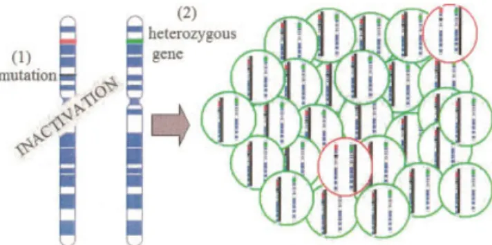
1.2.2.3. Skewed X-chromosome inactivation
The sex chromosomes differ in males and females. Males are hemizygous for X-chromosome, while females inactivate one of their X-chromosomes for dosage compensation. Therefore, females are mosaics for X-chromosome inactivation (XCI), as a result of random inactivation of one of the X-chromosomes. Disturbed XCI was used to explain the female predominance in autoimmune diseases by Kast in 1977 and developed by Stewart in 1998. According to Kast and Stewart, the disturbance of the mosaicism could lead to differences of the antigen presentation, causing autoimmunity. Although Chitnis et al. was failed to show the association of several autoimmune diseases with skewed XCI in 2000, the association was found with SSc, autoimmune thyroid disease (AITD), and pre-eclampsia (Ozbalkan et al., 2005; Ozcelik et al., 2006; Uz et al., 2007; Uz et al., 2008).
9
1.3. X-inactivation 1.3.1. History
X-chromosome inactivation was first proposed by Lyon in 1961 to be the dosage compensation mechanism in mice. Upon several unexpected results in her analysis of mutations affecting the coat color of female mice, she suggested that one of the two X-chromosomes in each cell of a female is inactivated randomly leading females to be mosaics. She suggested that the X-inactivation event occurred early in development that causes formation of large patches of different color (Lyon, 1961).
After these proposals, a hypothesis came from Ohno and colleagues, who demonstrated, first in mice then in humans, each Barr body (Barr et al., 1949) was a single X-chromosome, and the other X was euchromatic like the autosomes (Ohno et al., 1960, Ohno et al., 1961).
1.3.2. Mechanism
There are different mechanisms for sex development in different species. The presence of a Y chromosome is necessary for male development in mammals, while in fruit flies and worms, it is dependent on the ratio of the X-chromosome to autosomal chromosomes. In order to compensate the dosage problem between males and females, different species have developed different mechanisms. In Drosophila melanogaster, XY males increase the expression of their single X-chromosome, while in
Caenorhabditis elegans, XX hermaphrodites reduce each X chromosome’s
expression by half, in order to achieve similar transcription levels in XO males. However, in mammals, females inactivate one of the X chromosomes (Pontier and Gribnau, 2011).
10 There is a random XCI in eutherian mammals, while in marsupials, X-chromosome inactivation is imprinted - the X-X-chromosome coming from the father is always inactivated (Cooper et al., 1971). However, it was shown that eutherian mammals also have imprinted XCI, which is limited to extra-embryonic tissues-the placenta. In four-cell stage of mouse embryos, paternally-derived X-chromosomes undergo an early imprinted inactivation, leaving the maternal X-chromosome active only. The extraembryonic tissues retain this early imprinted inactivation, but in the early blastocyst, initial imprinted X-inactivation is reversed in the inner cell mass that give rise to the embryo. Each of these cells then independently and randomly inactivates one of the X-chromosomes irreversibly, leading to mosaicism (Huynh and Lee, 2004). The existence of imprinted XCI in humans remains controversial. Recently, it was shown that XCI in human extraembryonic tissues is also random (Moreira de Mello et al., 2010).
X-inactivation is a multistep process including counting, choice, silencing and maintenance. In the counting step, the X-chromosome number relative to autosomes is determined, while in choice, X-chromosome that remain active (Xa) is chosen. Silencing is then initiated and spread on the inactive X-chromosome (Xi), and maintained in daughter cell lineages (Chow et al., 2005).
XCI is regulated by the X-inactivation center (XIC), which is mapped to Xq13. XIC consists of several genes such as, XIST, TSIX, and XITE (Brown
et al., 1991; Lee et al., 1999; Kalantry 2011). XIST (X-inactive specific
transcript), which is a non-coding RNA, is expressed from the future Xi. During the inactivation process, XIST is regulated by TSIX, which is the antisense complementary transcript of XIST. TSIX, which is regulated by
XITE, is expressed from Xa and prevents XIST expression in the Xa
11
1.4. Autoimmune disorders that were selected in this study 1.4.1. Rheumatoid Arthritis (RA)
Rheumatoid arthritis (RA) is an inflammatory disease that causes joint destruction, resulting in pain, stiffness and swelling of peripheral joints (Aletaha et al., 2010). RA affects 1% of the population, worldwide, with a female predominance (Gabriel and Michaud, 2009). There are multicellular inflammation in the joint, including infiltration of lymphocytes and granulocytes into the articular cartilage, and proliferation of macrophages. These processes not only cause pain and stiffness but also joint destruction and reduction of bone density (Nandakumar and Holmdahl, 2006).
1.4.1.1. Classification
In 1987, criteria for RA were developed by the American Rheumatology Association (ARA). According to these criteria four of the seven features are needed to be present: morning stiffness, arthritis of 3 or more joint areas, arthritis of hand joints, symmetric arthritis, rheumatoid nodules, positive serum rheumatoid factor (RF) and radiographic changes (Arnett et al., 1988). Recently, new criteria was developed and introduced by American College of Rheumatology/European League Against Rheumatism (ACR/EULAR). According to the new criteria, patients that have a point score of 6 or higher are called RA patient. The point scores were developed by ACR/EULAR. The points are given according to four areas of diagnosis: joint involvement, serological parameters, acute-phase reactants and duration of symptoms (Aletaha et al., 2010).
12
1.4.1.2. Prevalence, incidence
During last two decades, several incidence and prevalence studies of RA have been reported. According to these studies, the frequency changes with the different ethnic and racial groups. The prevalence of RA is between 0.5% and 1% and the incidence of RA decreased from 61.2 per 100,000 population in the period 1955–1964 to 32.7 per 100,000 in the period 1985– 1994 (Gabriel and Michaud, 2009). In south European countries, the occurrence of RA is relatively lower according to north European and north American countries (Alamanos and Drosos, 2005).
1.4.1.3. Causes
The cause of the RA is not well known, but genetic and environmental factors are thought to be involved. Smoking is the most reported risk factor for RA (Silman et al. 1996, Harrison 2002). It is reported that genetics determines 50-60% of susceptibility, severity and phenotype of RA (MacGregor et al., 2000; Deighton et al., 1989). Monozygotic twins have concordance rates of 15%, while dizygotic twins have rates of 4% (Silman
et al., 1993; Aho et al., 1986).
1.4.1.3.1. Associated genes
In 1978, HLA-DRB1 was first to be associated with RA (Stastny, 1978). Later on, shared epitope common to all HLA-DRB1 alleles were found to be associated with RA in different populations (Imboden, 2009).
In a large scale screening of 16,000 non-synonymous SNPs, PTPN22 R620W SNP was found to be associated with RA (Begovich et al., 2004).
13 Like PTPN22, CTLA-4 negatively regulates T-cell activation (Sharpe and Freeman, 2002). A SNP of CTLA-4 was found to be associated with RA in German (Seidl et al., 1998), Japanese (Yanagawa et al., 2000), and British (Vaidya et al., 2002) populations. Recently STAT4 was also found to be associated with RA in different populations (Remmers et al., 2007; Korman
et al., 2008).
1.4.1.3.2. Antibodies
RA is characterized by the presence of autoantibodies, such as rheumatoid factor (RF) and anti-cyclic citrullinated peptide (CCP) antibody. Although RF was never proved to cause arthritis in experimental systems, it was the only antibody that is used for classification of RA (Arnett et al., 1988). However, anti-CCP was found to be present in 70% of RA patients, and rare in healthy poeple (Agrawal et al., 2007). In the new classification criteria, anti-CCP is also included for diagnosis of RA (Aletaha et al., 2010).
1.4.2. Juvenile Idiopathic Arthritis (JIA)
Juvenile idiopathic arthritis (JIA), which is the inflammation (cellular damage) of the synovium (the lining of joints), is the most prevalent pediatric rheumatic disease that is seen in children with onset before 16 years of age. JIA patients have swollen, painful joints, which may be stiff and difficult to move. (Cuccurullo, 2004).
14
1.4.2.1. Classification
Juvenile idiopathic arthritis (JIA) term (Petty et al., 1998; Petty et al., 2003; Petty et al., 2004) was first proposed in 1994 and later revised in 1997.It is now used instead of the American term ‘juvenile rheumatoid arthritis’ (JRA) as defined by American College of Rheumatology (ACR) and the European classification ‘juvenile chronic arthritis’ (JCA) as defined by the European League Against Rheumatism (EULAR) (Wood et al., 1978). The American and European classifications of the disease were confusing (Table 1.3), and was difficult to use them interchangeably (disease duration is 6 weeks for ACR, while it is 12 weeks for EULAR). In order to improve research and treatment, the International League Against Rheumatism (ILAR) unified the criteria, using the term ‘juvenile idiopathic arthritis’. The word ‘idiopathic’ means ‘of unknown cause’.
Table 1.3. Comparison of the classification systems of arthritis in children
Classification ACR EULAR ILAR ____________ _____ _______ _____ Designation JRA JCA JIA
Types Systemic Systemic Systemic Pauciarticular Pauciarticular Oligoarticular Polyarticular RF-negative polyarticular RF-negative
polyarthritis RF-positive polyarticular RF-positive
polyarthritis Psoriatic Psoriatic
JAS Enthesitis-related
Undefined
ACR=American College of Rheumatology; EULAR=European League against Rheumatism; ILAR=International League of Associations for Rheumatology; JRA= juvenile rheumatoid arthritis; JCA= juvenile chronic arthritis; JIA= juvenile idiopathic arthritis; JAS= juvenile ankylosing spondylitis (Petty et al., 2003).
15
1.4.2.2. Types
According to ILAR, the subtypes of JIA are oligoarticular JIA, which may be persistent or extended, polyarticular rheumatoid factor (RF)–negative JIA, polyarticular RF-positive JIA, systemic JIA, enthesitis-related arthritis (ERA), psoriatic JIA, or a classification of “other JIA” when the criteria for more than one subtype of JIA or none of the criteria were met (Petty et al., 1998).
1.4.2.2.1. Oligoarticular JIA
Oligoarthritis, which is the most common type that affect about 50% of all children with JIA, is mostly seen in females. This term is used when there are four or fewer affected joints during the first 6 months of disease. According to the ILAR classification, children who have psoriasis/a family history of psoriasis, a human leukocyte antigen (HLA) B27-associated disease in a first-degree relative, and a positive rheumatoid factor (RF) test are excluded from the oligoarthritis category (Petty et al., 1998).
This form of JIA is the only one that is not seen in adults, and characterized by asymmetric arthritis, early age of onset (before 6 years of age), female predominance, and high frequency of positive antinuclear antibodies (ANAs).
According to the ILAR classification, oligoarthritis subtype is divided to persistent oligoarthritis, in which the disease consists of four or fewer joints, and extended oligoarthritis, in which arthritis extends to more than four joints after the first 6 months of disease (Petty et al., 1998; Ravelli et al., 2007).
16
1.4.2.2.2. Polyarticular JIA
Polyarticular arthritis affects 35% children with JIA, more girls than boys. This kind of JIA usually involves small joints of the hands and feet. Polyarticular JIA is often symmetrical. There are two types of polyarticular JIA: rheumatoid-factor-positive and rheumatoid-factor-negative polyarthritis (Cuccurullo, 2004; Ravelli et al., 2007).
1.4.2.2.2.1. Rheumatoid-factor-positive polyarthritis
This disease, comprises 10% of all patients with JIA, and is characterized by age of onset greater than 11 years of age with female predominance (Cuccurullo, 2004). Although it is same as adult RF-positive rheumatoid arthritis, the disease phenotype between children and adults is different as the children’s skeleton is still growing. It is mainly seen in adolescent girls (Cuccurullo, 2004; Ravelli et al., 2007).
It is characterized as a symmetrical polyarthritis and affects small joints of the hands and feet (Ravelli et al., 2007).
1.4.2.2.2.2. Rheumatoid-factor-negative polyarthritis
It is the most heterogeneous subtype (Ravelli et al., 2007). It affects 25% of all patients with JIA (Cuccurullo, 2004). There are at least three subsets of RF-negative polyarthritis. The first form resembles early-onset oligoarticular juvenile idiopathic arthritis because of the asymmetric arthritis, and early age of onset (Martini, 2003; Ravelli et al., 2007). The second subset is similar to adult onset RF-negative rheumatoid arthritis, because of the characteristics of symmetric synovitis of large and small
17 joints. The third form is known as dry synovitis. This subset is often poorly responsive to treatment and could follow a destructive progress (Ravelli et
al., 2007).
1.4.2.2.3. Systemic JIA
It usually begins in early childhood. Researchers sometimes call this Still’s disease. This type accounts for about 10-20% of cases of JIA. It affects both boys and girls almost equally. It is characterized by suddenly occurring fever. Anemia and weight loss may also occur (Ravelli et al., 2007).
1.4.2.2.4. Enthesitis-related arthritis
Enthesitis-related arthritis, which is characterized by the association of enthesitis (inflammation of the entheses) and arthritis, mainly affects male patients after the age of 6 years. The joints of the lower extremities are affected. It resembles oligoarthritis because of the hip involvement (Petty et
al., 1998; Petty et al., 2003; Ravelli et al., 2007).
1.4.2.2.5. Psoriatic arthritis
According to ILAR, in order to diagnose juvenile psoriatic arthritis, arthritis and psoriatic rash need to be present. If a rash is absent, dactylitis (sausage-shaped swelling of the fingers and toes that can be painful), nail pitting and the presence of psoriasis in a first degree relative must be present. The symptoms are similar to the subset of RF-negative polyarthritis, and oligoarthritis (Petty et al., 1998; Ravelli et al., 2007).
18
1.4.2.2.6. Undifferentiated arthritis
This subtype includes the patients that do not fulfil the inclusion criteria for any category (Petty et al., 2004; Ravelli et al., 2007).
1.4.2.3. Prevalence and incidence
The incidence and the prevalence of the disease differ among different ethnicity. It has an incidence of 2–20 cases per 100 000 population and a prevalence of 16–150 cases per 100 000 population (Ravelli et al., 2007).
The frequency of the subtypes differs. There is female predominance, except in the systemic and enthesitis-related arthritis (Table 1.4). In systemic arthritis female-male ratio is equal. In enthesitis-related arthritis, there is male predominance.
Table 1.4 International League of Associations for Rheumatology (ILAR)
categories of juvenile idiopathic arthritis
Frequency Onset age Sex ratio
Systemic arthritis 4-17% Throughout childhood
F=M
Oligoarthritis 27-56% Early childhood F>>>M Rheumatoid factor (+) polyarthritis 2-7% Late childhood F>>M Rheumatoid factor (-) polyarthritis 11-28% F>>M Enthesitis related arthritis 3-11% Late childhood M>>F Psoriatic arthritis 2-11% F>M Undifferentiated arthritis 11-21%
19
1.4.2.4. Causes
Although the cause of JIA is not well-understood, it is believed that JIA is caused by a combination of genetic and environmental factors such as viral or bacterial infections that may trigger the autoimmune process (Cuccurullo, 2004).
1.4.2.4.1. Associated Genes
There are both MHC-associated and Non-MHC genes that are found to be associated with JIA. The class I gene, HLA-B27, was the first HLA association found in JIA. It is found that HLA-B27 is a risk factor for oligoarthritis, particularly in older male patients (Rachelefsky et al., 1974; Reviewed in Borchers et al., 2006).
SNPs in PTPN22 and CTLA4 were also found to be associated with JIA in different populations (reviewed in Phelan et al., 2006).
1.4.2.4.2. Antibodies
Although wide variety of autoantibodies has been described in JIA patients, RF and antinuclear antibodies (ANA) are routinely used to provide serological support for the diagnosis of JIA.
ANA are detected in ~30–50% of patients with JIA (Berntson et al., 2003) the prevalence vary from 38% to 85% in oligoarthritis, ~30–50% in polyarthritis and 0–17% in systemic onset disease (Al-Matar et al., 2002; Moroldo et al., 2004).
20 In more recent studies, anti-CCP antibodies were reported in 77% of patients with JIA overall, 93% in RF-negative polyarthritis, 84% in oligoarthritis and 62% in systemic arthritis (Borchers et al., 2006).
1.5. Aim and Strategy
Most of the autoimmune diseases have high female predominance (Whitacre, 2001). Although the female prevalence is often attributed to the effect of estrogen, it is stated that other sex differences might have as much or more relevance to autoimmune disease, that is X-inactivation (Stewart, 1998). Recently, it has been shown that high proportion of scleroderma, AITD and pre-clampsia patients has extremely skewed X-inactivation in their blood cells (Ozbalkan et al., 2005; Ozcelik et al., 2006; Uz et al., 2007; Uz et al., 2008).
RA is an autoimmune disease, with unknown cause. Like other autoimmune diseases there is female predominance. Hormonal levels and pregnancy-related microchimerism was previously hypothesized to play role in autoimmunity. However our group and others observed that skewed XCI could be a factor (Ozbalkan et al., 2005; Brix et al., 2005; Ozcelik et al., 2006; Uz et al., 2007; Uz et al., 2008). Therefore we included the pediatric form of RA, JIA, in which the onset of the disease is before puberty, therefore hormonal status would not be effective. There is also a female predominance in JIA.
Here we hypothesize that skewed XCI might play a role in the pathogenesis of JIA and RA. In order to test our hypothesis, we analyzed the methylation status of a highly polymorphic CAG repeat in the androgen receptor (AR) gene. In this study we used JIA patients within the subgroups that have female predominance: oligoarthritis and polyarthritis from Turkish population, and RA from French population.
21
CHAPTER II
MATERIALS AND METHODS
2.1. MATERIALS
2.1.1. PEDIATRIC SAMPLES
2.1.1.1. Turkish Children Control Samples
In this study, 211 healthy, unrelated Turkish children with no history of autoimmune disorders or cancer were involved. The mean age at analysis was 13±4 (mean±SD). Informed consent was obtained from all subjects (or legal guardians of subjects who had not reached age of majority). The ethics committee of the participating institutions approved the study protocol.
2.1.1.2. Turkish Juvenile Idiopathic Arthritis Patients
Caucasian children diagnosed with juvenile idiopathic arthritis (n=81) were included in this study. All patients fulfilled the International League of Associations for Rheumatology diagnostic criteria for juvenile idiopathic arthritis (Petty et al. 2004). The mean±SD age of the patients was 10±5
22 years, and the mean age at the time of disease onset was 6±4 years. 21 of the patients were diagnosed with polyarthritis, while 60 were having oligoarthritis. Informed consent was obtained from all subjects (or legal guardians of subjects who had not reached age of majority). The ethics committee of the participating institutions approved the study protocol.
2.1.2. ADULT SAMPLES 2.1.2.1. French Control Samples
Healthy, unrelated French females (n=100) with no history of autoimmune disorders or cancer were involved in this study. Informed consent was obtained from all subjects. The ethics committee of the participating institutions approved the study protocol.
2.1.2.2. French Rheumatoid Arthritis Patients
French females diagnosed with rheumatoid arthritis (n=86) were involved. Informed consent was obtained from all subjects. The ethics committee of the participating institutions approved the study protocol.
2.1.2.3. Turkish Adult Control Samples
For determination of frequency of PTPN22 genotypes in Turkish population 68 unrelated healthy women were genotyped. Informed consent was obtained from all subjects. The ethics committee of the participating institutions approved the study protocol.
23
2.1.2.4. Turkish Systemic Sclerosis Patients
For determination of frequency of PTPN22 genotypes in Turkish population 71 SSc patients were enrolled in the study. The mean age of the patients was 47 years. Informed consent was obtained from all subjects. The ethics committee of the participating institutions approved the study protocol.
2.1.2.5. Turkish Autoimmune Thyroid Disease Patients
In order to determine the frequency of PTPN22 genotypes 104 AITD patients were enrolled in the study. The mean±SD age of the patients was 49±14 years. Informed consent was obtained from all subjects. The ethics committee of the participating institutions approved the study protocol.
2.1.3. CHEMICALS, REAGENTS AND ENZYMES 2.1.3.1. Primers
The primers used in polymerase chain reaction (PCR) were purchased from IONTEK (Istanbul, Turkey).
The primer sequences for AR are:
RS6, 5'- GTCCAAGACCTACCGAGGAG -3';
RS7, 5'- CCAGGACCAGGTAGCCTGTG -3'
The primer sequences for PTPN22 are:
PTPN22F, 5’-GATAATGTTGCTTCAACGGAATTTA -3’;
24
2.1.3.2. Enzymes
Taq DNA polymerase enzymes and the restriction digestion enzymes XcmI, RsaI and methylation sensitive HpaII were purchased from MBI Fermentas
(Amherst, NY, USA).
2.1.3.3. Thermal cyclers
For PCR reactions, the thermal cycler The GeneAmp System 9600 and 2400 (Perkin-Elmer, USA) were used.
2.1.3.4. Standard Solutions and Buffers
1X TAE (Tris-acetic acid-EDTA): 40mM Tris-acetate,
2 nM EDTA, pH 8.0
Ethidium bromide: 10mg/ml in water
(stock solution)
30 ng/ml (working solution)
Agarose gel loading buffer (6X): 15% ficoll
0.05% bromophenol 0.05% xylene cyanol
Acrylamide:bisacrylamide (30%): 29.5 gr acrylamide 0.44 gr bisacrylamide ddH2O to 100 ml
25
2.1.3.5. Chemicals and Reagents
Table 2.1. Chemicals, reagents, and kits used in this study
Reagent/Chemical Company
Agarose Basica LE, EU
Acetic acid Sigma, St Lois, MO, USA
Acrylamide Sigma, St Lois, MO, USA
Ammonium persulfate Carlo Elba, Milano, Italy
Bisacrylamide Sigma, St Lois, MO, USA
Bromophenol blue Sigma, St Lois, MO, USA
dNTPs MBI Fermentas, Amherst, NY, USA
EDTA Carlo Elba, Milano, Italy
Ethanol Merck, Frankfurt, Germany
Ethidium bromide Sigma, St Lois, MO, USA
Nucleospin® Blood Kit Macherey-Nagel Duren, Germany
TEMED Sigma, St Lois, MO, USA
Tris-Base Bio-Rad, CA, USA
2.1.3.6. Nucleic Acids
As a DNA marker, pUC Mix8 was used, which is purchased from MBI Fermentas (Amherst, NY, USA) (Figure 2.1).
26
Figure 2.1. Sizes of the fragments of PUC mix marker, 8, and appearance
on both agarose and polyacrylamide gel electrophoresis (MBI Fermentas web site)
2.2. METHODS
2.2.1. Sample Collection
Venous blood that was obtained from controls and patients were collected in EDTA-containing tubes. They were divided into 1 ml aliquots in 1.5 ml eppendorf tubes. 200 μl of blood was used for DNA isolation; the remaining bloods were stored at -80oC for later use.
27
2.2.2. Determination of X chromosome inactivation status
For dosage compensation in mammals, females randomly inactivate one of their X-chromosome, maternal or paternal (Lyon et al., 1961). The inactivation initiates upon transcription of XIST RNA from the X-chromosome that will be inactivated. The untranslated XIST RNA coat the X-chromosome that it is produced and with the help of histone modifications, such as hypermethylation of CpG islands (Wolf et al., 1984; Heard and Disteche, 2006). HpaII is a methyl sensitive restriction enzyme, which recognizes CpG dinucleotides. Therefore, X-linked genes that contain
HpaII recognition sites and silences during inactivation process can be used
for determination of X-chromosome inactivation. Allen et al. first used androgen receptor (AR), which has highly polymorphic CAG repeats, in order to determine the XCI pattern (Allen et al., 1992). In this assay, the DNA is digested with HpaII and the region that contains highly polymorphic CAG repeats flanked by restriction enzyme recognition site is amplified by PCR (Figure 2.2). After amplification only the inactive allele would give a product as the active one would be digested by HpaII. Therefore, in women informative for CAG repeat, methylation patterns of maternal and paternal X chromosomes can be identified.
28
>ref|NC_000023.9|NC_000023:66680599-66860844 Homo sapiens chromosome X, reference assembly
GGGAAAAAGGGCCGAGCTAGCCGCTCCAGTGCTGT ACAGGAGCCGAAGGGACGCACCACGCCAGCCCCA GCCC GGCTCCAGCGACAGCCAACGCCTCTTGCAGCGCGGCGGCTTCGAAGCCGCCGCCC GGAGCTGCC CTTTCCTCTTCGGTGAAGTTTTTAAAAGCTGCTAAAGACTCGGAGGAAGCAAGGAAAGTGCCTGGTAGGA CTGACGGCTGCCTTTGTCCTCCTCCTCTCCACCCCGCCTCCCCCCACCCTGCCTTCCCCCCCTCCCCCGT CTTCTCTCCCGCAGCTGCCTCAGTCGGCTACTCTCAGCCAACCCCCCTCACCACCCTTCTCCCCACCCGC CCCCCCGCCCCCGTCGGCCCAGCGCTGCCAGCCCGAGTTTGCAGAGAGGTAACTCCCTTTGGCTGCGAGC GGGCGAGCTAGCTGCACATTGCAAAGAAGGCTCTTAGGAGCCAGGCGACTGGGGAGCGGCTTCAGCACTG CAGCCACGACCCGCCTGGTTAGGCTGCACGCGGAGAGAACCCTCTGTTTTCCCCCACTCTCTCTCCACCT CCTCCTGCCTTCCCCACCCCGAGTGCGGAGCCAGAGATCAAAAGATGAAAAGGCAGTCAGGTCTTCAGTA GCCAAAAAACAAAACAAACAAAAACAAAAAAGCCGAAATAAAAGAAAAAGATAATAACTCAGTTCTTATT TGCACCTACTTCAGTGGACACTGAATTTGGAAGGTGGAGGATTTTGTTTTTTTCTTTTAAGATCTGGGCA TCTTTTGAATCTACCCTTCAAGTATTAAGAGACAGACTGTGAGCCTAGCAGGGCAGATCTTGTCCACCGT GTGTCTTCTTCTGCACGAGACTTTGAGGCTGTCAGAGCGCTTTTTGCGTGGTTGCTCCCGCAAGTTTCCT TCTCTGGAGCTTCCCGCAGGTGGGCAGCTAGCTGCAGCGACTACCGCATCATCACAGCCTGTTGAACTCT TCTGAGCAAGAGAAGGGGAGGCGGGGTAAGGGAAGTAGGTGGAAGATTCAGCCAAGCTCAAGGATGGAAG TGCAGTTAGGGCTGGGAAGGGTCTACCCTCGGCCGCCGTCCAAGACCTACCGAGGAGCTTTCCAGAATCT GTTCCAGAGCGTGCGCGAAGTGATCCAGAACCC GGGCCCCAGGCACCCAGAGGCCGCGAGCGCAGCACC TCCC GGCGCCAGTTTGCTGCTGCTGCAGCAGCAGCAGCAGCAGCAGCAGCAGCAGCAGCAGCAGCAGCA GCAGCAGCAGCAGCAGCAGCAGCAAGAGACTAGCCCCAGGCAGCAGCAGCAGCAGCAGGGTGAGGATGGT TCTCCCCAAGCCCATCGTAGAGGCCCCACAGGCTACCTGGTCCTGGATGAGGAACAGCAACCTTCACAGC CGCAGTCGGCCCTGGAGTGCCACCCCGAGAGAGGTTGCGTCCCAGAGCCTGGAGCCGCCGTGGCCGCCAG CAAGGGGCTGCCGCAGCAGCTGCCAGCACCTCC GGACGAGGATGACTCAGCTGCCCCATCCACGTTGTC CCTGCTGGGCCCCACTTTCCCC GGCTTAAGCAGCTGCTCCGCTGACCTTAAAGACATCCTGAGCGAGGC CAGCACCATGCAACTCCTTCAGCAACAGCAGCAGGAAGCAGTATCCGAAGGCAGCAGCAGCGGGAGAGCG AGGGAGGCCTCGGGGGCTCCCACTTCCTCCAAGGACAATTACTTAGGGGGCACTTCGACCATTTCTGACA ACGCCAAGGAGTTGTGTAAGGCAGTGTCGGTGTCCATGGGCCTGGGTGTGGAGGCGTTGGAGCATCTGAG TCCAGGGGAACAGCTTCGGGGGGATTGCATGT ACGCCCCACTTTTGGGAGTTCCACCCGCTGTGCGTCC
Figure 2.2. The sequence of AR, exon 1, downloaded from http://www.ncbi.nlm.nih.gov. The region amplified by PCR, the HpaII recognition sites and CAG repeat regions are shown.
2.2.2.1. DNA Isolation
The DNA isolation was carried out from 200 μl venous blood via Nucleospin® Blood kit (Macherey-Nagel Inc., PA, USA) according to manufacturer’s instructions. The remaining bloods were stored at -80o
C for later use. After isolation, DNA was stored at 4 oC. The quantity of DNA samples were measured using nanodrop. Ratio of 260/280 reading of 1.8 ± 0.2 was accepted as high quality DNA. The integrity of DNA samples were controlled by using 1% agarose gel electrophoresis.
RS7 (Reverse Primer) CAG Repeats RS6 (Forward Primer) RsaI RsaI HpaII HpaII HpaII HpaII HpaII HpaII
29
2.2.2.2. Restriction Enzyme Digestion
Two sets of reactions were prepared for each subject in restriction digestion protocol. Both sets contain 150-250 ng genomic DNA, 1X reaction buffer and 2 Units of RsaI. RsaI is a restriction enzyme with the recognition sites outside the PCR product. It digests active and inactive X alleles equally, and was used to improve the PCR. The only difference between these two sets is the presence of methylation sensitive restriction enzyme HpaII in the tube labeled as “digested (D)” (8 Units per 20 μL reaction). The tubes that do not contain HpaII were labeled as “undigested (U)”. Both reaction sets were completed to a final volume of 20 μL with deionized water. Restriction digestion reaction tubes were incubated at 37°C overnight. One control male sample that was cytogenetically verified as 46, XY was involved in this study as a control for complete digestion.
2.2.2.3. Polymerase Chain Reaction (PCR)
For each sample, two PCR were performed. Tubes that contain restriction digestion product of undigested reaction (without HpaII) as a template were labeled as “U”, and the tubes containing the product of digested reaction as a template were labeled as “D”. PCRs were carried out in a total volume of 25 μL. Restriction digestion products were amplified with 10 pmol of each primers, 0.12 mM of dNTP, 1X Taq polymerase buffer, 1.0 mM MgCl2, and 1 Unit Taq polymerase.
In order to amplify the desired region on AR locus, 94°C 5 min initial denaturation step was followed by 30 cycles of 94°C for 30 sec (denaturation), 58°C for 30 sec (annealing), 72°C for 30 sec (extension) amplification step. Final extension was performed at 72°C for 5 min.
30
2.2.2.4. Agarose Gel Electrophoresis
PCR products were run in the 1.5% agarose gel by using 1X TAE. Agarose was completely dissolved in 1X TAE electrophoresis buffer to required percentage in microwave and ethidium bromide was added to final concentration of 30ng/ml. The samples were loaded onto agarose gel with 1/5 volume of loading buffer. The gel was run in 1X TAE at 100V for 30 minutes at room temperature.
2.2.2.5. Polyacrilamide Gel Electrophoresis (PAGE)
The working PCR products were resolved by using 8% PAGE. In order to prepare PAGE, 30% acrylamide:bisacrylamide solution (29:1) was mixed with 10X TAE, 10 % Ammonium persulfate (APS), and TEMED and ddH2O. 10 μL from each sample was loaded and each gel was run at 15W for 2 hours. The gels were stained with EtBr for 5 min and destained in ddH2O for 5 min.
2.2.2.6. Densitometric Analysis
Densitometric analyses of the bands were performed twice for each sample using the MultiAnalyst version 1.1 software. The skewing ratio was calculated according to the formula in figure 2.3. A corrected ratio (CrR) was calculated by dividing the ratio of the predigested sample (upper/lower allele) by the ratio of the non-predigested sample for normalization of the ratios that were obtained from the densitometric analyses. The use of CrR compensates for preferential amplification of the shorter allele when the number of PCR cycles increases (Delforge et al., 1995). A skewed
31 population is defined as a cell population with greater than 80% expression of one of the AR alleles. This corresponds to CrR values of <0.25 or >4.
D1(U1+U2) Skewing ratio =
2U1(D1+D2)
Figure 2.3. The formula for calculation of skewing ratio
2.2.3. Determination of frequency of PTPN22 genotypes
PTPN22 encodes lymphoid protein tyrosine phosphatase (LYP), which
suppresses T-cell activation by bind to negative regulatory kinase Csk. More common allele 1858C can binding Csk, while 1858T does not (Bottini et al., 2004). 1858C→T SNP, which is in the first proline-rich motif in human LYP, is on exon 14 of PTNP22 gene. The C > T transition creates a restriction site for XcmI (figure 2.4). Therefore, in order to identify the polymorphism, PCR amplified fragment is digested by XcmI.
U1=intensity of upper allele of undigested sample
U2=intensity of lower allele of undigested sample
D1= intensity of upper allele of digested sample
32
>ref|NG_011432.1| Homo sapiens protein tyrosine phosphatase, non-receptor type 22 (lymphoid) (PTPN22), RefSeqGene on chromosome 1 TTATTATTATTATTTTTTGAGACGGAGTGTCCTGCTGCCACCCAGGCTGGAATGCAGTGGTGCAATCTCGGCTCAC TGCAAGCTCTGCCTTCCGGGTTCACGCCATTCTCCCACCTCAGCCTCCCGAGTAGCTGGGACTACAGGGGCCCGCC ACCACGCCCGGCTAATGTTTTGTGTTTTTAGTAGAGACACGGTTTCACCACGTTAGCCAGGATGGTTTCGATCTCC TGACCTTGTGATCTGCCCGCCTCGGCCTCCGAAAGTGCTGGGATTACAGGCGTGAGCCACCGCGCCCAGCCCTACT TTTGAGCTTTTAAAACAATAAATGTTAAAGAATAAGCAAAAACCTCCTGGGTTTGTACCTTAAGAGAATTTATTTT GCTTTTTCCTTGAATGAACAAGTGTCAACTTTACTGATAATGTTGCTTCAACGGAATTTAAATATAAATTATGGTA AATTTATATTTAATATTAGAATATAAGAATTTCCTTTGGATTGTTCTAATTAACAATTGTTACAATATTTTTGACA TTTTGGATAGCAACTGCTCCAAGGATAGATGATGAAATCCCCCCTCCACTTC CTGTACGGACACCTGAATCATTT ATTGTGGTTGAGGAAGCTGGTGAGTACAGTTCAGTAAGTATAAAATAAAGTGTGGGATGGGCATGGTGGCTCATGC CTTTAATTCCAGCACTTTGGGAAGCTGATGTGTGAGCCTTGAGTTTGAGGAGTTCATTGAGGCCAGGAGTTCAAGA CTAGCCTGCGCAACATAGTGAGACCTCATCTCTAATTTTTTTTTTTTAATTTAGCAGAGCAATAGCAGCATGCATG TGTAGTCCCAACTATTTGGATGGTGGAGGTGAGAGGATCACTTGAGCCCAGGAGTTGGGGGCTGCAGTAAGCCATG ATTGTGCCACTGCACTCCAGTCTGGGTGACAGAGCAAGACCCTGTCTCAAAAA
Figure 2.4. The sequence of PTPN22, exon 14, downloaded from http://www.ncbi.nlm.nih.gov. The region amplified by PCR, and the XcmI recognition sites are shown. The C > T transition at nucleotide 1858, shown in red, creates in the T allele a restriction site for XcmI.
2.2.3.1. Polymerase Chain Reaction (PCR)
PCRs were carried out in a total volume of 25 μL. DNAs products isolated from venous blood were amplified with 10 pmol of each primer, 0.2 mM of dNTP, 1X Taq polymerase buffer, 2.0 mM MgCl2, and 1 Unit Taq polymerase.
In order to amplify the desired region on PTPN22 locus, 95°C 5 min initial denaturation step was followed by 35 cycles of 95°C for 30 sec (denaturation), 59°C for 30 sec (annealing), 72°C for 40 sec (extension) amplification step. Final extension was performed at 72°C for 5 min.
PCR products were resolved on a 1.5% agarose gel by using 1X TAE. Ethidium bromide was added to final concentration of 30ng/ml. The samples were loaded onto agarose gels with 1/5 volume of loading buffer. The gels were run in 1X TAE at 100V for 30 minutes at room temperature.
PTPN22 F
PTPN22 R
33
2.2.3.2. Restriction Enzyme Digestion
The PCR amplified fragments are digested by XcmI, which recognizes the T allele. The sets contain PCR fragments, 1X reaction buffer and 2 Units of
XcmI. Reaction sets were completed to a final volume of 20 μL with
deionized water and were incubated at 37°C overnight. Each digestion was resolved on 3% agarose gel by using 1X TAE. Ethidium bromide was added to final concentration of 30ng/ml. The samples were loaded onto agarose gel with 1/5 volume of loading buffer, and the fragments were visualized by U.V. When the individual is homozygous for C allele, the product is 215 bp, while the T/T homozygous individuals have the bands of 169, and 46 bp.
2.2.4. Statistical Analysis
The results from case and control groups were compared by Fisher’s Exact Test. In addition, odd’s ratio value for control and case groups were performed in 95% confidence interval.
34
CHAPTER III
RESULTS
Only the samples whose alleles were adequately resolved were labeled as ‘informative’. Extremely skewed XCI was described as the inactivation of one allele more than 90%. If one of the alleles was preferentially inactivated more than 80%, the XCI pattern was named as ‘skewed’.
3.1. PCR-based X inactivation study of peripheral blood of Turkish pediatric control samples
XCI was found to be informative in 155 of 211 Turkish pediatric controls (73.5%). Skewed XCI was observed in 11 of 155 informative controls (7.1%) and extremely skewed XCI was observed in only 2 of 155 control individuals (1.3%). Extremely skewed XCI is a very rare event in the healthy individuals. Our results displayed consistency with the previously published results (Amos-Landgraf et al., 2006). The gel image of representative control samples is exhibited on Figure 3.1.
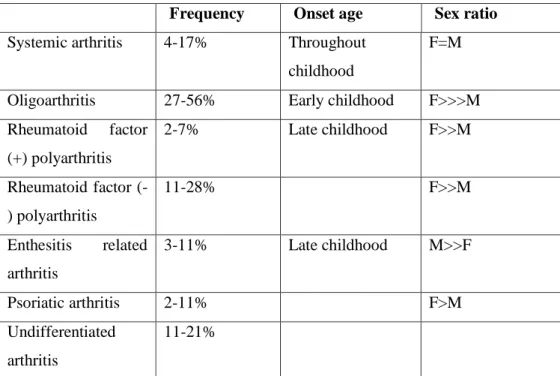
35 Figure 3.1. Gel image of XCI patterns of 3 controls. Polymerase chain reaction
products from the androgen receptor methylation assay demonstrate X chromosome inactivation patterns in samples P-81 (allele ratio: 75%:25%), P-82 (85%:15%), and P-85 (67%:33%). For each sample, DNA was either undigested (-) or digested (+) with the methylation-sensitive restriction enzyme HpaII. Marker M; pUC mix, 8. 331-bp and 242- bp fragments are visible.
3.2. PCR-based X-inactivation study of peripheral blood of Turkish juvenile idiopathic arthritis patients
81 children diagnosed with JIA were genotyped for XCI pattern. 62 of them display informative XCI (76.5%). Extremely skewed XCI was observed in 8 of the 62 informative patients (12.9%). The same pattern was observed in only 2 of 155 informative pediatric controls (P=0.0008; odds ratio=11.33 (95%CI: 2.62-48.48). Skewed XCI was observed in 14 of 62 informative patients (22.6%) and only in 11 of 155 control group (7.1%) (P=0.0036; odds ratio=3.81 (95%CI: 1.65-8.83). The gel image of representative JIA samples is exhibited on Figure 3.2. The overall XCI pattern of JIA samples and control group children are summarized in Table 3.1. Skewed and extremely skewed pattern difference between JIA patients and control individuals were statistically significant. To the best of our knowledge this is the first study that investigates XCI pattern in the blood cells of the pediatric samples.
M P-81 P-82 P-85 . HpaI - + - + - +
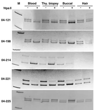
36
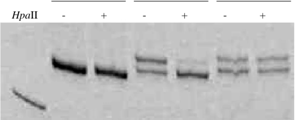
Figure 3.2. Gel image of XCI patterns of 3 JIA patients. Polymerase chain reaction products from the androgen receptor methylation assay demonstrate X chromosome inactivation patterns in samples 06-15 (allele ratio: not informative), 06-16 (93%:7%), and 06-17 (51%:49%). For each sample, DNA was either undigested (-) or digested (+) with the methylation-sensitive restriction enzyme HpaII. Marker M; pUC mix, 8. 242- bp fragment is visible.
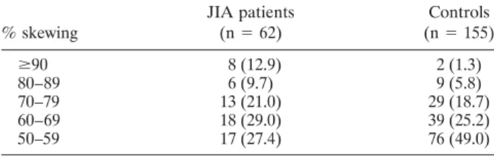
Table 3.1 Proportion of the JIA patients and controls with skewed XCI
Degree of skewing (%) No. (%) observed with skewing
JIA patients Controls
(n=62) (n=155) 90+ 8 (12.9)* 2 (1.3) 80-89 6 (9.7) * 9 (5.8) 70-79 13 (21.0) 29(18.7) 60-69 18 (29.0) 39 (25.2) 50-59 17 (27.4) 76 (49.0) * P < 0.005 versus controls
For comparison by Fisher’s Exact Test, For ≥80% skewing, P= 0.0036 (odds Ratio=3.81 [95% CI 1.65-8.83]); for ≥90% skewing, P=0.0008 (odds ratio=11.33 [95% CI: 2.62-48.48]),
M 06-15 06-16 06-17 .
37 The clinical data of JIA (Table 3.2) was used in order to construct a correlation between skewed XCI and any clinical state of the disease. The only attractive correlation was established between nonrandom XCI and the clinical classification of JIA. It was interesting that only one patient was polyarticular in the group with skewed XCI pattern (7.1%) whereas 12 in 48 patients with random XCI pattern were polyarticular (25.0%). Although the number of patients with skewed XCI was small, most probably skewed XCI may play a role more in the etiology of oligoarthritis JIA.
Previously it has been reported that after years of immunosuppressive agent exposure, skewed XCI may occur in feline hematopoietic cells (Abkowitz et
al. 1993). Therefore, we investigated the correlation between XCI ratios and
the treatment of patients. At the time of sample collection, all of the patients were receiving some treatment. Fourteen were receiving nonsteroidal anti-inflammatory drugs (NSAIDs), 11 were receiving methotrexate (MTX), 10 were receiving MTX plus NSAIDs, and 9 were receiving intraarticular corticosteroids plus NSAIDs, while the remaining 18 were receiving various combinations of these and other drugs. Seven (50%) of the patients that had skewed XCI had received treatment with immunosuppressive agents for 6– 10 years, and 1 (7%) had received immunosuppressive treatment for 2 years. The remaining 6 had received antiinflammatory treatment alone. Twenty-seven (56.3%) of the patients with random XCI had received immunosuppressive treatment for more than 2 years. These results indicate that it is unlikely that immunosuppressive therapy caused skewed XCI in the patients.