Joint Diseases and
Related Surgery Original Article / Özgün Makale
2017;28(3):142-151 doi: 10.5606/ehc.2017.55186
Does leukocyte-poor or leukocyte-rich platelet-rich plasma applied with
biopolymers have superiority to conventional platelet-rich plasma
applications on chondrocyte proliferation?
Biyopolimerler ile uygulanan lökositten fakir veya lökositten zengin trombositten
zengin plazmanın kondrosit proliferasyonunda konvansiyonel trombositten
zengin plazma uygulamalarına üstünlüğü var mıdır?
Duygu Yaşar Şirin, PhD.,1 İbrahim Yılmaz, MD.,2 Mehmet İsyar, MD.,3
Kadir Öznam, MD.,4 Mahir Mahiroğulları,MD.5
1Department of Biology, Namık Kemal University, Faculty of Arts and Science, Tekirdağ, Turkey 2Department of Medical Pharmacology, İstanbul Medipol University School of Medicine, İstanbul, Turkey
3Department of Orthopaedic and Traumatology, Acıbadem Hospitals Group, İstanbul, Turkey
4Department of Orthopaedic and Traumatology, İstanbul Medipol University School of Medicine, İstanbul, Turkey 5Department of Orthopaedic and Traumatology, Memorial Health Group, İstanbul, Turkey
• Received: February 10, 2017 Accepted: July 25, 2017
• Correspondence: Duygu Yaşar Şirin, PhD. Namık Kemal Üniversitesi, Fen Edebiyat Fakültesi, Biyoloji Bölümü, 59030 Tekirdağ, Turkey. Tel: +90 505 - 650 76 36 e-mail: [email protected]
ÖZ
Amaç: Bu çalışmada trombositten zengin plazma (TZP) içeriğindeki lökosit konsantrasyonunun ve TZP’nin ilaç taşıyıcı sistem kullanılarak uygulanmasının in vitro koşullarda kondrosit proliferasyonu üzerine
olası etkileri araştırıldı.
Hastalar ve yöntemler: Medikal veya konservatif tedaviye yanıt vermemiş ve total diz artroplastisi geçirmiş dokuz erkek hastanın (ort. yaş 65 yıl; dağılım 49-81 yıl) kanı iki formülasyon hazırlamak üzere kullanıldı: Düşük konsantrasyon lökositli TZP (2000-4000 lökosit/µL) saf TZP (S-TZP) olarak, yüksek konsantrasyon lökositli TZP (9000-11000 lökosit/µL) ise lökositten zengin TZP (L-TZP) olarak belirlendi. Numuneler kontrol grubu (grup 1), direkt S-TZP uygulanan kondrosit kültürleri (grup 2), direkt L-TZP uygulanan kondrosit kültürleri (grup 3), S-TZP uygulanan hidrojel ile kültürlenen kondrositler (grup 4) ve L-TZP uygulanan hidrojel ile kültürlenen kondrositler (grup 5) olmak üzere beş gruba ayrıldı. Tüm gruplarda hücre morfolojisi, canlılık ve proliferasyon bir prekondrosit belirteci olan evreye özgü embriyonik antijen 1’in (SSEA-1) ifadesi ile karşılaştırıldı.
Bulgular: Maksimum hücre proliferasyonu ve SSEA-1 ifadesi grup 4’te gerçekleşti; SSEA-1 ifadesi ve hücre proliferasyonu arasında istatistiksel olarak anlamlı ilişki vardı.
Sonuç: Çalışmamız TZP’nin lökosit konsantrasyonunun ve hidrojel gibi taşıyıcı sistemlerin önemini ve taşıyıcı sistem ile uygulanan L-TZP’nin kıkırdak hasarının tedavisinde TZP’nin konvansiyonel uygulamalarından daha etkin olduğunu gösterdi.
Anahtar sözcükler: Eşkültür teknikleri; hidrojel; trombositten zengin plazma; evreye özgü embriyonik antijenler.
ABSTRACT
Objectives: This study aims to investigate the possible effects of leukocyte concentration in the content of platelet-rich plasma (PRP) and the administration of PRP using a drug delivery system on chondrocyte proliferation in vitro conditions.
Patients and methods: Blood from nine male patients (mean age 65 years; range 49 to 81 years) with advanced stage osteoarthritis who had not responded to medical or conservative treatments and underwent total knee arthroplasty was used to prepare two formulations: PRP with low concentration leukocytes (2000-4000 leukocytes/µL) was designated as pure PRP (P-PRP), whereas PRP with high concentration leukocytes (9000-11000 leukocytes/µL) as leukocyte-rich PRP (L-PRP). Samples were divided into five groups as control group (group 1), chondrocyte cultures with P-PRP applied directly (group 2), chondrocyte cultures with L-PRP applied directly (group 3), chondrocytes co-cultured with P-PRP applied hydrogel (group 4), and chondrocytes co-cultured with L-PRP applied hydrogel (group 5). In all groups; cell morphology, viability and proliferation were compared with the expression of stage-specific embryonic antigen-1 (SSEA-1), a precondrocyte marker. Results: Maximum cell proliferation and SSEA-1 expression occurred in group 4, with a statistically significant correlation between SSEA-1 expression and cell proliferation.
Conclusion: Our study showed the importance of leukocyte concentration of PRP and efficiency of delivery systems such as hydrogel and that L-PRP administered with a delivery system is more efficient than conventional applications of PRP in the treatment of cartilage damage. Keywords: Co-culture techniques; hydrogel; platelet-rich plasma; stage-specific embryonic antigens.
Osteoarthritis (OA), a degenerative joint disease, is a major cause of physical disability which results in impaired quality of life and which cannot be remediated spontaneously. Unfortunately, current pharmacological and surgical treatment approaches are still not sufficient to achieve a complete treatment.[1]
Platelet-rich plasma (PRP), which has important functions in blood coagulation and homeostasis, is used as a non-pharmaceutical agent supporting the normal healing process by releasing growth factors to the local environment for the treatment of cartilage damage or OA occurred after damage.[2] Platelet-rich plasma has been reported as effective in treatment and holds great promise in almost all fields of
medicine.[3] However, PRP has been prepared with
different contents and concentrations or by means of different ready-to-use commercial kits, leading to a questionable success of PRP therapy.[4,5] After all, it appears from the current literature that more in-depth studies are needed to reveal the effects of PRP containing different concentrations of platelets and also leukocytes, particularly on chondrocytes and chondrocyte proliferation.[5]
Additionally, there are at least two more important issues that have not been fully elucidated in the PRP-related literature. The first is peptide growth factors in PRP, which have short half-life of two minutes at most, in that therapeutic efficiency of these growth factors is still a question unless they are delivered with a drug carrier matrix.[6] A drug carrier system such as biocompatible hydrogels has great capacity to maintain and prolong the half-life of peptide growth factors in PRP. Secondly, the expression of stage-specific embryonic antigen-1 (SSEA-1), a prechondrocyte differentiation marker, has not been investigated in chondrocyte cell cultures subjected to PRP. SSEA-1 expression may indicate maturated chondrocyte proliferation, which is a desired marker for proper treatment of cartilage.
In this respect, the following questions are required to be answered: (i) Can we increase the bioactivity of PRP using a drug delivery system? (ii) Does leukocyte concentration of PRP have any effect on chondrocyte proliferation? (iii) If PRP has such an effect, then what should be the appropriate leukocyte concentration of PRP? (iv) Does PRP administration support extracellular matrix formation in the tissue? Therefore, in this study, we aimed to investigate the possible effects of leukocyte concentration in the content of PRP and the administration of PRP using a drug delivery system on chondrocyte proliferation
in vitro conditions.
PATIENTS AND METHODS
This study was conducted at Medipol Istanbul University, Faculty of Medicine between January 2015 and August 2016. Blood from nine male patients (mean age 65 years; range 49 to 81 years) with advanced stage osteoarthritis who had not responded to medical or conservative treatments and underwent total knee arthroplasty was used to prepare two formulations: PRP with low concentration leukocytes (2000-4000 leukocytes/µL) was designated as pure PRP (P-PRP), whereas PRP with high concentration leukocytes (9000-11000 leukocytes/µL) as leukocyte-rich PRP (L-PRP). Samples were divided into five groups as control group (group 1), chondrocyte cultures with P-PRP applied directly (group 2), chondrocyte cultures with L-PRP applied directly (group 3), chondrocytes co-cultured with P-PRP applied hydrogel (group 4), and chondrocytes co-cultured with L-PRP applied hydrogel (group 5). The experimental design of groups was given in Table I. The study protocol was approved by the Medipol Istanbul University, Faculty of Medicine Ethics Committee (10840098-296/24.11.2014). A written informed consent was obtained from each patient. The study was conducted in accordance with the principles of the Declaration of Helsinki.
To minimize experimental inaccuracies, similar analyses were performed by the same researcher and a blinding method was applied in the preparation of hydrogel and PRP, which were prepared in a closed system under aseptic conditions to avoid contamination. The experiments were repeated six times for each experimental group.
Cell viability, toxicity and proliferation in all experimental and control groups were determined by MTT (3-[4,5-dimethylthiazol-2-yl]-2,5-diphenyltetrazolium bromide; Thiazolyl blue) analysis after 24 hours and on days 10 and 21, and SSEA-1 expression with commercial enzyme-linked immunosorbent assay (ELISA) kit. Simultaneously, environmental scanning electron microscope (ESEM) and inverted light microscope images were recorded.
Patients had stage 4 gonarthrosis according to Kellgren-Lawrence scale.[7] The hematocrit levels were 42%-52%. In order to prepare the PRP, patients were invited to the clinic on the 21st day after surgery for autologous blood collection.
Patients who used non-steroid anti-inflammatory, anti-thrombotic or anti-coagulant drugs in the last two weeks or consumed alcohol and/or cigarettes in the last 48 hours were excluded. Also, complete blood count and electrocardiogram test results were evaluated and patients with arrhythmia, anemia,
polycythemia, and thrombotic disease were excluded. Finally, standard primary chondrocyte cultures were performed from osteochondral tissues obtained from the remaining nine eligible patients.
Tissues from lateral and medial femoral condyles as well as the tibial plateau are routinely removed during total knee arthroplasty and treated as surgical waste. These osteochondral tissues were retained during surgery and transferred to our cell culture laboratory under sterile conditions. Standard primary culture protocols were followed to obtain a monolayer human primary chondrocyte culture.
Chondrocytes were detached from the culture vessel surfaces using trypsin- ethylenediaminetetraacetic, and then stained with Trypan blue and counted with
a Neubauer Chamber. Approximately 3.2x10⁴ cells
were placed in each well in a 96-well plate for MTT analysis (Vybrant MTT Cell Proliferation assay) (Cat# V-13154), Cell Biolabs Inc., USA), and 9.5x10⁵ cells in each well in six-well plate for hydrogel applications
and 8x10⁵ cells in 35 mm petri dishes for ESEM
evaluations. All cultures were incubated for 24 hours for cells to attach and proliferate. Cell culture media supplemented with 6.5% PRP in addition to standard ingredients were refreshed every other day in P-PRP and L-PRP experimental groups.
For photoactivated PRP preparation, autologous blood (24 mL) was taken from patients and aliquoted into 3×8.5 mL vacutainer tubes containing trisodium citrate (22.0 g/L), citric acid (8.0 g/L), and dextrose (24.5 g/L) (BD Vacutainer®, Becton Drive, Franklin Lakes, NJ, USA). After centrifugation at 1000 rpm for 10 minutes, platelet-poor plasma, buffy coat layer (containing PRP and leukocytes) and a red blood cell layer were separated. Platelet-poor plasma was withdrawn from each tube to the level of 10 mm above the buffy coat and discarded. The remaining plasma and the buffy coat were withdrawn into a single sterile tube and exposed to photoactivation for 10 minutes[8] which were achieved using the commercial device Adi-Light 2 (AdiStem Ltd., Hong Kong). Total cell
count was performed for the same remaining plasma and the buffy coat using a hematology analyzer (Sysmex XT-2000i; Roche Diagnostics, Basel, Switzerland) and white blood cells were counted based on morphological characteristics of the cells after Giemsa staining with light microscope.
Obtained P-PRP and L-PRP were added to the hydrogel at a concentration of 6.5%. Since PRP cryopreservation is a safe procedure,[9] the remaining PRP was stored for use on days 10 and 21.
For the preparation of hydrogel with PRP, 12% polyvinyl alcohol solution and 4% starch solution were mixed and stirred until they become homogeneous so that weak cross-links could be formed in this mixture. Autologous PRP obtained from patients under aseptic conditions was added to this mixture at a final concentration of 6.5% and mixed well with the vortex. Afterwards, a saturated solution of sodium tetraborate (16%) was added by mixing. Thus, this biocompatible hydrogel with more stable cross-links was obtained, which behaves like an osmotic pump and maintains the controlled release of PRP.[10] Hydrogel prepared with PRP in this manner is suitable for surgical applications, and at the application area, this hydrogel lasts 21 days with no degradation.
TABlE I
Experimental design of groups
Group Description Application
Group 1 Control (-)
Group 2 Monolayer chondrocyte culture P-PRP Group 3 Monolayer chondrocyte culture L-PRP Group 4 Hydrogel and chondrocyte co-culture P-PRP Group 5 Hydrogel and chondrocyte co-culture L-PRP P-PRP: Pure platelet-rich plasma; L-PRP: Leukocyte-platelet-rich plasma.
Figure 1. Graphic representation of platelet-rich plasma application with hydrogel delivery system showing controlled released platelet-rich plasma migrating from upper chamber to lower chamber through a porous filter. PRP: Platelet-rich plasma
Transwell chamber Cell culture media Hydrogel Porous membrane Relased PRP Monolayer chondrocyte culture
Hydrogels prepared in equal volumes was placed on the upper part of transwell chambers. Schematic representation of the experimental design was given in Figure 1. The chamber was placed on the monolayer chondrocyte culture, and the culture was monitored regularly by changing the nutrient medium every two days.
Cell morphology and confluency were analyzed using an inverted CKX41 Olympus microscope (Olympus Optical Co. Ltd. Hatagaya, Shibuya-ku Japan). Microphotographs of the cell organizations were obtained and analyzed at 4¥, 10¥, 20¥ and 40¥ magnifications in the confocal/contrast phase before and after PRP applications with the Olympus Cell Soft Imaging System software. A Quanta 250 Field emission gun (Fei Company, Hillsboro, Oregon, USA) ESEM was used for electron microscopy. ESEM analysis was carried out to assess the surface topography and composition of the samples. A device with a lifting system and the ability to transfer the electron beam in a high vacuum was used. This enabled us to obtain images of the extracellular matrix, as well as the characteristic cellular structures.[11]
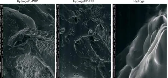
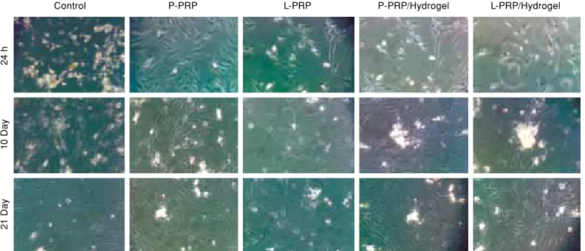
Images of hydrogels were taken at a pressure of 100 Pa in ESEM vacuum mode, under magnification of 2,000¥, at a resolution depth (horizontal field width [HFW]) of 207 µm, at an operating voltage of 5.00 kV, and at a wavelength-dispersive (WD) of 9.1-9.5 mm (Figure 2). Images of chondrocytes were taken at a pressure of 220-221 Pa in ESEM vacuum mode, under magnification of 5,000¥, at a resolution depth (HFW) of 82.9 µm, at an operating voltage of 5.00-10.00 kV, and at a WD of 8.3-10.7 mm (Figure 3).
The ELISA analyses were performed after 24 hours and on days 10 and 21 using a MR-96A, Mindray microplate reader (Mindray Bio-Medical Electronics Co. Ltd., Shenzhen, PRC) according to the commercial kit manual. For ELISA analysis, 3.2¥104 cells were placed into each well of the black 96-well microplates provided by kit and incubated overnight at 37°C in a cell culture incubator. Cells were fixed with 50 µL of fixing solution and the assay was performed immediately after cell fixation. Cells were washed
Figure 2. Environmental scanning electron microscope images of hydrogels. P-PRP: Pure platelet-rich plasma; L-PRP: Leukocyte-platelet-rich plasma.
Hydrogel/L-PRP Hydrogel/P-PRP Hydrogel
Figure 3. Environmental scanning electron microscope images of chondrocytes. P-PRP: Pure platelet-rich plasma; L-PRP: Leukocyte-platelet-rich plasma. 24 h C on tr ol P -P R P L-P R P P -P R P/ H yd ro ge l L-P R P / H yd ro ge l 10 Day 21 Day
three times with 50 µL of wash buffer for five minutes with gentle shaking. Cell dehydration solution of 50 µL was added to each well and incubated. Cells were washed again with wash buffer. After incubating one hour with 50 µL of blocking buffer, cells were washed and labeled in order with primary and secondary antibodies for two hours. Substrate MN1 of 50 µL was added to each well and incubated for 20 minutes at room temperature. The plates were read using a fluorescence plate reader with excitation at 540 nm and emission at 600 nm.
Viability tests were carried out by a commercial MTT kit according to the manufacturer’s instructions. A 12 mM MTT stock solution was prepared by adding 1 mL of sterile phosphate buffered saline to a 5 mg vial of MTT. After removing cell culture medium, 90 µL of fresh culture medium and 10 µL of MTT stock solution
were added per well and incubated at 37°C for two hours, protected from light. Afterwards, 50 µL of dimethyl sulfoxide was added to each well and mixed thoroughly with a pipette, then incubated at 37°C for an additional 10 minutes prior to its photometric measurement at a 570 nm wavelength. The viability of the control group was assumed to be 100% prior to addition of PRP (at 0 hours) to the culture medium.
Stage-specific embryonic antigen-1 is up-regulated during the differentiation of human mesenchymal stem cells and controversially downregulated in undifferentiated cells. Therefore, SSEA-1 expression is considered as a differentiation marker. In drug trials, increased or decreased SSEA-1 in cultured cells is determined without the need to prepare cell lysates. For this purpose, we used a commercial human prechondrocyte characterization kit (Cat#K36094-21A,
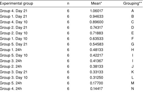
Figure 4. Inverted microscopy of chondrocyte cultures. P-PRP: Pure platelet-rich plasma; L-PRP: Leukocyte-platelet-rich plasma.
Control P-PRP L-PRP P-PRP/Hydrogel L-PRP/Hydrogel
24 h 10 D ay 21 D ay TABlE II
Comparison of difference between experimental groups: variance analysis of stage-specific embryonic antigen-1, enzyme-linked immunosorbent assay and variance analysis of MTT analyses
Variance analysis of SSEA-1 ELISA analysis
Source Degree of freedom Adjusted sum of squares Adjusted mean squares F-value p
Method 14 6.51729 0.465521 22752.72 0.000
Error 75 0.00153 0.000020
Total 89 6.51882
Variance analysis of MTT analysis
Source Degree of freedom Adjusted sum of squares Adjusted mean squares F-value p
Method 14 14.6339 1.04528 134971.53 0.000
Error 75 0.0006 0.00001
Total 89 14.6345
Celprogen, USA).[12] Based on the manufacturer’s protocol, measurements were carried out using a fluorescence plate reader with excitation at 540 nm and emission at 600 nm.
Statistical analysis
Descriptive statistics were expressed as mean±standard deviation. Minitab R16 (Minitab Inc., State College, Pennsylvania, USA) program was used
for statistical evaluation. Assessments were carried out at 95% confidence interval. The significance of the difference between groups was evaluated with the analysis of variance.
A posthoc test with multiple pairwise comparisons, namely the Tukey Honestly Significant Difference (HSD) test, was performed in case of difference between groups to determine the group in which
TABlE III
Comparison of difference between experimental groups: Tukey Pairwise Comparisons for Stage-specific embryonic antigen-1 and MTT analysis Tukey Pairwise Comparisons for SSEA-1 (Grouping information using the Tukey Method and 95% confidence interval)
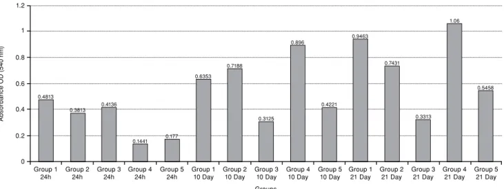
Experimental group n Mean* Grouping**
Group 4. Day 21 6 1.06017 A Group 1. Day 21 6 0.94633 B Group 4. Day 10 6 0.89600 C Group 2. Day 21 6 0.74317 D Group 2. Day 10 6 0.71883 E Group 1. Day 10 6 0.63533 F Group 5. Day 21 6 0.54583 G Group 1. 24h 6 0.48133 H Group 5. Day 10 6 0.42217 I Group 3. 24h 6 0.41367 I Group 2. 24h 6 0.38133 J Group 3. Day 21 6 0.33133 K Group 3. Day 10 6 0.31250 L Group 5. 24h 6 0.17700 M Group 4. 24h 6 0.14417 N
Tukey Pairwise Comparisons for MTT-cell viability toxicity and proliferation (Grouping information using the Tukey Method and 95% confidence interval)
Experimental group n Mean* Grouping**
Group 4. Day 21 6 1.69133 A Group 4. Day 10 6 1.25367 B Group 5. Day 10 6 1.09800 C Group 5. Day 21 6 1.08800 D Group 2. Day 21 6 0.94583 E Group 2. Day 10 6 0.84150 F Group 1. Day 21 6 0.60700 G Group 1. Day 10 6 0.50100 H Group 1. 24h 6 0.47600 I Group 3. 24h 6 0.44033 J Group 2. 24h 6 0.42850 K Group 3. Day 21 6 0.38450 L Group 3. Day 10 6 0.33483 M Group 5. 24h 6 0.32867 N Group 4. 24h 6 0.30833 O
MTT: 3-(4,5-dimethylthiazol-2-yl)-2,5-diphenyltetrazolium bromide; SSEA-1: Stage-specific embryonic antigen-1; * Difference of means. ** Groups that do not share a letter are significantly different.
differences arose and to prevent false positive results. So, the differences between the averages of the same sample groups were evaluated.
RESUlTS
The viability of the chondrocytes in all groups was evaluated after 10 and 21 days of culture. Chondrocytes proliferated well in experimental groups, including the control in a time-dependent manner (Figure 4). P-PRP and P-PRP-containing hydrogels enhanced cellular viability particularly after 10 and 21 days of culture (Figure 4). Besides, P-PRP influenced the maturation of chondrocytes, increased the mitotic and adhesion activity and enhanced the formation of larger elliptical cells (Figure 3).
The difference between groups identified with variance analysis and Tukey HSD test was statistically significant (Tables II and III). Tukey pairwise comparisons showed a statistically significant difference between subgroups except for 10th day of group 5 and 24th hour of group 3, which were similar (Table III).
Compared to the control group, minimum SSEA-1 expression was detected at 24 hours in group 4 and group 5. Minimum SSEA-1 expression was detected in groups 3 and 5 on the 10th day. However, on the
21st day, minimum SSEA-1 expression was detected
in group 3 compared to control group (Figure 5). Notably, maximum SSEA-1 expression was observed
Figure 5. Bar graph of enzyme-linked immunosorbent assay analysis: Stage-specific embryonic antigen-1 expression of all groups. OD: Optical density.
1.2 1 0.8 0.6 0.4 0.2 0 Group 1
24h Group 224h Group 4 24h Group 110 Day Group 310 Day Group 510 Day Group 221 Day Group 4 21 Day Groups A bs or ba nc e O D ( 54 0 n m ) 0.4813 0.3813 0.4136 0.1441 0.177 0.6353 0.7188 0.3125 0.896 0.4221 0.9463 0.7431 0.3313 1.06 0.5458 Group 3
24h Group 5 24h Group 2 10 Day Group 410 Day Group 1 21 Day Group 321 Day Group 521 Day
Figure 6. Bar graph of MTT analysis. OD: Optical density; MTT: 3-(4,5-dimethylthiazol-2-yl)-2,5-diphenyltetrazolium bromide. 1.8 1.6 1.4 1.2 1 0.8 0.6 0.4 0.2 0 Group 1
24h Group 224h Group 4 24h Group 110 Day Group 310 Day Group 510 Day Group 221 Day Group 4 21 Day Groups A bs or ba nc e O D ( 54 0 n m ) 0.476 0.4285 0.4403 0.3083 0.3287 0.501 0.8415 0.3348 1.2536 1.098 0.607 0.9458 0.3845 1.6913 1.088 Group 3
in group 4 on the 21st day, followed by the 10th day of group 4.
MTT assay results were consistent with SSEA-1 ELISA results. Maximum viability was observed in group 4 on the 21st day. Minimum viability was observed in groups 4 and 5 at 24 hours, followed by group 2 on the 10th and 21st days (Figure 6). SSEA-1 and MTT analyses showed statistically significant correlations (p=0.000, Pearson product moment correlation coefficient=0.745).
DISCUSSION
Cartilage injuries resulting from osteoarthritis are clinical problems reducing the quality of life and which may cause severe disabilities.[13] The self-repair capacity of cartilage is very limited. Besides, there is no admitted application or treatment protocol for present commercial drugs, and therefore different clinics use distinct treatment strategies. Consequently, the success rates of treatment are not high enough and usually patient satisfaction is not achieved.[14] The consideration of all these reasons in the treatment of cartilage damage through replacement of tissue by biocompatible drug delivery systems represents the focus of recent research.[15]
Platelet-rich plasma as a storage vehicle of growth factors, such as platelet-derived growth factor, transforming growth factor-b, platelet-derived endothelial growth factor, vascular endothelial growth factor, and platelet factor 4, is used in therapy since 1990s,[16] especially in wound healing.[17] Many reports suggest that PRP activates the autocrine and paracrine system, inflammatory response, regulates chemotaxis, and induces the regulation of coagulation and cell differentiation.[18]
Therefore, PRP has been put into practice for treatment in almost all areas of medicine including sports medicine and orthopedics.[19] Unfortunately, growth factors in PRP have a very short half-life. These growth factors are quickly metabolized by surrounding tissue and removed from the injection area.[6,20] Because of this reason, high and repeated doses are used in intraarticular or systemic administration of PRP for an effective treatment. A great number of researches also reported that PRP diffuses micro-environment simply because of the difference in concentration and causes undesirable side effects.[21]
The goal of many researches is to improve delivery strategies or drug carrier systems for products like PRP in order to prevent degradation
and maintain longer biological activity.[20]
A controlled drug delivery system is suitable to transport small amounts of growth factors, provide long-term release within a certain time period, eliminate the side and/or adverse effects caused by systemic administrations, as well as enhance the in vivo activity of growth factors. In this study, we have used a hydrogel prepared with non-toxic natural materials as a delivery system. As a result of our study, SSEA-1 expression was most abundant in the group treated with P-PRP applied with hydrogel (Day 21), besides cell viability and proliferation. This finding indicates that the hydrogel we have used maintained the stability and extended the half-life of growth factors in PRP.
We have also investigated the effect of the amount of leukocytes present in the PRP prepared by conventional methods. Many researches reported that leukocytes in the PRP interact with bone marrow-derived progenitor cells and support healing processes.[17,22] Others ignore the effects of leukocytes or, in other words, the need for the removal of
leukocytes in PRP.[23] More importantly, to our
knowledge, there are no clinical and/or preclinical studies revealing the amount of leukocyte and platelet content of PRP clearly.
In our study, proliferation, extracellular matrix formation and SSEA-1 expression were found to be lower in L-PRP-treated groups with a high leukocyte ratio than in groups treated with P-PRP. These results obtained with in vitro experiments need to be confirmed with animal studies. The influence of leukocytes on the biology of chondrocytes and their potential benefits should be carefully analyzed since this might shed light to many controversial data from the literature.[24]
A great number of researches published in the literature represent studies that were conducted on rats or rabbits as a model of mammalian. Although animal models are suitable to observe the systemic effects of PRP, animal tissue physiology and sensitivity differ from those of human tissue.[25-27] Concerning the small amount of studies demonstrating the in vitro effects of different leukocyte concentrations of PRP, it was observed that these studies are on synoviocytes.[28] or other tissues such as human ligament fibroblasts.[26] To the best of our knowledge, in the literature, only one
in vitro experimental study tested the effect of PRP
on chondrocytes isolated from osteoarthritis tissue. Unfortunately, the cell cultures in this single study were established with tissues isolated only from four male patients.[4] In our study, we used primary chondrocyte cultures obtained from both female
and male patients who underwent surgery. There are many studies performed on cell lines; however, it is well documented that cell line physiology and even genetic differs from the original cell.
In this study, we have used primary chondrocyte cultures obtained from the chondral tissue comprised of cartilage cells and also the extracellular matrix components to investigate the effect of various PRP formulations on human chondrocytes in vitro. In vitro effects of two different formulations of PRP, P-PRP and L-PRP were tested by exposing chondrocytes with or without the presence of hydrogel. Cell proliferation, matrix production, and SSEA-1 expression were evaluated. All formulations that stimulated chondrocyte proliferation throughout the culture period were evaluated, and it was revealed that P-PRP applied with hydrogel induced greater cell growth and SSEA-1 expression compared with other formulations.
Stage-specific embryonic antigen-1, also known as CD15, is an antigenic epitope associated with cell adhesion, migration, and differentiation. It is expressed on differentiated adult human cartilage chondrocytes but not on dedifferentiated adult human cartilage chondrocytes.[29]
It is not sufficient to improve the proliferation of chondrocytes in a proper treatment. Ensuring the integrity of the tissue also requires the achievement of an intact extracellular matrix. Our results showed that the expression of SSEA-1 correlates with cell proliferation. Increased SSEA-1 expression was evaluated as maturated chondrocyte presence.
Limitations of our study is that we have have performed the experiments in vitro so the results cannot be generalized to the in vivo effects of PRP administration on cartilage tissue.
In conclusion, administration of P-PRP in the presence of a drug delivery system that supports controlled release not only increases cell proliferation but also supports extracellular matrix development. The identification of the optimal amounts and ratios of these blood components may ideally lead to a formulation more suitable for the treatment of cartilage lesions. Our findings have demonstrated that P-PRP should be used with a drug delivery system such as hydrogel. We believe that the results of our work may provide a significant contribution to the literature. However, it should be noted that our findings have been obtained from in vitro experiments. Further clinical research is needed to improve the effectiveness of PRP treatment in cartilage regeneration.
Declaration of conflicting interests
The authors declared no conflicts of interest with respect to the authorship and/or publication of this article.
Funding
This study was supported by grants (Project no: NKUBAP.00.10.AR.15.08) from Namik Kemal University Scientific Research Administration Division.
REFERENCES
1. Lu YC, Evans CH, Grodzinsky AJ. Effects of short-term glucocorticoid treatment on changes in cartilage matrix degradation and chondrocyte gene expression induced by mechanical injury and inflammatory cytokines. Arthritis Res Ther 2011;13:142.
2. Xie X, Zhang C, Tuan RS. Biology of platelet-rich plasma and its clinical application in cartilage repair. Arthritis Res Ther 2014;16:204.
3. Nurden AT, Nurden P, Sanchez M, Andia I, Anitua E. Platelets and wound healing. Front Biosci 2008;13:3532-48. 4. Atik OS. Is the evidence behind platelet-rich plasma
therapies strong enough? Eklem Hastalik Cerrahisi 2012;23:1.
5. Cavallo C, Filardo G, Mariani E, Kon E, Marcacci M, Pereira Ruiz MT, et al. Comparison of platelet-rich plasma formulations for cartilage healing: an in vitro study. J Bone Joint Surg [Am] 2014;96:423-9.
6. Getgood A, Brooks R, Fortier L, Rushton N. Articular cartilage tissue engineering: today's research, tomorrow's practice? J Bone Joint Surg [Br] 2009;91:565-76.
7. Kellgren JH, Lawrence JS. Radiological assessment of osteo-arthrosis. Ann Rheum Dis 1957;16:494-502.
8. Perut F, Filardo G, Mariani E, Cenacchi A, Pratelli L, Devescovi V, et al. Preparation method and growth factor content of platelet concentrate influence the osteogenic differentiation of bone marrow stromal cells. Cytotherapy 2013;15:830-9.
9. Roffi A, Filardo G, Assirelli E, Cavallo C, Cenacchi A, Facchini A, et al. Does platelet-rich plasma freeze-thawing influence growth factor release and their effects on chondrocytes and synoviocytes? Biomed Res Int 2014;2014:692913.
10. Yilmaz I, Gokay NS, Gokce A, Tonbul M, Gokce C. A novel designed chitosan based hydrogel which is capable of consecutively controlled release of TGF-Beta 1 and BMP-7. Turkiye Klinikleri J Med Sci 2013;33:18-32.
11. Isyar M, Yilmaz I, Guler O, Mahirogullari M. Could platelet rich plasma be soaked to polymeric hydrogel which could be used at biological repair of cartilage? OTSHD 2015;4:40-51. 12. Available from: http://www.celprogen.com/details.
php?pid=10765.
13. Atik OŞ, Erdoğan D, Seymen CM, Bozkurt HH, Kaplanoğlu GT. Is there crosstalk between subchondral bone, cartilage, and meniscus in the pathogenesis of osteoarthritis? Eklem Hastalik Cerrahisi 2016;27:62-7.
14. Das BR, Roy A, Khan FR. Cartilage oligomeric matrix protein in monitoring and prognostication of osteoarthritis and its utility in drug development. Perspect Clin Res 2015;6:4-9.
15. Mardones R, Jofré CM, Minguell JJ. Cell Therapy and Tissue Engineering Approaches for Cartilage Repair and/or
Regeneration. Int J Stem Cells 2015;8:48-53.
16. Gehring S, Hoerauf H, Laqua H, Kirchner H, Klüter H. Preparation of autologous platelets for the ophthalmologic treatment of macular holes. Transfusion 1999;39:144-8. 17. Eppley BL, Pietrzak WS, Blanton M. Platelet-rich plasma: a
review of biology and applications in plastic surgery. Plast Reconstr Surg 2006;118:147-59.
18. Marx RE. Platelet-rich plasma (PRP): what is PRP and what is not PRP? Implant Dent 2001;10:225-8.
19. Jo CH, Shin JS, Lee YG, Shin WH, Kim H, Lee SY, et al. Platelet-rich plasma for arthroscopic repair of large to massive rotator cuff tears: a randomized, single-blind, parallel-group trial. Am J Sports Med 2013;41:2240-8. 20. Mall NA, Tanaka MJ, Choi LS, Paletta GA Jr. Factors affecting
rotator cuff healing. J Bone Joint Surg Am 2014;96:778-88. 21. Arora NS, Ramanayake T, Ren YF, Romanos GE. Platelet-rich
plasma: a literature review. Implant Dent 2009;18:303-10. 22. “Wrotniak M, Bielecki T, Gaździk TS. Current opinion about
using the platelet-rich gel in orthopaedics and trauma surgery. Ortop Traumatol Rehabil 2007;9:227-38.
23. Everts PA, van Zundert A, Schönberger JP, Devilee RJ, Knape JT. What do we use: rich plasma or platelet-leukocyte gel? J Biomed Mater Res A 2008;85:1135-6.
24. Dohan Ehrenfest DM, Rasmusson L, Albrektsson T. Classification of platelet concentrates: from pure platelet-rich plasma (P-PRP) to leucocyte- and platelet-platelet-rich fibrin (L-PRF). Trends Biotechnol 2009;27:158-67.
25. Dolkart O, Chechik O, Zarfati Y, Brosh T, Alhajajra F, Maman E. A single dose of platelet-rich plasma improves the organization and strength of a surgically repaired rotator cuff tendon in rats. Arch Orthop Trauma Surg 2014;134:1271-7.
26. Halpern BC, Chaudhury S, Rodeo SA. The role of platelet-rich plasma in inducing musculoskeletal tissue healing. HSS J 2012;8:137-45.
27. Isyar M, Yilmaz I, Nusran G, Guler O, Yalcin S, Mahirogullari M. Safety of bioabsorbable implants in vitro. BMC Surg 2015;15:127.
28. Braun HJ, Kim HJ, Chu CR, Dragoo JL. The effect of platelet-rich plasma formulations and blood products on human synoviocytes: implications for intra-articular injury and therapy. Am J Sports Med 2014;42:1204-10.
29. de la Fuente R, Abad JL, García-Castro J, Fernández-Miguel G, Petriz J, Rubio D, et al. Dedifferentiated adult articular chondrocytes: a population of human multipotent primitive cells. Exp Cell Res 2004;297:313-28.