An Unusual Location of Intra-Abdominal Desmoid Tumor
Batın İçi Desmoid Tümörün Beklenmedik Bir Yerleșim Yeri
Ahmet Hakan Haliloğlu
1, Nuray Haliloğlu
2, Ömür Ataoğlu
3, Sümer Baltacı
4, Cemil Yağcı
41 Ufuk University School of Medicine Department of Urology 2 Ankara University Faculty of Medicine Department of Radiology 3 Micro-Pat Pathology Laboratory
4 Ankara University Faculty of Medicine Department of Urology
Desmoid tümörler ağırlıklı olarak genç kadınlarda bulunan lokal agresif benign tümörlerdir. Biz burada histopatolojik olarak ispatlanmıș renal desmoid tümörü olan 36 yașında bir kadın has-tayı sunuyoruz. Beklenmedik yerleșim yerlerindeki izole desmoid tümörlerin tanısı histopatolojik inceleme yapılmadan mümkün olmayabilir fakat üreme çağındaki bir kadın hastada komșu yapılarda bası ya da yer değiștirmeye neden olan büyük bir batın içi kitle sap-tandığında ayırıcı tanıda düșünülebilir.
Anahtar Sözcükler: Desmoid, Fibromatosis, Abdominal, Agresif fibromat
Desmoid tumors are locally aggressive benign neoplasms which are predominantly found in young women. Here we present a 36-year-old woman with histopathologically proven renal desmoid tumor. Diagnosis of isolated desmoid tumors in extraordinary locations may not be possible without histopathological examination but they may be considered in the differential diagnosis when a huge intraabdominal mass causing compression or displacament of neighboring structures is detected in a female patient at reproductive age.
Key Words: Desmoid, Fibromatosis, Abdominal, Aggressive fibromat
Desmoid tumors are rare, benign tumors of musculoaponeurotic fibrous tissue, which are predominantly found in young females (1, 2). They are locally aggressive tumors with no predisposition for distant metastasis and have a strong tendency to recur after surgical removal (2, 3). Desmoid tumors may be found in extra-abdominal, abdominal and intra-abdominal sites, and less commonly in the extremities and trunk. Intraabdominal desmoids can arise from the mesentery, retroperitoneum, or pelvis (2, 4). This case report demonstrates an unusual location of a histopathologically proven desmoid tumor in a young woman
at reproductive age.
Case Report
A 36-year-old woman was admitted to the department of urology with right flank pain which existed in the last 10 days. The patient’s own
and her family’s medical history were unremarkable. She had only taken oral contraceptive pills for two months and she was not taking them recently. She also had no previous history of abdominal surgery except for two cesarean sections, the latest performed 5 years ago.
On physical examination the only abnormal finding was right flank tenderness. The results of routine blood tests and urine analysis were within normal limits. Ultrasonography (US) revealed an egzophytic solid mass in the lower pole of the right kidney. Abdominal computed tomography (CT) was performed in order to delineate the nature of the mass and to investigate it’s relation with the neighboring anatomical structures. On pre-contrast and contrast enhanced axial CT scans, a 11x10 cm, minimally enhanced, lobulated solid mass with well-circumscribed margins was
Ankara Üniversitesi Tıp Fakültesi Mecmuası 2012, 65 (3)
DOI: 10.1501/Tıpfak_000000831
CERRAHİ TIP BİLİMLERİ/SURGICAL SCIENCES
Olgu Sunumu / Case Reports
Received: 23.02.2011 Accepted: 19.06.2013 İCorresponding author
Doç.Dr. Nuray Haliloğlu Phone : 595 67 81
E-mail : [email protected]
Ankara University School of Medicine Department of Radiology
Ankara Üniversitesi Tıp Fakültesi Mecmuası 2012, 65 (3)
An Unusual Location of Intra-Abdominal Desmoid Tumor 176
.
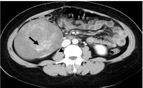
Figure 1: On contrast enhanced axial CT scan, a renal mass with smooth contours and
minimally enhanced internal components (arrow) is demonstrated. The mass is neighboring the inferior vena cava and compressing the right ureter (arrowhead).
Figure 2: Panoramic view of the tumor. H&E x100 (A). Histologic specimen showing spindle
cells seperated by collagen. Cells are uniform, lacking nuclear hyperchromosia and atypia. H&E x200 (B).
detected in the inferior pole of the right kidney which compressed the right ureter and caused mild hydronephrosis. The mass was also neighboring the inferoposterior aspect of the liver, the right iliopsoas muscle and inferior vena cava but there were no signs of invasion (Figure 1). Neither abdominal lymphadenopathy nor thrombosis of the renal veins/inferior vena cava were present. Radical nephrectomy was performed and no intraoperative or postoperative complications occured.
Examination of the nephrectomy material revealed a mass measuring 9 cm. in its longest diameter and localized in the lower pole of the
kidney. The lesion was
well-circumscribed, apparently encapsulated and when cut; , it was
grayish white in color with focal hemorrhagic areas, with a firm and rather rubbery in consistence.. Microscopically, the tumor composed of fibroblast-like spindle cells and exhibited rare mitotic figures. Nuclei were bland. There was no obvious cellularity but bundles of collagen fibers and rare mixoid changes were present within the tumor (Figure 2). The mass was clearly marginated and there were no signs of invasion of the perirenal fat, renal vein or ureter.
The immunohistochemistry analysis revealed no positive stainings with
cytokeratin, S-100, desmin, actin , myoglobulin, CD 34 and HMB-45. The final histopathological diagnosis was consistent with desmoid tumor
Discussion
The exact etiology of desmoid tumors is unknown but these tumors are usually associated with surgical trauma or endocrine factors including pregnancy and use of oral contraceptives. It is also shown that patients with Gardner’s syndrome may develop desmoid tumors as well (1, 5). Histopathologically desmoid tumors
are characterized by proliferation of well-differentiated fibroblasts and bundles of collagen with an infiltrative growth pattern and absence of cytological features of malignancy (1).
Site of surgical scars and the bowel mesentery are common locations of desmoid tumors and retroperitoneum and abdominal wall are less common sites of occurence (5). Isolated intraabdominal desmoid tumors are rare and are mostly seen in females at reproductive age (6). It is known that desmoid tumors may cause hydronephrosis secondary to compression or displacement of ureters (6-8). Nevertheless as far as we are concerned there is no published report in the literature describing an intraabdominal desmoid tumor originating from kidneys.
Patients with isolated intraabdominal desmoid tumors may present with a palpabl mass or with symptoms due to desmoid-induced complications of adjacent organs including bowel, ureter and abdominal blood vessel obstruction (4). When a mass located in the abdominal wall or in the abdominal cavity which does
Journal of Ankara University Faculty of Medicine 2012, 65 (3)
Ahmet Hakan Haliloğlu, Nuray Haliloğlu, Ömür Ataoğlu, Sümer Baltacı, Cemil Yağcı 177
not arise from the surrounding organs is detected, the possibility of an intra-abdominal desmoid tumor must be considered in the differential diagnosis (9).
When a solid renal mass is detected, the extensions of the tumor and it’s relations with adjacent structures such as vessels and ureter must be documented preoperatively in order to distinguish whether the tumor has truly invaded the adjacent structures suggesting malignant neoplasms or compressed them suggesting benign neoplasms. This may not always allow the clinician to differentiate an isolated desmoid from other retroperitoneal and pelvic neoplasms, but may help an appropriate theraphy planning (2, 10).
Desmoid tumors can be seen as relatively hypodense, solitary or multiple encapsulated masses or poorly circumscribed nodules on CT scans (9). Since they are composed of well-differentiated fibrous tissue, rapid enhancement may not be seen after intravenous
contrast medium injection. For atypical masses in unexpected locations, dynamic enhanced scans can be performed, as late phase postcontrast images may be useful to demonstrate the gradual enhancement of a fibromatous mass.
The patient we presented here did not have clinical and radiological findings suggesting renal cell carcinoma. As CT scan demonstrated a minimally enhanced, clearly marginated mass, primary renal lymphoma could be considered in the differential diagnosis but there were no clinical or laboratory findings or lympadenopathy to support the diagnosis. Lack of endocrinological factors, history of abdominal surgery or Gardner’s syndrome and the location of the tumor, were unfavorable for the diagnosis of desmoid tumor. Wide surgical resection and histopathological examination was obligatory for the diagnosis.
Although surgical excision is the main strategy for treatment of desmoid
tumors there are also options of systemic treatment with steroids,
antiinflammatory drugs, chemotheraphy and tamoxifen
when complete excision of the tumor is not possible due to the potential risk of damaging neighboring vital structures (1, 5). As the mass in our patient was widely resected, no adjuvant systemic theraphy was administered. The patient is under follow-up.
Conclusion
Diagnosis of isolated desmoid tumors in extraordinary locations may not be possible without histopathological examination. Nevertheless desmoid tumors can be considered in the differential diagnosis when a huge intraabdominal mass causing compression or displacament of neighboring structures is detected in a female patient at reproductive age.
REFERENCES
1. Fujita K, Sugao H, Tsujıkawa K, Itoh
Y. Desmoid tumor in a scar from radical nephrectomy for renal cancer. Int J Urol 2003;10:274–275.
2. Kawashima A, Goldman SM, Fishman
EK, et al. CT of intraabdominal desmoid tumors: is the tumor different in patients with Gardner’s Disease? Am J Roentgenol 1994;162:339–342.
3. Einstein DM, Tagliabue JR, Desai RK.
Abdominal desmoids: CT findings in 25 patients. Am J Roentgenol 1991;157:275–279.
4. Pho LN, Coffin CM, Burt RW.
Abdominal desmoid in familial adenomatous polyposis presenting as apancreatic cystic lesion. Fam Cancer 2005;4:135–138.
5. Richard HM, Thall EH III, Mıtty H,
Gribetz ME, Gelernt I. Desmoid tumor–ureteral fistula in Gardner’s Syndrome. Urology 1997;49:135–138.
6. Collins D, Myers E, Kavanagh D,
Lennon G, McDermott E. Mesenteric desmoid tumor causing ureteric obstruction. Int J Urol 2008;15:261– 262.
7. Porru D, Stancati S, Giliberto GL, et
al. Bilateral ureteral compression by multiple abdominal desmoid tumors in Gardner Syndrome: case report and literature review. Urology 2004;64:1031.
8. Soon JL, Lau WKO, Seow–Choen F,
Cheng CWS. Unresectable desmoid tumours causing obstructive uropathy in familial adenomatous polyposis. Asian J Surg 2005;28:233–237.
9. Kreuzberg B, Koudelova J, Ferda J, et
al. Diagnostic problems of abdominal desmoid tumors in various locations. Eur J Radiol 2007;62:180–185.
10. Toprak U, Erdoğan A, Gulbay M, et
al. Preoperative evaluation of renal anatomy and renal masses with helical CT, 3D–CT and 3D–CT angiography. Diagn Interv Radiol 2005;11:35–40.
Ankara Üniversitesi Tıp Fakültesi Mecmuası 2012, 65 (3)
An Unusual Location of Intra-Abdominal Desmoid Tumor 178