Case Report
Introduction
Stromal tumors are the prominent mesenchymal tumors of gastrointestinal tract (GIT) (1). They are more apparent in upper mid-dle age, mostly in 5th and 6th decades. They occur equally in both sexes (1). Gastrointestinal Stromal Tumors (GISTs) are most
com-monly localized in the stomach (40-70%) and may be found in the intestine (20-32%), colon (10%) and esophagus (5%) (1, 2). They also can be seen in extra-GIT sites like mesentery, omentum and retroperitoneum (2). Major patient complaints regarding GISTs are upper abdominal pain, GIT bleeding and abdominal masses (1). Interstitial cells of Cajal (ICC), which GISTs are thought to originate from, both express tyrosine kinase receptors (c-kit) and stain with CD34 and Vimentin. KIT (CD117) is a transmem-brane tyrosine kinase receptor, which is expressed in hematopoietic stem cells, germ cells, mast cells, melanocytes and ICC (2). C-Kit positivity is an important parameter in diagnosis of GIST. Recently, a specific marker, namely discovered on GIST1 (DOG-1), has been discovered for GIST diagnosis. It is shown to be positive on ICCs and gastric surface epithelium. DOG-1 is also expressed in high amounts in KIT mutation-negative cases besides typical GIST cases (3). Immunohistochemical diagnosis of GIST is adopted when mesenchymal tumors stain positive with c-Kit (CD117) and the DOG-1 where the rest of mesenchymal cell markers were negative. Demonstration of CD117 and recently DOG-1 positivity immunochemically stands for the standard technique of definitive diagnosis of GIST. CD34 is positive in 70-80% of the cases. Vimentin is diffuse and strong positive in many stromal tumors. SMA is positive in 30-40% and S100 in 5% of GISTs. Staining with desmin is rare (1). Malignant stromal tumors mostly metastasize to liver, peritoneum, lungs and bones (4). Brain and lymph node metastases are unusual (4). No breast metastasis was presented in literature before.
Hereby, we present a case of GIST with liver, bone, lymph node and breast metastases and aimed to discuss its unusual metastatic review-ing literature.
Case Presentation
A 46 year-old woman was referred to us from another health facility with the complaint of back pain. The radiological imaging studies revealed multiple liver metastases with the largest being 5.5 cm in diameter as well as multiple bone involvements. Positron Emission
An Unusual Gastrointestinal Stromal Tumor
Presentain: Breast, Liver and Lymph Node Metastasis
Bermal Hasbay
1, Hüseyin Özgür Aytaç
2, Fazilet Kayaselçuk
1, Neşe Torun
31Department of Pathology, Başkent University Turgut Noyan Hospital, Adana, Turkey 2Department of General Surgery, Başkent University Turgut Noyan Hospital, Adana, Turkey 3Department of Nuclear Medicine, Başkent University Turgut Noyan Hospital, Adana, Turkey
Address for Correspondence :
Bermal Hasba , e-mail: [email protected] Accepted: 13.06.2017Received: 01.03.2017
Eur J Breast Health 2017; 13: 216-8
DOI: 10.5152/ejbh.2017.3492
216
ABSTRACT
Gastrointestinal Stromal Tumors (GIST) are the common mesenchymal tumors of gastrointestinal tract. They can display benign and malignant clinical behavior. The most common metastasis sites of malignant stromal tumor are liver, peritoneum, lung and bones. Metastasis to breast is ex-tremely rare. Here, we present a case of GIST with liver, bone, lymph node and breast metastasis by reviewing the literature.
Keywords: Gastrointestinal stromal tumor, metastasis, breast, lymph node
Cite this article as: Hasbay B, Aytaç HÖ, Kayaselçuk F, Torun N. An Unusual Gastrointestinal Stromal Tumor presentain: Breast, Liver and Lymph Node Metastasis. Eur J Breast Health 2017; 13: 216-218.
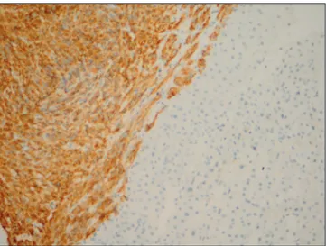
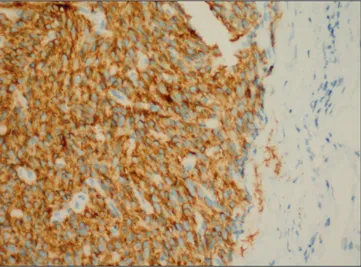
Computed Tomography (PET-CT, General Electric Medical System, Milwaukee, USA) demonstrated elevated F-18-fluoro-2-deoxyglucose (FDG) uptake in her left breast in addition to the above-mentioned lesions. Histologic samples taken from abdominal lymph nodes and liver revealed neoplastic lesions composed of cells with prominent hyperchromatic nuclei and spindle-shaped cytoplasm. Immunohisto-chemistry studies showed strong positive staining with CD117 (Code A4502, Dako, California) in Figure 1 and DOG-1 (Clone SP31, Thermo Scientific, USA) in Figure 2, positive staining with Vimen-tin (Code-Nr. M 0725, Dako, Denmark) and negative staining with CD34 (Clone QBEnd-10, Dako, Denmark), S100 (Code ZO311, Dako, Denmark) and Desmin (Clone D33, Dako, Denmark) in both liver and lymph node tissues. The patient was diagnosed to have GIST metastasis and therefore was started on imatinib therapy. A control PET-CT obtained on the second month of imatinib treatment dem-onstrated progression in liver metastases, variable response in bone me-tastases and partial regression in breast and lymph node meme-tastases. No mass was observed on the physical examination of either of the breasts and the axillae. Breast ultrasound and Magnetic Resonance Imaging showed two small lesions of 5 and 3 mm in close location, which both were thought to be benign.
As for the fact that nuclear regression had been experienced in the left breast under imatinib therapy, tissue diagnosis was planned to rule out
malignancy. After the suspected area was marked with needle localiza-tion by ultrasound, a wide excisional biopsy was performed. When the specimen of 10x4x4 cm yellow color fatty tissue was examined macroscopically in serial sections, no significant mass was observed in cross-sectional surfaces. Complete sampling of the specimen dem-onstrated fibroadipose and acinar tissue, as well as two lesions in 3x2 mm and 2x2 mm size composed of spindle cells with prominent nuclei and spindle-shaped cytoplasm (Figure 3). These cells stained strongly with CD117 (Figure 4) and DOG1 (Figure 5) and did not stain with S100, CD34 and SMA (Clone 1A4, Code M0851, Dako, Denmark) immunohistochemically. Microscopy in 10x High Power Magnifica-tion (HPM) of these lesions showed 4-5 mitoses. Ki67 (Clone MIB-1, Coce M7240, Dako, Denmark) proliferation index was calculated as 3-4%. The case was interpreted as a GIST metastasizing to liver, lymph nodes, bones and breast when evaluated immunohistochemically with its histomorphological findings.
A lumbar MRI scan was obtained due to increased bone pain, which revealed progressive bone metastases. The imatinib dose was doubled; however, she was lost to follow-up. Patient gave orally informed consent.
Discussion and Conclusion
Gastrointestinal stromal tumors (GISTs) can occur anywhere along
217
Hasbay et al. An Unusual Gastrointestinal Stromal Tumor
Figure 1. CD117 positivity by immunohistochemistry in liver
Figure 2. DOG 1 negative in lymphoid cells in lymph node, while
positive in infiltration areas
Figure 3. Tumor infiltration well separated from mammary stroma,
as well as ductal structures (H&E stain, x100)
the Gastrointestinal tract (GIT) from esophagus to anus besides in omentum, mesentery, retroperitoneum deriving from Interstitial cells of cajal (ICCs) (3, 5). C-kit and DOG-1 positivity in used in the pathologic diagnosis of GISTs (3). Stromal tumors like leiomyomas, leiomyosarcomas, desmoid tumors, mesenteric fibromatosis and fibro-sarcomas are considered in differential diagnosis. Tumor size, number of mitosis and localization are the most important prognostic factors of GISTs (1). GISTs are structured as spindle-like in 70%, epitheli-oid in 20% and mixed in 10%. Spacing signet-ring-like areas can also be observed. Major route of metastases of malignant stromal tumors are liver, peritoneum, lungs and bones (4). Brain and lymph node metastases are extraordinary (4). No breast metastasis of GIST was declared before. There are cases in the literature presenting invasive breast cancer of the breast accompanying GIST as a second primary tumor (6, 7). Subash et al. (6) presented a case with GIST in which Position Emission Computed Tomography (PET-CT) demonstrated an increased uptake in her left breast and breast biopsy proved an inva-sive ductal breast cancer . Obinna et al. (4) presented a case with GIST who developed an intramammary lymph node metastasis and intesti-nal metastasis . Our case presentation seems to be the first in literature to demonstrate a breast metastasis of GIST. CD117 is expressed in Phyllodes tumors (PT) of breast (8). This study showed weak staining with CD117 in 5 of 17 PTs (8).
Gastrointestinal stromal tumors rarely can contain signet-ring-like components. Epithelioid type GISTs comprising signet-ring-like cells must be differentiated from invasive lobular cancers of breast in terms of diagnosis. In the study of Yared et al. (9) CD117 was applied to the tissue samples obtained from 21 normal breast epithelia, 24 benign breast lesions, 29 in situ breast carcinomas and 41 invasive carcino-mas. Positive staining was demonstrated in 100% of normal breast epithelia, 88% of benign breast lesions, 20% of in situ carcinomas and 18% of invasive breast carcinomas. Of the 6 invasive breast cancers which stained strong positive, two were lobular, one was ductal and one was signet ring cell cancers . Also, CD117 positivity was shown in a case of gastric epithelioid GIST presented in the literature, which later presented an axillary mass and ER and GCDFP- 15 positivity in breast and gastric samples resulting in the diagnosis of primary lobular carcinoma of the breast and a gastric metastasis (10).
Therefore, especially before diagnosing epithelioid-type stromal tu-mors, lobular and signet ring cell carcinomas of the breast must be
dif-ferentiated by immunohistochemical and radiological examinations. Studies concerning GIST in the literature commonly present liver, lung and bone metastases. Hereby, we presented the first case of GIST metastasizing to breast, which we think is a very extraordinary condi-tion and discussed according to the literature. It must be kept in mind that demonstration of a spindle cell neoplasia can either belong to a primary tumor or a metastasis. Therefore, CD117, which is stained both in GIST and most spindle cell bearing conditions of the breast such as Phyllodes tumors, and DOG1 staining studies must be added to foresee GIST metastasis.
Informed Consent: İnformed consent was obtained from patient who partici-pated in this study.
Peer-review: Externally peer-reviewed.
Author Contributions: Concept - B.H., H.Ö.A., F.K.; Design - B.H., H.Ö.A., F.K.; Supervision - B.H., F.K.; Resources - B.H., F.K.; Materials – B.H., H.Ö.A.; Data Collection and/or Processing - B.H., N.T.; Analysis and/ or Interpretation - B.H., F.K.; Literature Search - B.H., Writing Manuscript - H.Ö.A., B.H., Critical Review - F.K.,B.H.; Other - B.H.,N.T.
Conflict of Interest: No conflict of interests was declared by the authors.
Financial Disclosure: The authors declared that this study has received no financial support.
References
1. Sturgeon C, Cheifec G, Espat NJ. Gastrointestinal stromal tumors: a spectrum of disease. Surg Oncol 2003; 12: 21-26. (PMID: 12689667)
[CrossRef]
2. Miettien M, Lasota J. Gastrointestinal stromal tumors. Definition, clini-cal, histologiclini-cal, İmmünhistochemical and molecular genetic features and differantial diagnosis. Virchows Arch 2001; 438; 1-12. (PMID: 11213830) [CrossRef]
3. Miettinen M, Wang ZF, Lasota J. DOG 1 Antibody in the Differential Diagnosis of Gastrointestinal Stromal Tumors A Study of 1840 Cases. Am J Surg Pathol 2009; 33: 1401-1408. (PMID: 19606013) [CrossRef]
4. Igwilo OC, Byrne MP, Nguyen KD, Atkinson J. Malign gastric stromal tumor: Unusual metastatic patterns. South Med J 2003;96: 512-515. (PMID: 12911195) [CrossRef]
5. Deitos AP. The reapraisal of gastrointestinal stromal tumors: from stout to the KIT revolution. Virchows Arch 2003; 442: 421-428.
6. Ramani SK, Basu S, Parmar V, Gujral S , Bibte S. Second primary malig-nancy of breast in a patient of gastrointestinal stromal tumor presenting as fluoro-deoxyglucose-avid breast incidentaloma in fluoro-deoxyglucose- positron emission tomography. J Cancer Res Ther 2011; 7: 387-389. (PMID: 22044838) [CrossRef]
7. Takeuchı H, Hıroshıge S, Hashımato K, Kusumoto T, Yoshıkawa Y, Muto Y. Synhronous Double Tumor of Breast Cancer and Gastrointestinal stro-mal tumor in a Patient with Neurofibromatosis Type 1 Report of a case. Anticancer Res 2011; 31: 4481-4484. (PMID: 22199319)
8. Bose P, Dunn ST, Yang J, Allen R, El- Khoury C, and Tfaylı A. C-kit Expression and Mutations in Phylloides Tumors of the Breast. Anticancer Res 2010; 30: 4731-4736. (PMID: 21115932)
9. Yared MA, Middleton LP, Meric F, Cristofanilli M, Sahin AA. Expression of c-kit proto-oncogene product in breast tissue. Breast J. 2004; 10: 323-327. (PMID: 15239791) [CrossRef]
10. Vennapusa B, Oman S A, Prasher G, Cerilli LA. C-Kit positive gastric metastasis of lobular carcinoma of the breast masquerading as gastro-intestinal stromal tumor. Breast Cancer 2010; 17: 303-305. (PMID: 19466511) [CrossRef]
218
Eur J Breast Health 2017; 13: 216-8