Effect of Distal Masseter to Facial Nerve Transfer in
Paralytic Patients with Preserved Facial Nerve Continuity
on Improving Scaled Measurement of Improvement in
Lip Excursion (SMILE): A Vectoral Analysis
Berke Özücer , Osman Halit Çam
Department of Otorhinolaryngology, Başkent University School of Medicine, İstanbul, Turkey Original Investigation
Corresponding Author:
Berke Özücer; [email protected]
Received Date: 29.07.2020 Accepted Date: 2.11.2020
Content of this journal is licensed under a Creative Commons Attribution 4.0 International License. Available online at www.turkarchotolaryngol.net
DOI: 10.5152/tao.2020.5823
Abstract Objective: Distal masseter-to-facial neurorrhaphy is an option to improve smile excursion in facial paraly-sis patients in the early period without truncating the facial nerve truncus and by ensuring the continuity of the facial nerve. This study aimed to study the effect of distal masseter-to-facial neurorrhaphy on smile ex-cursion.
Methods: Charts of eight patients were retrospective-ly examined. Screenshots showing the best possible smiles were taken from preoperative videos. Screen-shots were taken from postoperative videos showing the best combination of a natural smile on the healthy side and a smile with clenched teeth on the paralytic side. Emotrics and Photoshop software were used for computing vertical, horizontal, and overall excursion from facial landmarks. Scaled measurements of im-provement in lip excursion and lip angle was evaluat-ed. Symmetry was evaluated by accepting the healthy side as 100 percent, and the paralytic side was calcu-lated as a percentage of the healthy side.
Results: Five patients had total facial paralysis and three had facial paresis. Mean postoperative fol-low-up period was 15.0±10.2 months. The average
interval between facial denervation and nerve repair was 14.0±4.1 months (range, 11-23). All neurorrha-phies were coapted end-to-end to either the zygo-matic or the buccal branch without an interposition graft. Mean postoperative initial movement occurred at 95.5±20.5 days (range, 72-138). Paralytic side to healthy side horizontal excursion changed from pre-operative 72.5±17.4% to postpre-operative 93.4±6.9%. Vertical excursion changed from preoperative 38.4±24.6% to postoperative 89.3±11.8%. Overall excursion changed from preoperative 68.4±19.6% to postoperative 92.9±10.4%. Paralytic side to healthy side mean lip angle changed from 64.7% preoperative to 95.2% postoperatively. All changes were statistical-ly significant (p<0.05).
Conclusion: Facial paralysis patients with an asym-metric smile benefit from distal masseter-to-facial nerve transfer and it improves smile excursion dra-matically. This effect was especially prominent in the vertical component of the smiling vector.
Keywords: Facial paralysis, facial palsy, facial paresis, masseter nerve, smile excursion, facial reanimation ORCID iDs of the authors:
B.Ö. 0000-0001-7798-6793; O.H.Ç. 0000-0002-2785-4474.
Cite this article as: Özücer B, Çam OH. Effect
of Distal Masseter to Facial Nerve Transfer in Paralytic Patients with Preserved Facial Nerve Continuity on Improving Scaled Measurement of Improvement in Lip Excursion (SMILE): A Vectoral Analysis. Turk Arch Otorhinolaryngol 2020; 58(4): 249-53.
Introduction
Facial paralysis is a disabling disease associated with depression and severe decrease in life quality (1). Numerous facial reanimation techniques are de-scribed to address facial paralysis. Recently, paralytic patients with anatomically and physically preserved nerve continuity present as a dilemma; two practical approaches are possible: (a) conservatively waiting for spontaneous recovery, (b) or early intervention by connecting additional nerve input from oth-er cranial noth-erves. Denoth-ervation time is a factor that
makes the timing of the intervention as crucial as the intervention itself. While recent studies have shown that early intervention resulted in more sat-isfactory results, current literature tends to stress the importance of early intervention (2). Nerve transfer to the facial nerve trunk offers a trade-off of giving up natural facial nerve healing. Neurorrhaphy to dis-tal branches without truncating avoids this trade-off and makes early nerve transfer a viable option. The masseter nerve, a motor branch of the trigemi-nal nerve, is a donor nerve described relatively more
recently by Spira (3) in 1978. It was first described as a donor nerve for free gracilis muscle transfer, but its utilization solely in nerve grafting was reported numerously in the literature (4, 5). Its proximity to the surgical field eliminates the need for an inter-position graft, and thereby prevents extra morbidity, allowing for theoretically faster and better healing due to single-anastomosis. This study aimed to evaluate the effects of distal masseter-to-fa-cial nerve transfer in famasseter-to-fa-cial palsy patients with preserved famasseter-to-fa-cial nerve continuity on improving smile excursion.
Methods
This study was approved by Başkent University Institutional Review Board (Approval Date: June 16, 2020; Approval Num-ber: KA20/241) and supported by Başkent University Research Fund. Informed consent was obtained from all patients. Records of patients who were operated on from January 2018 to January 2020 were examined consecutively. All patients were operated on by the first author. Truncation of the facial nerve truncus was a criterion for exclusion from the study. Patients were consecu-tively included, and patient records and surgical notes were ret-rospectively reviewed. Denervation time and follow-up period were noted for each patient.
Operative Technique
The operative technique was based on the cadaveric studies by Col-lar et al. (6) and Borschel et al. (7). In all cases, the masseteric nerve was identified through a limited, minimally invasive dissection fol-lowing a limited preauricular incision rather than a parotidecto-my-like approach. Landmarks of the subzygomatic triangle were marked 3 to 1 cm measurement as explained by Borschel et al. (7).
A preauricular incision was carried out with a 15-blade, and sub-cutaneous dissection was carried out bluntly. Buccal and zygomatic branches that approximate the origin of zygomatic muscle on the zygomatic bone were identified with blunt dissection of a delicate hemostat parallel to the distal branches of the facial nerve. After the recipient branch was identified, the masseter nerve was explored. Basic nerve stimulator (Stimuplex® HNS12 Nerve Stimulator, Braun Medical Inc.; Melsungen, Germany) was used to identify the nerve branches. The masseter nerve in the masseter muscle was dissected; abundant masseter nerve dissection was ensured for a tension-free coaptation without the need for an interposition graft. Approximately 4 to 5 nylon sutures (10-0) were used for end-to-end coaptation. All cases were finalized with fibrin sealant (Baxter International Inc.; Illinois, USA). Minivac drains were removed and all patients were discharged on the first postoperative day.
Evaluation
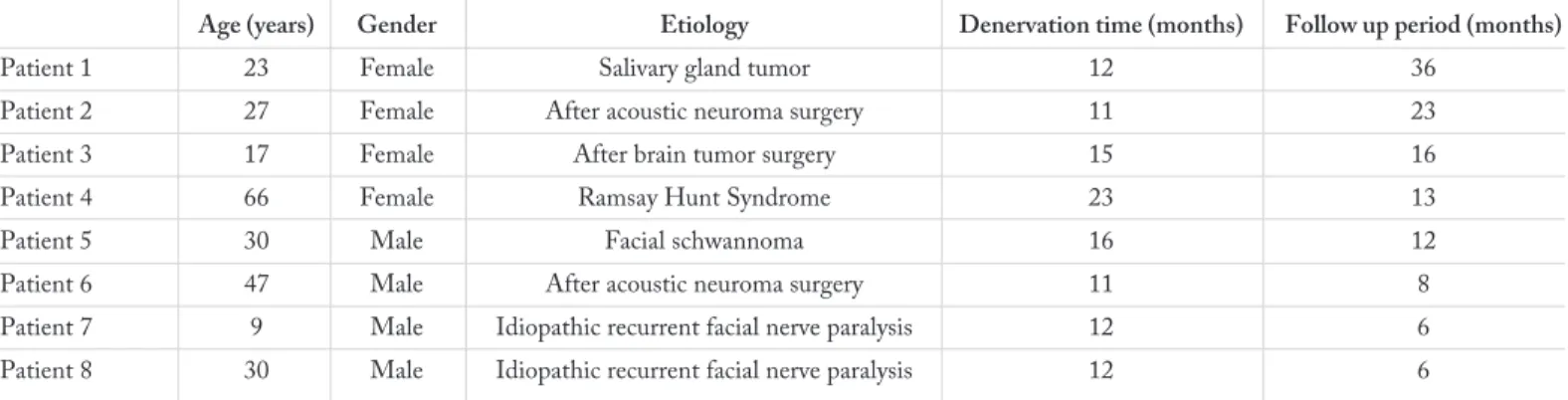
Evaluations were based on the screenshots taken from the standardized preoperative and postoperative videos of patients. Screenshots of the best possible smiles were taken from the preoperative videos. Screenshots of the best possible combined smiles with a natural smile on the healthy side and a smile with clenched teeth on the paralytic side were taken from the postop-erative videos. The Emotrics sofware (Emotrics Software, Mass Eye and Ear; Boston, MA, USA) was used to automatically mark the facial landmarks such as the iris and the facial midline with the software’s artificial intelligence algorithm (8) (Figure 1). Best possible smile photos were marked, and scaled measure-ments of improvement in lip angle and lip excursion (SMILEs) were calculated by vectorial analysis as previously described (9).
Statistical Analysis
On the healthy side, vertical excursion was marked (y), horizon-tal excursion was marked (x), and overall excursion was marked (z). On the paralytic side, vertical excursion was marked (y’), horizontal excursion was marked (x’), and overall excursion was marked (z’). Vectors on the healthy side were accepted as 100%, and the paralytic side was scale measured relatively as (x/x’) x100. Preoperative and postoperative values, and change rates were compared with the Wilcoxon Signed Ranks test. IBM Sta-tistical Package for the Social Sciences version 24 (IBM SPSS Corp.; Armonk, NY, USA) was used for statistical analysis. P values lower than 0.05 were accepted as statistically significant.
Main Points
• Distal masseter-to-facial nerve transfer is a viable option for early reinnervation without truncating the main facial nerve truncus.
• Mean postoperative initial movement occurred at the third postoperative month.
• Facial paralysis patients with asymmetric smile benefit from distal masseter-to-facial nerve transfer, which improves smile excursion; this improvement is especially dramatic in the ver-tical vector.
Table 1. Patient data
Age (years) Gender Etiology Denervation time (months) Follow up period (months)
Patient 1 23 Female Salivary gland tumor 12 36
Patient 2 27 Female After acoustic neuroma surgery 11 23
Patient 3 17 Female After brain tumor surgery 15 16
Patient 4 66 Female Ramsay Hunt Syndrome 23 13
Patient 5 30 Male Facial schwannoma 16 12
Patient 6 47 Male After acoustic neuroma surgery 11 8
Patient 7 9 Male Idiopathic recurrent facial nerve paralysis 12 6
Results
Five patients had total facial paralysis, and three had facial pa-resis; etiologies are detailed in Table 1. Four patients were male and four were female (Table 1). Mean time between facial de-nervation and nerve repair was 14.0±4.1 months (range, 11-23). Mean postoperative follow-up period was 15.0±10.2 months. All neurorrhaphies were coapted end-to-end to either the zygo-matic or the buccal branch without an interposition graft. Two of the eight patients received additional static suspension with tensor fascia lata graft for a better resting facial symmetry. Three of the eight patients had undergone upper eyelid gold weight placement at another center, and two had adjunctive upper eye-lid golden weight placement. Two patients (25%) complained about temporary painful and limited oral opening in the early postoperative period, and these were resolved without interven-tion. Slight atrophy of the masseter was present in some pa-tients, but none opted for filler or fat augmentation.
The mean time to postoperative initial movement was 95.5±20.5 days (range, 72-138) (Figure 2). Paralytic side to healthy side horizontal excursion percentage changed from preoperative 72.5±17.4% to postoperative 93.4±6.9%. Vertical excursion per-centage changed from preoperative 38.4±24.6% to postoperative 89.3±11.8%. Overall excursion percentage changed from preop-erative 68.4±19.6% to postoppreop-erative 92.9±10.4% (Figure 3). Para-lytic side to healthy side mean lip angle changed from 64.7% pre-operative to 95.2% postpre-operatively. All changes were statistically significant (p<0.05); results are further detailed in Table 2.
Discussion
End-to-end hypoglossal (XII) nerve transfers have historical-ly been the workhorse technique in for facial animation. Jump and other modified hypoglossal nerve transfer procedures were Figure 1. Marking of facial landmarks in Emotrics software and
vectoral analysis in pixels via Photoshop
Figure 2. Preoperative and postoperative images of best possible smile
Figure 3. Evaluation of smile excursion vectors and lip angles Table 2. SMILE vectoral analysis before and after distal
masseterofacial anastomosis
Preoperative Postoperative
(%) (%) p z
Horizontal lip excursion 72.5 93.4 0.036 -2.100 Vertical lip excursion 38.4 89.3 0.012 -2.521 Overall lip excursion 68.4 92.9 0.036 -2.100
Lip angle 64.7 95.2 0.012 -2.521
Table 3. Pros and cons of using the masseter nerve
Pros Cons
Proximity to surgical field Masseter muscle atrophy associated asymmetry
Eliminating the need for an interposition graft Tonus
Fast healing Need for adaptation and physical therapy Strong excursion
introduced to eliminate (a) the need for an interposition graft, (b) a possible hemiglossal atrophy, and (c) the synkinetic mass movement of the face (10).
These three complications and extra donor morbidity can be avoided with masseterofacial anastomosis. Comparison of mas-seterofacial and hypoglossofacial nerve anastomosis revealed superiority for masseterofacial nerve anastomosis in dynamic movements, whereas, at rest, the hypoglossofacial anastomo-sis was found slightly better (11). Our study results show that masseterofacial anastomosis of distal facial branches provided a powerful and symmetric smile (Table 2). Nevertheless, it should be noted that adjunct procedures may be required in complete flaccid paralytic patients with a chubby face to achieve symmet-ric resting tonus. A combination of tensor fascia lata static sling is a viable option.
Our study included the patients with intact facial nerve and branches and those with truncated facial nerve were excluded. This is to say that our results represent a very homogeneous pop-ulation, and this makes our results reliable. We compared pre- and postoperative maximum smile images of each patient. To better understand the differences between patients with intact facial nerve and truncated facial nerve, further investigations with a larger sample size are needed.
Masseteric nerve input ensures fast healing due to proximity, which is essential for patients with long denervation time. Nerve transfer to distal facial branches also ensures the continuation of spontaneous healing of the anatomically preserved nerve conti-nuity. Unlike crossfacial nerve grafting, which ensures synchro-nous facial movements, using the new masseterofacial anasto-mosis for harmonious mimicry requires learning, practice, and cortical adaptation (Table 3). Rehabilitation and functional re-covery after masseterofacial nerve anastomosis is a process that can be expedited with physical therapy (12).
Nevertheless, there are reports in the literature that show that masseter muscle co-activation occurs naturally when smiling. This was shown with electromyography measurements (13). Compared to those performed with non-facial nerve donors, masseterofacial nerve anastomosis seems to be an advantageous option in terms of spontaneity and synchronicity.
There are many surgical treatment strategies for paretic patients about which they were informed during their preoperative visits. The decision-making process was carried out together with the patient. From the patient’s aspect, masseterofacial anastomosis alone has the advantages of being a single-stage surgery and not involving the healthy side of the face.
Hontanilla and Marre (14) reported about their experience with masseterofacial anastomosis in nine incomplete paralysis patients that were comparable to our patient series. Frey et al. (15) reported the “facial upgrading” in paretic patients with a procedure which they called distal end-to-side cross-facial nerve grafting (CFNG). In this procedure, distal facial nerve branch of
the donor side is transected and coapted end-to-end to the sural nerve graft. Distally, the sural nerve graft is coapted end-to-side following an epineural window. With this procedure, native fa-cial nerve axonal input is neither lost nor at risk on the paretic side (15).
Biglioli et al. (16) reported 20 midface paresis cases treated with dual innervation in two stages. The first stage is CFNG to the preauricular region. The second stage is end-to-end masseter-ofacial anastomosis accompanied by end-to-side coaptation of CFNG distal to the coaptation site of masseterofacial anasto-mosis.
Besides the debates on the surgical management of facial par-alysis cases, follow-up and objective recovery measurements are not well described. Outcome measures in facial palsy are mainly evaluated by patient-reported outcome measures, cli-nician-graded scoring systems, and objective software analysis. Patient-reported measures are subjective, but an excellent way to understand the social burden of the effects of facial paraly-sis. Clinician-graded scoring systems are almost subjective, but none of the scaling systems satisfies all (17).
House-Brackmann (HB) staging is a quick clinical grading system and has been used widely worldwide for many years. However, the HB grading system is mainly dependent on the evaluator and remains insufficient, such as the mouth assess-ment subpart is far from being adequate in evaluating changes in smile excursion. Thus, the use of a computer-based objective evaluation system has been described in the literature (8, 15, 18). Many evaluation systems were introduced in the late 1990s, and there has been a focus on the use of objective metrics in dynamic facial reanimation (19-23). In 2000s, computer analysis software have been used to analyze facial movements (20-22). Develop-ments in artificial intelligence and high-resolution photogra-phy/video technology made it possible to evaluate the effects of any facial intervention with more precision.
In 2012 Hadlock and Urban (23) reported using software to de-tect total facial weakness and zonal facial weakness. They found the software very useful in distinguishing healthy subjects from facial paralysis patients and concluded that the software could be used in rehabilitation follow-ups. Hontanilla et al. (18) re-ported FACIAL CLIMA evaluation, mainly an optic camera system that recognizes facial movements such as smiling, mouth puckering, and eye closing.
This optical system automatically defines and calculates the vec-toral changes with three cameras. However, we have used the Emotrics software that allows user verification for the reference points and calculates the vectors on a static image. That Emo-trics software was developed using the standard face databases allow the results to be more realistic, whereas FACIAL CLIMA calculates the dynamic vectoral changes in the same patient. The disadvantage of Emotrics is the need for high-quality photo-graphs (18).
Since objective was to address and repair the symmetry, the SMILE system which evaluates the smile relative to the healthy side was used. One of the limitations of this study is the limited sample size, but on the other hand, it brings novelty in terms of reporting quantitative outcomes of an intervention using novel outcome measures. Using quantitative outcome measures and artificial intelligence software is crucial for the comparison of different interventions. These tools have the potential to offer a universal and automated outcome measures in the future.
Conclusion
Facial paralysis patients with asymmetric smile benefit from distal masseter-to-facial nerve transfer, which improves smile excursion dramatically. Further studies are required to confirm these findings.
Ethics Committee Approval: Ethics committee approval was received for this study from the Başkent University Institutional Review Board (Approval Date: June 16, 2020; Approval Number: KA20/241). Informed Consent: Informed consent was obtained from the patients who participated in this study.
Peer-review: Externally peer-reviewed.
Author Contributions: Concept - B.Ö., O.H.Ç.; Design - B.Ö., O.H.Ç.; Supervision - B.Ö., O.H.Ç.; Resources - B.Ö., O.H.Ç.; Materials - B.Ö., O.H.Ç.; Data Collection and/or Processing - B.Ö., O.H.Ç.; Analysis and/or Interpretation - B.Ö., O.H.Ç.; Literature Search - B.Ö., O.H.Ç.; Writing - B.Ö., O.H.Ç.; Critical Reviews - B.Ö., O.H.Ç.
Conflict of Interest: The authors have no conflicts of interest to de-clare.
Financial Disclosure: This study was supported by Başkent University Research Fund.
References
1. Nellis JC, Ishii M, Byrne PJ, Boahene KDO, Dey JK, Ishii LE. Association among facial paralysis, depression, and quality of life in facial plastic surgery patients. JAMA Facial Plast Surg 2017; 19: 190-6.
2. Albathi M, Oyer S, Ishii LE, Byrne P, Ishii M, Boahene KO. Early nerve grafting for facial paralysis after cerebellopontine angle tu-mor resection with preserved facial nerve continuity. JAMA Facial Plast Surg 2016; 18: 54-60.
3. Spira M. Anastomosis of masseteric nerve to lower division of fa-cial nerve for correction of lower fafa-cial paralysis. Preliminary re-port. Plast Reconstr Surg 1978; 61: 330-4.
4. Klebuc MJ. Facial reanimation using the masseter-to-facial nerve transfer. Plast Reconstr Surg 2011; 127: 1909-15.
5. Murphey AW, Clinkscales WB, Oyer SL. Masseteric nerve trans-fer for facial nerve paralysis. JAMA Facial Plast Surg 2018; 20: 104-10.
6. Collar RM, Byrne PJ, Boahene KDO. The subzygomatic triangle: rapid, minimally invasive identification of the masseteric nerve for facial reanimation. Plast Reconstr Surg 2013; 132: 183-8. 7. Borschel GH, Kawamura DH, Kasukurthi R, Hunter DA, Zuker
RM, Woo AS. The motor nerve to the masseter muscle: an ana-tomic and histomorphometric study to facilitate its use in facial reanimation. J Plast Reconstr Aesthetic Surg 2012; 65: 363-6. 8. Guarin DL, Dusseldorp J, Hadlock TA, Jowett N. A machine
learning approach for automated facial measurements in facial palsy. JAMA Facial Plast Surg 2018; 20: 335-7.
9. Bray D, Henstrom DK, Cheney ML, Hadlock TA. Assessing outcomes in facial reanimation: evaluation and validation of the SMILE system for measuring lip excursion during smiling. Arch Facial Plast Surg 2010; 12: 352-4.
10. Le Clerc N, Herman P, Kania R, Tran H, Altabaa K, Huy PT, et al. Comparison of 3 procedures for hypoglossalfacial anastomosis. Otol Neurotol 2013; 34: 1483-8.
11. Altamami NM, Zaouche S, Vertu-Ciolino D. A comparative ret-rospective study: hypoglossofacial versus masseterofacial nerve anastomosis using Sunnybrook facial grading system. Eur Arch Otorhinolaryngol 2019; 276: 209-16.
12. Pavese C, Cecini M, Lozza A, Biglioli F, Lisi C, Bejor M, et al. Re-habilitation and functional recovery after masseteric-facial nerve anastomosis. Eur J Phys Rehabil Med 2016; 52: 379-88.
13. Lenz Y, Kiefer J, Dietrich F, Stark GB, Eisenhardt SU. Pre-op-erative masseter muscle EMG activation during smile predicts synchronicity of smile development in facial palsy patients under-going reanimation with the masseter nerve: a prospective cohort study. J Plast Reconstr Aesthet Surg 2019; 72: 505-12.
14. Hontanilla B, Marre D. Masseteric-facial nerve transposition for reanimation of the smile in incomplete facial paralysis. Br J Oral Maxillofac Surg 2015; 53: 943-8.
15. Frey M, Giovanoli P, Michaelidou M. Functional upgrading of par-tially recovered facial palsy by cross-face nerve grafting with distal end-to-side neurorrhaphy. Plast Reconstr Surg 2006; 117: 597-608. 16. Biglioli F, Soliman M, El-Shazly M, Saadeldeen W, Abda EA, Al-levi F, et al. Use of the masseteric nerve to treat segmental midface paresis. Br J Oral Maxillofac Surg 2018; 56: 719-26.
17. Dusseldorp JR, van Veen MM, Mohan S, Hadlock TA. Outcome tracking in facial palsy. Otolaryngol Clin North Am 2018; 51: 1033-50.
18. Hontanilla B, Marre D, Cabello A. Facial reanimation with grac-ilis muscle transfer neurotized to cross-facial nerve graft versus masseteric nerve: a comparative study using the FACIAL CLIMA evaluating system. Plast Reconstr Surg 2013; 131: 1241-52. 19. Revenaugh PC, Smith RM, Plitt MA, Ishii L, Boahene K, Byrne
PJ. Use of objective metrics in dynamic facial reanimation: a sys-tematic review. JAMA Facial Plast Surg 2018; 20: 501-8.
20. Linstrom CJ, Silverman CA, Colson D. Facial motion analysis with a video and computer system after treatment of acoustic neu-roma. Otol Neurotol 2002; 23: 572-9.
21. Sargent EW, Fadhli OA, Cohen RS. Measurement of facial move-ment with computer software. Arch Otolaryngol Head Neck Surg 1998; 124: 313-8.
22. Linstrom CJ. Objective facial motion analysis in patients with fa-cial nerve dysfunction. Laryngoscope 2002; 112: 1129-47. 23. Hadlock TA, Urban LS. Toward a universal, automated facial
mea-surement tool in facial reanimation. Arch Facial Plast Surg 2012; 14: 277-82.