Turk J Anaesth Reanim 2015; 43: 126-9
Case R
epor
t
Compartment syndrome of the extremities is a rare but potentially devastating condition. Anaesthetic and analgesic drugs used in the perioperative period may cause a delayed diagnosis by preventing the symptoms from appearing, and irreversible complications can occur. In this report, a case of compartment syndrome secondary to vascular access and its treatment in a morbidly obese patient who underwent abdominoplasty was presented.
Keywords: Compartment syndrome, general anaesthesia, complication
Abstract
Address for Correspondence: Dr. Coşkun Araz, Başkent Üniversitesi Tıp Fakültesi, Anesteziyoloji Anabilim Dalı, Ankara, Türkiye Phone: +90 312 212 68 68-4816 E-mail: [email protected]
©Copyright 2015 by Turkish Anaesthesiology and Intensive Care Society - Available online at www.jtaics.org
Received: 16.05.2014 Accepted: 21.07.2014 Available Online Date: 05.02.2015
126
Introduction
C
ompartment syndrome (CS) is a clinical picture in which the pressure in the closed cavities that include veins and nerves increases to a point that it prevents tissue perfusion and impairment in the circulation of tissues disrupts their metabolisms and functions. Factors such as burns, fractures, pressure dressing, plaster casting, artery damage and bone or soft tissue damage, such as extrinsic pressure, frequently contribute in its pathophysiology (1-3). Even though it is usually observed in the volar forearm and anterior and deep posterior compartments of the lower extremity, it can be observed in all areas where the skeletal muscle of the body is covered with fascia (4).Compartment syndrome, whose main findings are pain, paraesthesia and loss of function, it is difficult to recognize under anaesthesia. In the postoperative period, the residual effects of anaesthesia or the usage of analgesics can prevent early peri-od diagnosis by masking the clinical findings (4). In this case report, we present a case of compartment syndrome, and its successful treatment, that developed because of extravasation of intravenous fluid that accumulated in the hand during the intervention in our patient who underwent abdominoplasty because of obesity.
Case Presentation
A 34-year-old female patient was taken for surgery to perform abdominoplasty because of obesity (body mass index: 45.8 kg m2-1). The patient, who had diabetes mellitus and hypothyroidism under control, was taken into the operation room. She was monitored by electrocardiography, pulse oximetry and non-invasive blood pressure, and an intravenous vascular access was established in the left hand with an 18 G cannula in the second attempt (the unsuccessful first attempt was also performed on the same hand). In the anaesthesia induction performed using this vascular access, 4 mg kg−1 thiopental sodium, 0.5 mg kg−1 rocuronium and 2 mcg kg−1 fentanyl were administered. Additionally, 2 g of cefazolin sodium was administered. Maintenance of anaesthesia was established by the administration of isoflurane 0.8–1.2 at a minimum al-veolar concentration in a mixture of 50%–50% N2O–O2. During the operation, which lasted 320 min in total, 500 mL colloid and 6000 crystalloid fluid replacements were performed intravenously. In line with the results of the spot blood gas analyses conducted during this period, a total of 20 mL calcium gluconate and 70 mL sodium bicarbonate replacements were sent inside the fluids. No noticeable swelling, colour, temperature change or a problem in the going rate of the fluid was observed during or at the end of the operation in the cannula entry point. Postoperative pain treatment was provided using patient-controlled analgesia prepared with tramadol. After the surgery, the patient was admitted to the service, and following her description of severe pain in her hand with vascular access, the place of the vascular access was changed within the first hour following her exit from the operation room. Upon observing a relatively mild swelling compared to the right
A Case of Compartment Syndrome in the Hand Secondary to
Intravenous Fluid Application
Coşkun Araz, Seçil Çetin, Melek Didik, Sevgi Ballı Seyhan, Özgür Kömürcü, Gülnaz Arslan
Department of Anaesthesiology, Başkent University Faculty of Medicine, Ankara, Turkey
hand, it was treated by elevation and diluted boric acid. After the patient continued to complain about the pain, and alter-ation in the tissue perfusion was observed, fasciotomy was performed at the left hand volar and dorsal surfaces, accom-panied with local anaesthesia and sedation, in the 10th post-operative hour with the diagnosis of compartment syndrome (Figure 1). Pain was observed to rapidly decrease following the fasciotomy. The patient was discharged four days after the op-eration. During discharge, no ischaemic findings in the hand and no motor or sensory function loss were present. In the sec-ond week following the operation, incision points at the hand were sutured. All lesions in the patient’s hand healed without problems, and apart from marks due to wound healing, no sensory and/or motor defects that could limit the daily life of the patient were observed.
Discussion
Compartment syndrome is a syndrome accompanied with a pressure increase that occurs inside the muscle or muscle groups that are wrapped with closed fascia tissues or an alter-ation in tissue perfusion. Even though there is more than one mechanism discussed in its pathophysiology, the arteriovenous gradient theory is the most commonly accepted theory. Ac-cording to this theory, when the inner compartment pressure, which is normally 10–15 mmHg, increases to over 30 mmHg, local vein pressure increases, and local arteriovenous pressure gradient and regional blood flow decrease. Because of this, oxygen delivery decreases and remains decreased. Muscle cell damage occurs because of ischaemia caused by prolonged cases, and intracellular toxic materials spread to the extracellular area. This situation leads to the occurrence of the clinical findings of compartment syndrome. Pain, pallor, paraesthesia, paralysis and pulselessness are accepted to be the five main findings of compartment syndrome (5P findings). During the advanced periods, this damaged tissue can heal with fibrosis, and con-tracture formation (3-9).
Compartment syndrome development during anaesthesia is rare, but it is a complication that can lead to serious results (4).
In particular, areas that remain under pressure because they remain in sustained lithotomy, recumbent or prone positions are under risk. Cases of compartment syndrome that also de-veloped during anaesthesia because of intra-arterial medicine administration, pressured liquid infusion or extravasation of hypertonic agents are reported (3, 4). Initially, the cause of compartment syndrome that developed in our patient was considered to be medicines and fluids that were administered via peripheral intravenous access. Administered fluids mostly displayed a crystalloid character, and they were administered to the patient from a height of 80 cm above the patient via grav-ity; devices that provided pressurised infusion were not used. However, because of the developing electrolyte and acid–base changes, calcium and sodium bicarbonate were added to the fluids. We maintain that these can also contribute to the aeti-ology. However, a local necrosis or a reactive lesion was not ob-served. Our patient was in the supine position during the sur-gery, and her left arm that had the vascular access was opened 90° sideways. Therefore, the position was not considered to be related to the developed complication.
Establishing intravenous access is a critical and essential aspect of anaesthesia applications. Anaesthesia application is resumed after establishing proper intravenous access(es) by evaluating many factors such as the surgery that will be performed, the patient and the medicines that are planned to be used. In our clinic, to ease the vascular access and to ensure that proximal veins stay strong, establishing vascular access from the dorsum of the hand in patients with suitable vein structure is frequently preferred. In our patient as well, vascular access was established from the dorsum of the hand with an 18 G cannula in the second attempt. All fluid and medicine administrations during the surgical operation, including the anaesthesia induction, were performed via this vascular access. No problems occurred regarding the going rate of the fluid during the surgery. Fur-thermore, no changes regarding subcutaneous leakage from the hand were observed either during or after the surgery. The patient’s hand anatomy of already being plump because of her being obese and the swelling being minor may be among the reasons for overlooking this complication. Additionally, we would like to point out that the fluid can exit the vein in the first attempt because of vascular access being established in the second attempt. It is known that fluids given from vascular accesses establish more distally after failed proximal attempts, especially in extremities, can leak out of the proximal entry ar-eas. Similarly, fluids can leak out of the failed proximal entry area. In our patient, there was no problem in the going rate of the fluids, and medicine administrations were performed without problems throughout the entire surgery. Nevertheless, compartment syndrome developed. It is thought that subcu-taneous leakage that occurred from the vicinity of the cannula or the initial entry area may have caused the development of compartment syndrome.
For diagnosis, it is sufficient to demonstrate that tissue perfu-sion is altered and that the pressure inside the compartment
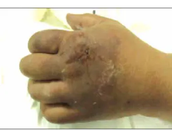
Figure 1. Patient’s hand prior to fasciotomy. Note the demar-cation line
Araz et al. Compartment Syndrome Secondary to Intravenous Fluid Application
is >30 mmHg. Direct pressure measurements can be taken with catheters placed inside the tissue, or auxiliary methods such as tis-sue oxygen saturation measurement, arterial Doppler examina-tion or near-infrared spectroscopy can be used. However, these methods are not common (2, 3, 5, 6). Because of the critical role of early diagnosis and treatment, it is reported that clinical picture should be prioritized over auxiliary diagnosis methods in diagnosing the syndrome. Classically, clinical findings, especially pain, being more severe than visible damage, should be a warn-ing sign (3). In our patient, compartment syndrome was not ini-tially considered. Because the pain in our patient remained and the appearance of a demarcation line belonging to an ischaemic area on the hand (Figure), emergency fasciotomy was performed after the diagnosis of compartment syndrome was made. Treatment of compartment syndrome is by establishing the normal perfusion of the tissues by decreasing the inner com-partment pressure to normal ranges as soon as possible (7). To establish this, increasing the compartment volume may be performed with methods such as increasing venous return, re-moval or emptying of the structures that take up space and fasciotomy (10). Early diagnosis and performing the planned treatment as soon as possible before tissue necrosis develops are important in preventing permanent complications. In patients whose diagnosis and treatment are delayed, loss of function and extremity amputation caused by advanced muscle and nerve damage can occur (11).
The diagnosis of compartment syndromes that occur after an-aesthesia is difficult. Because of the residual anaesthetic effect, patients are unable to convey the symptoms that they feel, which may delay the diagnosis, and analgesics added in the postop-erative period can mask the feeling of pain (3, 12). Dressings and bandages can also conceal visual findings. Therefore, it is paramount to closely follow-up the patients. In our patient, the first finding was the enduring pain in the hand, despite a high dose analgesic. A visual change was not detected during or at the end of the surgery. When the patient was admitted to the service and reported severe pain in her hand, the vascular access was removed, and her hand was treated by elevation and boric acid. The patient was closely followed-up, and because the pain still remained in the 10th hour and as tissue perfusion alteration con-tinued to increase, emergency fasciotomy was performed. The patient’s clinical condition quickly healed after fasciotomy, and she recuperated without a motor squeal. We maintain that we were late in diagnosing compartment syndrome and in perform-ing fasciotomy in our patient. The reason for this delay could be the unclear findings of the patient. The enduring pain despite analgesic treatment prompted us to consider compartment syn-drome. Right from the start, we took protective measures such as removing the vascular access, elevation and boric acid dressing. However, fasciotomy was performed without further delay after the condition worsened.
Conclusion
Compartment syndrome is a condition that can develop be-cause of various be-causes. It can lead to very serious compli-cations if its diagnosis and treatment are not performed in
the early stages. The major findings of compartment syndrome can vary in patients in whom anaesthesia and analgesics are given. Here we presented the case of a patient with compart-ment syndrome that developed because of intravenous fluid infusion and that was successfully treated. Thus, we would like to emphasize that the evaluation of the entry area of the vascu-lar access and tissue perfusion should be frequently performed while intravenous infusion is performed in patients under an-aesthesia and that one should be informed and careful about the possible complications of compartment syndrome.
Informed Consent: Verbal informed consent was obtained from patient who participated in this study.
Peer-review: Externally peer-reviewed.
Author Contributions: Concept - C.A., S.B.S.; Design - C.A., S.Ç., M.D.; Supervision - C.A., S.Ç., G.A.; Funding - C.A., Ö.K.; Materials - M.D., S.B.S., Ö.K.; Data Collection and/or Processing - C.A., M.D., S.B.S., Ö.K.; Analysis and/or Interpretation - C.A., S.Ç., M.D., S.B.S., Ö.K., G.A.; Literature Review - C.A., M.D., S.Ç.; Writer - C.A., S.Ç., M.D.; Critical Review - C.A., G.A., S.Ç.; Other - C.A., S.Ç., M.D.
Conflict of Interest: No conflict of interest was declared by the authors. Financial Disclosure: The authors declared that this study has re-ceived no financial support.
References
1. Tiwari A, Al H, Myint F, Hamilton G. Acute compartment syndromes. Br J Surg 2002; 89: 397-412. [CrossRef]
2. Elliott KG, Johnstone AJ. Diagnosing acute compartment syndrome. J Bone Joint Surg Br 2003; 85: 625-32.
3. Willsey DB, Peterfreund RA. Compartment syndrome of the upper arm after pressurized infiltration of intravenous fluid. J Clin Anesth 1997; 9: 428-30. [CrossRef]
4. Martin JT. Compartment syndromes: concepts and perspec-tives for the anesthesiologist. Anesth Analg 1992; 75: 275-83.
[CrossRef]
5. Kerrary S, Schouman T, Cox A, Bertolus C, Febrer G, Bertrand JC. Acute compartment syndrome following fibula flap harvest for mandibular reconstruction. J Craniomaxillofac Surg 2011; 39: 206-8. [CrossRef]
6. Shadgan B, Menon M, O’Brien PJ, Reid WD. Diagnostic tech-niques in acute compartment syndrome of the leg. J Orthop Trauma 2008; 22: 581-7. [CrossRef]
7. Ikegami Y, Hasegawa A, Tsukada Y, Abe Y, Shimada J, Tase C. Two cases of acute atraumatic compartment syndrome compli-cated with severe heat stroke. Fukushima J Med Sci 2010; 56: 129-33. [CrossRef]
8. Laframboise MA, Muir B. Acute compartment syndrome of the foot in a soccer player: a case report. J Can Chiropr Assoc 2011; 55: 302-12.
9. Botte MJ, Gelberman RH. Acute compartment syndrome of the forearm. Hand Clin 1998; 14: 391-403.
10. Garayoa SA, Romero-Munoz LM, Pons-Villanueva J. Acute compartment syndrome of the forearm caused by calcific
tendi-Turk J Anaesth Reanim 2015; 43: 126-9
nitis of the distal biceps. Musculoskelet Surg 2010; 94: 137-9.
[CrossRef]
11. Erdos J, Dlaska C, Szatmary P, Humenberger M, Vecsei V, Haj-du S. Acute compartment syndrome in children: a case series in
24 patients and review of the literature. Int Orthop 2011; 35: 569-75. [CrossRef]
12. Skjeldal S, Strømsøe K, Alho A, Johnsen U, Torvik A. Acute compartment syndrome: for how long can muscle tolerate inc-reased tissue pressure? Eur J Surg 1992; 158: 437-8.
Araz et al. Compartment Syndrome Secondary to Intravenous Fluid Application