Ankara Üniv Vet Fak Derg, 64, 345-348, 2017
Short Communication / Kısa Bilimsel Çalışma
Management of a complete stifle luxation with a hinged transarticular
hybrid external fixator in a dog
Cenk YARDIMCI, Ahmet ÖZAK, Taylan ÖNYAY, Kamil Serdar İNAL, Birsen Deniz ÖZBAKIR
Ondokuz Mayıs University, Faculty of Veterinary Medicine, Department of Surgery, Samsun, Turkey.
Summary: A 2 years old, 15 kg, male, mix breed dog that was presented with a right open tibial fracture and left complete stifle luxation. Cranial and caudal cruciate ligaments with medial and lateral collateral ligaments were completely torn. Stabilization of the stifle was established with a hinged transarticular external fixator. After fixator removal, radiographic findings were normal besides some periarticular osteophyte formations. Physical therapy was initiated postoperatively. Now the dog can perform all daily activities with mild lameness.
Keywords: Dog, external fixation, luxation, stifle.
Bir köpekte tam diz çıkığının transartiküler hibrid eksternal fiksatör ile sağaltımı
Özet: İki yaşlı, 15 kg ağırlığında, dişi, melez bir köpek sağ tibia’da açık kırık ve sol diz ekleminde çıkık şikayeti ile kliniğe getirildi. Sol diz ekleminde ön ve arka çapraz bağlar ile lateral ve medial kollateral ligamentlerin kopuk olduğu belirlendi. Diz ekleminin stabilizasyonu transartiküler eksternal fiksatör ile sağlandı. Fiksatörün uzaklaştırılmasından sonra radyografik bulgular periartiküler osteofit formasyonları dışında normal olarak gözlendi. Postoperatif olarak fizik tedaviye başlandı. An itibariyle köpeğin tüm günlük aktivitelerini hafif topallık bulguları ile gerçekleştirebildiği öğrenildi.
Anahtar sözcükler: Diz, eksternal fiksasyon, köpek, luksasyon.
The integrity of the stifle joint is provided mainly by cranial and caudal cruciate ligaments and supported with dual collateral ligaments. Meniscii support the joint primarily by providing stress transference like a pressure pad. If one or more of these components are damaged, the stability of the joint is compromised (2, 10). The most likely causes of such instabilities are cranial cruciate, caudal cruciate and medial collateral ligament injuries (2, 5).
Complete luxation of the stifle is rarely encountered in canines (2, 5) due to the extreme force required to damage multiple ligamentous attachments inside the joint (3, 5, 9). Treatment preferences vary from prosthetic reconstruction to arthrodesis of the joint (3, 5), depending on the surgeon's preference and the joint's structural integrity (1, 2, 3, 5). Both intra and extracapsular methods have been utilized in the past to treat complete stifle joint luxations (1, 3) mainly to retain mobility, promote weight bearing and provide pain relief (4, 5). The aim of this report is to present the extraarticular surgical repair and the following physical therapy outcomes of a dog suffering from complete stifle joint luxation.
The case presented was a 2 years old, 15 kg, male, mix breed dog that was brought to the clinics of Ondokuz Mayıs University, Faculty of Veterinary Medicine. The patient was unable to use its hind limbs properly and dragged them when trying to walk.
Clinical examination revealed an old puncture wound at the medial side of the right tibia consistent with a type I open fracture. There was swelling, severe pain and decrease in range of motion in the left stifle joint. No sensory or motor deficits were found in the neurological examination of the hind limbs.
According to the orthogonal radiographic evaluation of the right tibia and fibula, transverse fractures in the diaphyseal areas of both bones were detected. Complete luxation of the stifle joint was seen in the ventrodorsal (Figure 1a) and mediolateral (Figure 1b) radiographs of the left hind limb.
The left stifle joint was exposed and its components were examined to see if any were intact. Only the meniscii and patellar ligament was intact in the capsule. The torn ends of both cruciate ligaments were debrided and the joint was lavaged with sterile saline solution. The joint
Cenk Yardımcı - Ahmet Özak - Taylan Önyay - Kamil Serdar İnal - Birsen Deniz Özbakır 346
capsule was repaired using matress sutures to provide rigidity. The fascia was also closed with matress sutures for the same reason. The subcutaneous tissue and skin was routinely closed.
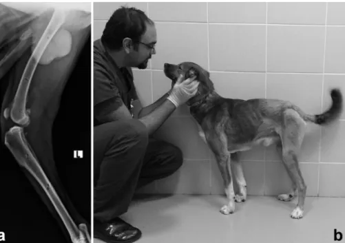
Figure 1. Ventrodorsal (a) radiograph of both hindlimbs at the initial clinical examination. Note the complete luxation of the left stifle joint and open diaphyseal transverse fracture of the right tibia. Femorotibial joint incongruity was also apparent in mediolateral (b) radiograph.
Şekil 1. Her iki arka bacağın ilk klinik muayenede ventrodorsal (a) radyografisi. Sol diz ekleminde tam luksasyon ve sağ tibiada açık diafizel transversal kırık. Mediolateral radyografide (b) femorotibial eklem uyumsuzluğu görülüyor.
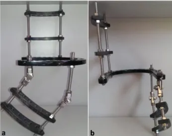
Figure 2. Lateral (a) and caudal (b) views of the frame. Frame was composed of a centered stretched ring connected with a 2 arched semicircular column at proximal and 2 arched hinged semicircular column at distal.
Şekil 2. Çerçevenin yandan (a) ve arkadan (b) görünümü. Ortada büyük bir gerilmiş halka ile bağlı, femurun lateraline ve açılı menteşe ile tibianın medialine gelecek şekilde organize edilmiş ikişer yarım daire halka.
The external fixator frame used was composed of a proximal and distal part, with two double semicircular arches that align with the lateral side of the femur and the
medial side of tibia (Figure 2a); the proximal and distal parts of the frame were connected to each other via a stretched ring 100 mm in diameter (Figure 2b). The natural anatomical shape of the hind limb was provided with hinges placed at a 135o angle between the distal part and
the omega ring. The frame was attached to the leg using 4 mm Ø negative profile end-threaded half pins, 2 at femur and 2 at tibia (Figure 3a) and further stabilized by use of 2mm ØK-wires on the stretched ring (with 60o of angle to
each other) running through both epicondyles of the femur (Figure 3b).
Figure 3. Ventrodorsal (a) and mediolateral (b) views of left stifle immediate after the operation. Note the complete congruity of the femorotibial joint despite the torn cruciate and collateral ligaments.
Şekil 3. Sol diz ekleminin operasyondan hemen sonraki ventrodorsal (a) ve mediolateral (b) görünümü. Kopan çapraz ve kollateral bağlara rağmen femorotibial eklemin tam uyumu.
The patient was able to bear weight on the operated limb at the first day after the surgery. There was lameness due to restriction of joint motion caused by the fixator. According to the radiological examination at the 4th week
after surgery; stifle joint anatomy was normal but mild periarticular osteophyte formation around the femoral trochlear groove, femoral condyle and tibial plateau was present. The hybrid external fixator was removed at this time (Figure 4a). There was no pain or drawer motion in the joint after fixator removal. The range of motion of the stifle was severely reduced to 40º. A physical therapy regimen was initiated, utilizing passive movements of the stifle after heating and massaging the surrounding areas. Electrotherapy, using trans cutaneous nerve stimulation was initiated at the 5th week using 3-4 mA currents
depending on the animal's comfort level during stimulation. After physiotherapy, the dog resumed normal walking and standing posture, and the masses of both quadriceps and gastrocnemius muscles visibly increased
Ankara Üniv Vet Fak Derg, 64, 2017 347
(Figure 4b). The clinical outcome of the patient at the 3rd
month was satisfactory, there was no finding of obvious pain or discomfort, no limp could be detected while walking. The dog still refrained from using the leg while running of climbing stairs.
Cranial migration of the tibia was seen on the 3rd month radiogram with no adverse clinical symptoms. The patient owner reported that the dog had mild lameness symptoms and could perform everyday activities with ease during telephone interview.
Complete stifle joint luxations happen with severe traumas caused by excessive force (3, 6), despite the patient history was unknown, there was evidence suggesting such cause, like the open fracture on the ipsilateral tibia.
Complete stifle joint luxation was described as the total loss of contact of articular surfaces and if there was even a slight contact between the surfaces, it was classified as a subluxation (8).
Some type of surgical intervention is always necessary in stifle joint luxations for a favorable outcome (2). There is some controversy in the treatment modality about utilizing intracapsular repair or periarticular fibrosis (1, 3). In the past, intra-, trans- and extraarticular repair techniques were met with clinical success (2). The meniscii and joint capsule must always be repaired as they are very important for stability (3, 11), the favorable outcome in our case confirms this, as we haven't done any intraarticular repair. It also suggests that periarticular fibrosis alonecreates adequate fixation for a relatively
rigid and robust stabilization. The cranial cruciate ligament is considered especially important for the stability of the joint (7), sole periarticular fibrosis did not prevent the migration of tibia in this case. In our point of view, body weight has great importance in terms of recovery and the slim build of the dog in our study had a positive impact in clinical outcome.
Though many techniques were utilized to stabilize the stifle joint, the end results of the repairs of such techniques are more or less the same, (2) and some loss in the range of motion is always expected (1, 3, 12). Transarticular external fixation is relatively less invasive than intracapsular techniques. A semicircular external fixator allows for versatile use of pins. Another advantage is the ease of revision, in case of a complication, unlike intracapsular techniques which may need another invasion of the joint capsule to be corrected, like misplaced femoral screws or ruptured sutures (5, 11). During the consolidation process we did not experience with any complication associated with the external fixation technique and the dog tolerated the frame perfectly.
After removal of the external fixator, the dog was reluctant to put its body weight on the relevant foot and there was serious impairment on joint motion. The stifle joint was at is normal angle while at rest but the total range of motion was 20o towards extension and flexion (Total
40o, with a 90o ROM loss). After physical therapy the loss
in ROM was 45o in the fourth week, and the dog was still
limping while running or climbing stairs, so the decision was made to start electrical therapy. Transcutaneous
Figure 4. Mediolateral radiograph of the left stifle after removal of the frame (a). Mild cranial displacement of tibia was seen but did not complicate with significant lameness. The dog was able to use the operated limb with mild lameness (b).
Şekil 4. Sol diz eklemin fiksatör uzaklaştırılmasından hemen sonraki mediolateral radyografik görünümü (a). Tibianın hafif kraniale yer değiştirmesi görüldü, fakat klinik yansıması belirgin değildi. Köpek hafif topallık ile opere edilen bacağını kullanabilmektedir (b).
Cenk Yardımcı - Ahmet Özak - Taylan Önyay - Kamil Serdar İnal - Birsen Deniz Özbakır 348
electrical nerve stimulation has the additional benefit of strengthening the muscles surrounding the joint without any strain to its structures. The muscles have a supportive role in providing stability to the stifle (2) and strengthening them resulted in a better posture and reduced severity of the limp.
The treatment of complete joint luxation using a hybrid transarticular external fixator, provided a satisfactory outcome for the dog. The importance of regular physical and electrotherapy sessions and the low body weight of the dog in clinical outcome cannot be underestimated.
References
1. Aron D (1988): Traumatic dislocation of the stifle
joint-treatment of 12 dogs and one cat. J Am An Hosp Assoc, 24,
333-340.
2. Bruce W (1998): Multiple ligamentous injuries of the
canine stifle joint: A study of 12 cases. J Small Anim Pract,
39, 333-340.
3. Hulse D, Shires P (1986): Multiple ligament injury of the
stifle joint in the dog. J Am An Hosp Assoc, 22, 105-110.
4. Jaeger GH, Wosar MA, Marcellin-Little DJ, et al. (2005): Use of hinged transarticular external fixation for
adjunctive joint stabilization in dogs and cats: 14 cases (1999-2003). J Am Vet Med Assoc, 227, 586-591.
5. Keeley B, Glyde M, Guerin S, et al. (2007): Stifle joint
luxation in the dog and cat: The use of temporary intraoperative transarticular pinning to facilitate joint reconstruction. Vet Comp Orthop Traumatol, 20, 198-203.
6. Parker R, Schubert T (1981): Repair of ligamentous joint
injuries in three dogs using spiked washers. J Am Anim
Hosp Assoc, 17, 45.
7. Pearson PT (1971): Ligamentous and meniscal injuries of
the stifle joint. Vet Clin North Am Small Anim Pract, 1,
489-501.
8. Pond M (1971): Normal joint tissues and their reaction to
injury. Vet Clin North Am Small Anim Pract, 1, 523-535.
9. Robins G (1990): The canine stifle joint. 693-760. In: WG Whittick (Ed), Canine Orthopedics. 2nd ed. Lea & Febiger, Philadelphia.
10. Slatter DH (2003): Stifle joint. 2090-2133. In: Slatter DH (Ed), Textbook of small animal surgery, Elsevier, Philadelphia.
11. Smith G, Torg J (1985): Fibular head transposition for
repair of cruciate-deficient stifle in the dog. J Am Vet Med
Assoc, 187, 375-383.
12. Welches C, Scavelli T (1990): Transarticular pinning to
repair luxation of the stifle joint in dogs and cats: A retrospective study of 10 cases. J Am An Hosp Assoc, 26,
207-214.
Geliş tarihi: 25.03.2016 / Kabul tarihi: 02.11.2016
Address for correspondence:
Assoc.Prof.Dr. Cenk Yardımcı Ondokuz Mayıs University,
Faculty of Veterinary Medicine, Department of Surgery, Samsun, Turkey.