Effects of in ovo injected bisphenol A on the testis of one day old
chickens
Osman Behzat Burak ESENER, Hasan Hakan BOZKURT
İstanbul University, Faculty of Veterinary Medicine, Department of Histology and Embryology, İstanbul, Turkey.
Summary: Bisphenol-A (BPA), an environmental estrogen, has adverse effects on the male reproductive tract in mammals and birds. In ovo administration of BPA at high doses have been reported to cause the left gonad to form an ovotestis in fowl and quail. However, there are no studies on morphometric effects of in ovo administration of BPA on testis. Therefore, the aim of present study was to investigate the morphometric effects of in ovo administration of low and high doses of BPA on hatched testis. For comparative purposes, well characterized synthetic estrogen diethylstilbestrol (DES) was also examined. Chicken eggs were treated (injection volume 100 µl/egg) on day 4 of incubation before the gonads start to differentiate. Doses were 67 (low BPA group) and 134 (high BPA group) µg BPA/g egg, and 0.02 (low DES group) and 0.2 (high DES group) µg DES/g egg. Sertoli cell and germ cell numbers were determined by stereological methods. Our results indicated that low doses of BPA had no negative effects, on the contrary it was at least increased germ cells proliferation in avian similar to mammals counterpart. However, as doses were titrated upwards, negative effects emerged on seminiferous tubules of testis.
Keywords: Bisphenol A, BPA, chicken, diethylstilbestrol (DES), testis.
In ovo enjekte edilen bisfenol A’nın bir günlük civciv testisleri üzerine etkisi
Özet: Çevresel östrojenlerden biri olan bisfenol A (BPA) nın memelilerde ve kuşlarda erkek genital sistemi üzerine olumsuz etkileri bulunmaktadır. Tavuk ve bıldırcınlarda in ovo olarak uygulanan yüksek dozda BPA nın sol gonadın ovotestise dönüşmesine sebep olduğu bildirilmiştir. Ancak, in ovo uygulanan BPA nın testiste morfometrik etkileri üzerine bir çalışma bulunmamaktadır. Bu çalışmanın amacı in ovo uygulanan düşük ve yüksek dozdaki BPA nın çıkım yapan civcivlerin testislerinde morfometrik etkilerini ortaya koymaktır. Çalışmada ayrıca karşılaştırma amacıyla iyi bilinen sentetik östrogen dietilstilbestrol (DES) de kullanılmıştır. Enjeksiyonlar (enjeksiyon hacmi 100 µl/yumurta) tavuk yumurtalarına gonadların farklılaşmaya başlamasından önce, kuluçkanın dördüncü gününde uygulanmıştır. Uygulanan dozlar 67 (düşük doz BPA) ve 134 (yüksek doz BPA) µg BPA/gr yumurta, 0.02 (düşük doz DES) ve 0.2 (yüksek doz DES) µg DES/gr yumurtadır. Sertoli ve germ hücresi sayıları stereolojik yöntemlerle belirlenmiştir. Çalışmamızın sonuçları düşük dozda uygulanan BPA nın olumsuz etki oluşturmadığını, tam tersine memelilerdekine benzer bir şekilde kanatlılarda germ hücresi proliferasyonunu arttırdığını göstermiştir. Ancak doz arttığında seminifer tubullerde olumsuz etkilerin açığa çıktığı belirlenmiştir.
Anahtar sözcükler: Bisfenol A, BPA, civciv, dietilstilbestrol (DES), testis.
Introduction
The hypothesis, that environmental contaminants can disrupt endocrine functions in wildlife and in humans has attracted much attention during recent years (26). One of the widely studied of these, bisphenol-A (BPA), has adverse effects on the male reproductive tracts in animals (1, 10, 16). Plastic monomer bisphenol A (BPA) has proved, in a number of in vitro test systems, to be one of the most potent environmental estrogens, generally being 104 to 105 of magnitude less potent than estradiol (3). BPA is used extensively as a component in polycarbonate plastics (14). It is also a component of epoxy resins used in dental sealants and in the lacquer lining of metal food cans. BPA has been shown to leak from lacquer lining in food cans (15), polycarbonate flasks (14), polycarbonate
baby bottles (8) and epoxy resin inner coating of canned foods and contaminate the contents (25). In mammals, its negative effects are on prostate development, epididymis size (10) and daily sperm count (19). In ovo administration of BPA at high doses has been reported to cause the left gonad to form an ovotestis in fowl and quail (5). However, this induced ovotestis does not persist until adulthood and does not result in any obvious morphological changes in adult testis (12). There are no studies on morphometric effects of in ovo administration of low dose BPA on testis. Therefore, the aim of present study was to investigate the morphometric effects of in ovo administrated low and high doses of BPA on hatched male chicken testis. For comparative purposes, well characterized synthetic estrogen diethylstilbestrol (DES) was also examined.
Materials and Methods
Animals: Fertile eggs of White Leghorn chickens (strain Lohmann LSL; Local breeder flock) were obtained from local hatcheries (HASTAVUK, Bursa, Turkey). The local Ethics Committee for Animal Research approved the animal experiments in this study (Istanbul University Faculty of Veterinary Medicine Ethics Committee, 2004-95).
Chemicals and preparation of emulsions: Bisphenol A (BPA; purity=99.4%) was purchased from BDH Lab. Inc. (London, UK), and diethylstilbestrol (DES; purity>99%) was purchased from Sigma Chemical (St. Louis, MO, USA). Eggs were incubated at 37.5ºC and 60% relative humidity and were turned every third hour. Because BPA did not dissolve readily in oil or water, propylene glycol was used as a solvent and an emulsion was prepared with peanut oil and lecithin (5). Diethylstilbestrol was also administered in the oil/lecithin/ propylene glycol emulsion. Control group only received the oil/lecithin/propylene glycol based emulsion.
Dosing and application of the emulsions: The compounds were injected into egg yolk using insulin syringes and disposable needles via a small hole punched at the blunt end of the egg. Chicken eggs were treated (injection volume 100 l/egg) on day 4 of incubation before the gonads start to differentiate (9, 13). After injection, the shell was sealed with paraffin wax. Doses were 67 (low BPA group) and 134 (high BPA group) g BPA/g egg, and 0.02 (low DES group) and 0.2 (high DES group) g DES/g egg. Doses were determined by considering the morphometric investigation possible in low ovotestis degree that would appear in low dose groups and in high ovotestis formation that would appear in high dose groups according to the data given in previous studies (5, 6) and also to keep the hatching rate high as the compounds were reported to cause embryotoxic effects (6).
Tissue preparation and sectioning: Hatching rates and male numbers were recorded (Table 1), chickens were decapitated within 24 h of hatching and left testes were removed immediately and fixed by immersion in 2.5% glutaraldehyde (in 0.15 M cacodylate buffer pH 7.4). After these steps, specimens were post fixed in 1 % osmium tetroxide for 2 hours, dehydrated in an ascending alcohol series and took into propylene oxide for 30 minutes. These procedures were completed by embedding the tissues in Epon 812. Semi-thin sections of 1μm thickness were cut using an ultramicrotome (LXB 2188 Ultramicrotome, NOVA, Bromma, Sweden). As the average length of the testis samples were 2.5 mm, four sections were cut from each testis with an approximately 500 µm section interval for sampling. Sections were stained with 1% toluidine blue.
Morphometric evaluation: The body and testis weights of all birds were recorded at death and the gonado-somato indices (testis weight/ body weight) and testis weight asymmetry (left testis weight/ body weight) were calculated. In the cortical area, the presence of at least five germ cells in meiotic prophase, was used for the identification of an ovotestis (6).
Volume density determination: Nuclear volume density and absolute volume of Sertoli cell, germ cell and seminiferous tubule volume density and absolute volume were determined by the standard point counting method (4, 7). This involved using a systematic clock-face sampling pattern from a random starting point at upper left corner of each section and evaluating 10 fields in each 4 sections, in a total of 40 fields, by using a 100-point eye-piece graducule (10x10=100 grid, a grid column=10 grid=100µm), inserted into a Nikon 100x objective fitted to a Nikon microscope (Figure 1A).
The grid points falling over each interested structure were counted. Total number of grid points over each structure were expressed as a percentage of the total number of grid points (=4000).
Absolute volume determination: Measured testis weight converted to testis volume with the equation; “Testis volume (cm3) = testis mass (g)/specific gravity” (17). For each animal, the percentage volume (volume density) was converted to absolute volume per testis by multiplying with testis volume (22). This was justified as shrinkage was minimal, that is, testis weights before and after fixation were comparable.
Nuclear volume determination: In brief, this involved initially obtaining the Sertoli cell and germ cell nuclear volumes for each bird by measuring 4 separate nucleolus to nuclear membrane radii (r1, r2, r3, r4) in 4 opposite directions for each nuclei of between 30 and 50 randomly selected Sertoli and germ cells in 4 sections, and then determining the nuclear volume of the inspected cell with the aid of the standard equation (22, 28):
4𝜋
3 ×
(𝑟1 3+ 𝑟2 3+𝑟3 3+ 𝑟4 3) 4
Mean value of the nuclear volume of Sertoli and germ cells of each animal were calculated for each cell type by collecting nuclear volumes and dividing to the number of cells used for nuclear volume measurement.
Cell number determination: Sertoli cell and germ cell numbers were determined by the nucleator method as described previously (22, 28) using absolute volumes. Sertoli and germ cell numbers of each animal were obtained by dividing absolute Sertoli and germ cell nuclear volumes by mean Sertoli cell and germ nuclei volumes in each testis. Statistical analyses were performed by using Duncan’s One -Way Anova method in Windows SPSS (Statistical Programme for Social Science
Ver.10.0). Average volumetric densities, absolute volumes and cell numbers of the components within a group were determined by calculation of the mean ± standard deviation (mean ± SD) for that group.
Results
Hatching rate: Highest hatching rate was on the control group. Experimental groups showed lower hatching rations. Lowest hatching ratio was seen on high dose BPA group (Table 1).
Body weight and testis weight: There were no statistical differences between groups in means of body weight (Table 1). Left testis weight and left testis weight/ body weight ratios were increased by the treatments.
These increases were statistically significant (P<0.05) in high dose BPA and DES groups (Table 2).
Testis weight asymmetry was higher in BPA groups and high dose DES group compared to control group. The increase in testis weight asymmetry (left testis/right testis weight ratio) in high dose DES group was statistically significant (P<0.01)(Table 2).
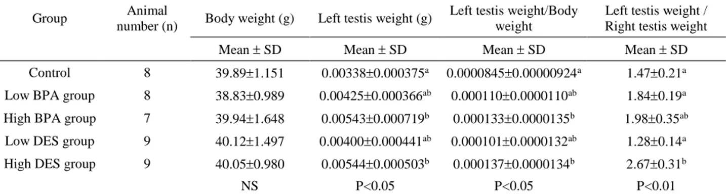
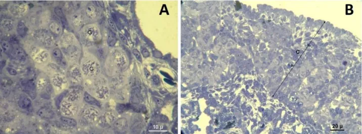
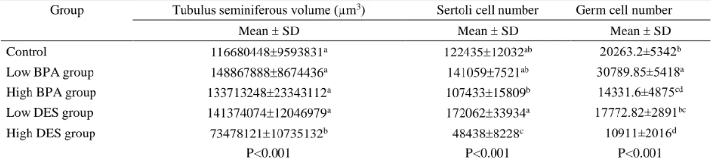
Morphological findings: In control group, testis was oval in shape and was surrounded with a thin layer of tunica albuginea (Figure 1B). Testis morphology and tubulus seminiferous appearance was normal and Sertoli cells, germ cells and Leydig cells were prominent in semi thin sections stained with Toluidine blue (Figures 1B-C-D). Neither lumen nor Sertoli cell only tubule formation was detected in control group (Figure 2). Dose-dependent increase of ovotestis frequency was detected in the testes of animals belonging to high DES and BPA and, low DES groups. Increase of ovotestis frequency in high dose groups was statistically significant (P<0.001) (Table 3). In low dose BPA group, testis morphologies were in normal appearance and neither ovotestis nor Sertoli cell only tubule formation was observed. Ovotestis formation was identified with the presence of five or more germ cells in the prophase stage in the cortex (5) (Figure 3A). Cortex region was thicker in ovotestis formed animals (Figure 3B). Sertoli cell only tubules were detected in low dose DES group and especially more in high dose DES and BPA groups (Figures 4A-B, 5A-B).
Table 1. Hatching rates. Tablo 1. Çıkım oranları.
Group In ovo injected egg
number
Hatched chicken number (total)
Hatched male chicken number
Hatching rates %
Low BPA group 41 27 8 65.9ab
High BPA group 42 17 7 40.5c
Low DES group 42 21 9 50.0bc
High DES group 42 29 9 69.0ab
Control 39 30 8 76.9a
Groups with different letters or doesn’t have a common letter (a,b,c,d) in the same column are significantly different P<0.01. Aynı sütunda farklı harf içeren ya da ortak harf (a,b,c,d) içermeyen gruplar arasındaki fark istatistiksel olarak anlamlıdır P<0.01.
Table 2. Body weight, left testis weight, left testis weight/body weight ratio and left testis/right testis weight ratio averages in groups (Mean ± SD, SD: Standart deviation).
Tablo 2. Grup içi vücut ağırlığı, sol testis ağırlığı, sol testis ağırlığı/vücut ağırlığı oranı, sol tesis ağırlığı /sağ testis ağırlığı oranı ortalamaları (Ortalama ± Standart sapma).
Group Animal
number (n) Body weight (g) Left testis weight (g)
Left testis weight/Body weight
Left testis weight / Right testis weight
Mean SD Mean SD Mean SD Mean SD
Control 8 39.891.151 0.003380.000375a 0.00008450.00000924a 1.470.21a
Low BPA group 8 38.830.989 0.004250.000366ab 0.0001100.0000110ab 1.840.19a
High BPA group 7 39.941.648 0.005430.000719b 0.0001330.0000135b 1.980.35ab
Low DES group 9 40.121.497 0.004000.000441ab 0.0001010.0000132ab 1.280.14a
High DES group 9 40.050.980 0.005440.000503b 0.0001370.0000134b 2.670.31b
NS P<0.05 P<0.05 P<0.01
Groups with different letters or doesn’t have a common letter (a,b,c,d) in the same column are significantly different. Degree of significance is indicated at the below of each column.
Aynı sütunda farklı harf içeren ya da ortak harf (a,b,c,d) içermeyen gruplar arasındaki fark istatiksel olarak anlamlıdır. İstatistiksel anlamlılığın derecesi her sütunun altında belirtilmiştir.
Table 3. Ovotestis frequency in groups. Tablo 3. Gruplarda ovotestis görülme oranı.
Group Low BPA group High BPA group Low DES group High DES group Control
Ovotestis frequency (%) 0b 75a 22.2b 100a 0b
Groups with different letters or doesn’t have a common letter (a,b,c,d) in the same row are significantly different P<0.001. Aynı satırda farklı harf içeren ya da ortak harf (a,b,c,d) içermeyen gruplar arasındaki fark istatistiksel olarak anlamlıdır P<0.001.
Figure 1. A-100-point eye-piece graducule. B- View of a testis section of one day old chicken in control group. C- General view of seminiferous tubule formation in control group. D- S= Germ cell (Spermatogonium), TS= Seminiferous tubule, L= Leydig cell, M= Myoepithelial cell, Arrow=Sertoli cell, control group (Toluidine blue).
Şekil 1. A-100-noktalı sayım gradikülü. B- Bir günlük civcivde testis kesitinin görünümü, kontrol grubu C- Seminifer tubul yapısının genel görünümü, kontrol grubu D- S= Spermatogonium (Germ hücresi), TS= Tubulus seminiferus, L= Leydig hücresi, M= Miyoepitel hücresi, Ok=Sertoli hücresi, kontrol grubu (Toluidine mavisi).
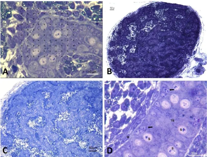
Figure 2. Seminiferous tubules in testis of one day old chicken, control group (Toluidine blue).
Şekil 2. Bir günlük civcivde seminifer tubul yapısı, kontrol grubu (Toluidine mavisi).
Figure 3. A- Germ cells in the prophase stage in the cortex of the testis, G= Germ cell (High dose DES group) B- Increased thickness of cortex region in high dose BPA group, C= Cortex (Toluidine blue).
Şekil 3. A- Testisin korteks bölgesinde profaz aşamasındaki germ hücreleri, G= Germ hücreleri, yüksek doz DES grubu B- Yüksek doz BPA grubunda korteks bölgesinde kalınlık artışı, C= Korteks bölgesi (Toluidine mavisi).
Figure 4. A and B -Ovotestis formation. Low dose DES group (Toluidine blue). Şekil 4. A ve B-Ovotestis oluşumu. Düşük doz DES grubu (Toluidine mavisi).
Figure 5. Sertoli cell only tubules (Shown with “ * ”), Low dose DES group. Black arrows= Sertoli cells (Toluidine blue).
Şekil 5. Sadece Sertoli hücresi içeren tubul görünümü (“*” ile gösterilmiştir), düşük doz DES grubu. Siyah ok= Sertoli hücresi (Toluidine mavisi).
Table 4. Tubulus seminiferous volume, Sertoli cell and germ cell numbers in groups (Mean ± SD, SD: Standart deviation). Tablo 4. Gruplardaki tubulus seminiferus hacmi, Sertoli hücresi ve germ hücre sayıları (Ortalama ± Standart sapma).
Group Tubulus seminiferous volume (µm3) Sertoli cell number Germ cell number
Mean SD Mean SD Mean SD
Control 1166804489593831a 12243512032ab 20263.2±5342b
Low BPA group 1488678888674436a 1410597521ab 30789.85±5418a
High BPA group 13371324823343112a 10743315809b 14331.6±4875cd
Low DES group 14137407412046979a 17206233934a 17772.82±2891bc
High DES group 7347812110735132b 484388228c 10911±2016d
P<0.001 P<0.001 P<0.001
Groups with different letters or doesn’t have a common letter (a,b,c,d) in the same column are significantly different. Degree of significance is indicated at the below of each column.
Aynı sütunda farklı harf içeren ya da ortak harf (a,b,c,d) içermeyen gruplar arasındaki fark istatistiksel olarak anlamlıdır. İstatistiksel anlamlılığın derecesi her sütunun altında belirtilmiştir.
Morphometric findings: In low dose groups and high dose BPA group there were no statistical differences (P>0.05) compared to control in means of tubulus seminiferous volume (Table 4). On contrary in high dose DES group tubulus seminiferous volume significantly decreased compared to other groups (P<0.001). Germ cell numbers significantly decreased (P<0.001) in high dose BPA and high dose DES groups (Table 4). In low dose BPA group there was a statistically significant increase in germ cell number (P<0.001).
There were no statistical differences (P>0.05) in Sertoli cell numbers between low dose groups and control group (Table 4). On the contrary, there was a decrease in Sertoli cell numbers in high dose groups. The prominent decrease in high dose DES group was statistically significant (P<0.001).
Discussion and Conclusion
In experimental groups, especially in high dose BPA group, hatching ratio of chickens was lower than control group. Berg et al. (6) also reported low hatching rate due to embryotoxic effects of BPA. Although in low BPA group all investigated parameters were increased, the only significant increase (P<0.001) was observed in germ cell number. In a rodent study stimulatory effect of weak and low dose potent estrogen was observed (4). In another study stimulatory effect of estrogen on the Sertoli cells of mammalian testis has been reported (27). Aktaş et al. (2) also reported this stimulatory effect on cockerel testis. However, as the dose was titrated upward, negative effect emerged for the seminiferous tubule of testis. Administration of 134 µg BPA/g egg, slowed down the formation of seminiferous tubule and caused ovotestis formation in the cortex of the gonad. Since DES is a more potent estrogenic substance than BPA (3), even its low dose caused ovotestis formation. Interestingly the testis weight was increased with ovotestis formation. The reason for this is that, oestrogens even in high doses may still
induce germ cell proliferation, but they undergo meiotic proliferation in the cortex of the gonad instead of mitotic proliferation in the seminiferous tubule. It must be a trigger point that affects the germ cell to stay in the cortex and undergo meiosis. The most reasonable candidate for this mechanism is oestrogen/testosterone balance, since the left gonad will differentiate into a testis or an ovary, depending on the relative concentrations of oestrogen and testosterone (24) and in intact embryonic chicken testes, testosterone levels are invariably higher than oestrogen levels, whereas the converse is true for the ovaries (20). Since there are no oestrogen receptors observed on the right testis (18) it can be speculated that, in the present study, observed effect on the left testis might have occurred via oestrogen receptors. Effect on the testis weight asymmetry by the treatment also supported this theory. The external estrogenic compounds administered in the present study seem to be altered the balance in the favour of oestrogen. However, this induced ovotestis does not persist until adulthood and does not result in any obvious morphological changes in adult testis (11). According to this results, either germ cells move into seminiferous tubules or the germ cell in the seminiferous tubules proliferate to the normal number and germ cells in the cortex somehow disappear. This phenomenon would appear to be worthy of further research. The number of the Sertoli cells was not affected in low doses but decreased significantly (P<0.001) in high doses of DES. Because the size of testis is related to the Sertoli cell number (21), its number at adulthood should be reaching its normal level. The reduced number of Sertoli cells in high dose group seems to catch up with later proliferation. There is time period for proliferation of Sertoli cells after hatching since Sertoli cells proliferate until week 8 after hatching (7).
Left testis weight increases in high dose groups because the estrogen receptor is expressed only in left gonad at embryonal stage and so the left gonad turns to ovotestis.
In conclusion, low dose BPA causes an increase in germ cell volume that means an increase in germ cell number so in lower doses BPA has positive effect on germ cell divisions. When the BPA dose is higher, positive effects disappear and negative effects occur due to the spoiled hormone balance. Primordial germ cells are localised at the cortex region of undifferentiated gonads at day 3.5 of embryogenesis and morphological differentiation begins between day 5.5 and 6.5 (23). In the testis of male embryo, seminiferous tubules are formed from medullary cords but at the presence of estradiol left gonad turns to ovary. As oestrogen/testosterone balance is disturbed, migration of germ cells from cortical region to seminiferous tubules is somehow negatively affected. This causes cortex development and Sertoli cell only tubule formation.
Acknowledgements
This study was recapitulated from the Ph.D. thesis of Osman Behzat Burak ESENER and was supported by the Research Fund of Istanbul University (Project No: T-517/21102004), Istanbul, Turkey.
Conflict of interest
I, Prof. Dr. H. Hakan BOZKURT, declare that I have no conflict of interest with the Research Fund of Istanbul University. I, Dr. Osman B. Burak ESENER, declare that I have no conflict of interest with the Research Fund of Istanbul University.
References
1. Akingbemi BT, Sottas CM, Koulova AI, et al. (2004):
Inhibition of testicular steroidogenesis by the xenoestrogen bisphenol A is associated with reduced pituitary luteinizing hormone secretion and decreased steroidogenic enzyme gene expression in rat Leydig cells. Endocrinology, 145,
592-603.
2. Aktaş A, Ulkay MB, Bozkurt HH (2008): The effects of
genistein on the cockerel testis during embryonic development. Kafkas Üniv Vet Fak Derg, 14, 157-160.
3. Andersen HR, Andersson AM, Arnold SF, et al. (1999):
Comparison of short-term estrogenicity tests for identification of hormone-disrupting chemicals. Environ
Health Perspect, 107, 89-108.
4. Atanassova N, Mckinnell C, Turner KJ, et al. (2000):
Comparative effects of neonatal exposure of male rats to potent and weak (environmental) estrogens on spermatogenesis at puberty and the relationship to adult testis size and fertility: Evidence for stimulatory effects of low estrogen levels. Endocrinology, 141, 3898-3907.
5. Berg C, Halldin K, Brunstrom B. (2001): Effects of
bisphenol A and tetrabromobisphenol A on sex organ development in quail and chicken embryos. Environ Toxicol
Chem, 20, 2836-2840.
6. Berg C, Halldin K, Fridolfsson A, et al. (1999): The avian
egg as a test system for endocrine disrupters: Effects of
diethylstilbestrol and ethynylestradiol on sex organ development. Sci Total Environ, 233, 57-66.
7. Bozkurt HH, Aktas A, Ulkay MB, et al. (2007): Sertoli
cell proliferation during the post hatching period in domestic fowl. J Vet Sci, 8, 219-222.
8. Brotons JA, Olea-Serrano MF, Villalobos M, et al. (1995): Xenoestrogens released from lacquer coatings in
food cans. Environ Health Perspect, 103, 608-612.
9. Bruggeman V, As PV, Decuypere E (2002):
Developmental endocrinology of the reproductive axis in the chicken embryo. Comp Biochem Physiol A Mol Integr
Physiol, 131, 839-846.
10. Gupta C (2000): Reproductive malformation of the male
offspring following maternal exposure to estrogenic chemicals. Proc Soc Exp Biol Med, 224, 61-68.
11. HalldinK, AxelssonJ, BrunstromB (2005): Effects of
endocrine modulators on sexual differentiation and reproductive function in male Japanese quail. Brain Res
Bull, 65, 211-218.
12. Halldin K, Berg C, Bergman A, et al. (2001): Distribution
of bisphenol A and tetrabromobisphenol A in quail eggs, embryos and laying birds and studies on reproduction variables in adults following in ovo exposure. Arch Toxicol,
75, 597-603.
13. Kirby JD, Froman DP. (1999): Reproduction in male birds. In, Whittow GC(Ed): Sturkie's Avian Physiology. 5th ed. pp. 597-615, Academic Press, New York.
14. Krishnan AV, Stathis P, Permuth SF, et al. (1993):
Bisphenol-A: An estrogenic substance is released from polycarbonate flasks during autoclaving. Endocrinology,
132, 2279-2286.
15. Lintelmann J, Katayama A, Kurihara N, et al. (2003):
Endocrine disruptors in the environment (IUPAC Technical Report). Pure Appl Chem, 75, 631-681.
16. Markey CM, Rubin BS, Soto AM, et al. (2003):
Endocrine disruptors: From Wingspread to environmental developmental biology. J Steroid Biochem Mol Biol, 83,
235-244.
17. Mori H, Christensen AK (1980): Morphometric analysis
of Leydig cells in the normal rat testis. J Cell Biol, 84,
340-354.
18. Nakabayashi O, Kikuchi H, Kikuchi T, et al. (1998):
Differential expression of genes for aromatase and estrogen receptor during the gonadal development in chicken embryos. J Mol Endocrinol, 20, 193-202.
19. Sakaue M, Ohsako S, Ishimura R, et al. (2001):
Bisphenol-A affects spermatogenesis in the mature rat even at a low dose. J Occup Health, 43, 185-190.
20. Scheib D, Guichard A, Mignot TM, et al. (1981):
Steroidogenesis by gonads of normal and of diethylstilbestrol-treated quail embryos: Radioimmunoassays on organ cultures. Gen Comp Endocrinol, 43, 519-526.
21. Sharpe RM, Mckinnell C, Kivlin C, et al. (2003):
Proliferation and functional maturation of Sertoli cells, and their relevance to disorders of testis function in adulthood.
Reproduction, 125, 769-784.
22. Sharpe RM, Walker M, Millar MR, et al. (2000): Effect
of neonatal gonadotropin-releasing hormone antagonist administration on Sertoli cell number and testicular development in the marmoset: Comparison with the rat.
23. Smith CA, Sinclair AH (2004): Sex determination:
Insights from the chicken. BioEssays, 26, 120-132.
24. Stevens L (1997): Sex chromosomes and sex determining
mechanisms in birds. Sci Prog, 80, 197-216.
25. Vandenberg LN, Maffini MV, Sonnenschein C, et al. (2009): Bisphenol-A and the great divide: A review of
controversies in the field of endocrine disruption. Endocr
Rev, 30, 75-95.
26. Vos JG, Dybing E, Greim HA, et al. (2000): Health effects
of endocrine-disrupting chemicals on wildlife, with special reference to the European situation. Crit Rev Toxicol, 30,
71-133.
27. Wistuba J, Brinkworth MH, Schlatt S, et al. (2003):
Intrauterine bisphenol A exposure leads to stimulatory effects on Sertoli cell number in rats. Environ Res, 91,
95-103.
28. Wreford NG (1995): Theory and practice of stereological
techniques applied to the estimation of cell number and nuclear volume in the testis. Microsc Res Tech, 32,
423-436.
Geliş tarihi: 09.06.2016 / Kabul tarihi: 03.04.2017
Address for correspondence:
Dr. Osman Behzat Burak ESENER İstanbul University, Faculty of Veterinary
Medicine, Department of Histology and Embryology, Avcilar, TR-34320 Istanbul, Turkey.
e-mail: [email protected] Tel: +90 212 4737070 / 17204 Fax: +90 212 4737241