CUAJ • November-December 2014 • Volume 8, Issues 11-12 © 2014 Canadian Urological Association
Cevahir Özer, MD;
*Mehmet Resit Gören, MD;
*Tulga Egilmez, MD;
*Nebil Bal, MD
†*Baskent University Adana Medical and Research Center, Department of Urology, Adana, Turkey; †Baskent University Adana Medical and Research Center, Department of Pathology, Adana, Turkey
Cite as: Can Urol Assoc J 2014;8(11-12):e928-30. http://dx.doi.org/10.5489/cuaj.1963 Published online December 15, 2014.
Abstract
Renal oncocytomas accounts for 3% to 9% of primary renal neo-plasms. The coexistence of renal cell carcinoma (RCC) within the oncocytoma is extremely rare. We report the case of an asy-ptomatic 74-year-old man with papillary RCC within oncocytoma managed with left radical nephrectomy.
Introduction
Renal oncocytomas are rare benign neoplasms accounting
for 3% to 9% of all primary renal neoplasms.1 Although
most are asymptomatic and discovered incidentally, a few symptomatic patients may complain of hematuria, flank pain, or palpable mass.1,2 Oncocytomas are usually
unifo-cal, but multifocal and bilateral appearances of the onco-cytomas and concomitant renal cell carcinoma (RCC) have been reported.3 To our knowledge, only 3 cases of
papil-lary subtype of RCC within a renal oncocytomahave been previously described in the literature.4-6 In this case report,
we present the fourth case of papillary RCC embedded in a renal oncocytoma.
Case report
A 74-year-old man with no medical history was referred to us due to a hyperecoic left kidney mass on routine abdomi-nal ultrasound. He had a 50-packets/year smoking history. Physical examination was unremarkable, and laboratory findings were within normal values, with hemoglobin of 14.8 g/dL and creatinine 1.02 mg/dL. His computed tomog-raphy scan of the abdomen confirmed a 6 × 5-cm left renal mass located between the renal hilum and lower pole.
He underwent a left radical nephrectomy through a left flank incision without perioperative complications. The patient fared well postoperatively and was discharged on postoperative day 3.
Specimen examination revealed a 5 × 5 × 4.5-cm hem-orrhagic, necrotic, gray, brown and red mass at the lower pole of the left kidney. There was another 1.7 × 1.5 × 1.5-cm yellow and orange colored mass within the tumoural mass. Microscopic examination revealed the outer zone of the tumoural mass as oncocytoma. There was a 1.5-cm uncapsulated, papillary and eosinophilic mass within the oncocytoma. Immunohistochemically, E-cadherin and Pax2 stained positively in oncocytoma areas. CD117 staining was weakly positive (Fig. 1). The papillary mass’ immunophe-notypic expression had positive vimentin and cytokeratin 7 – we concluded he had papillary RCC (Fig. 2).
The tumour stage was assessed as T1N0MX according to the American Joint Committee on Cancer (AJCC) classifica-tion. The patient did not receive adjuvant therapy. No recur-rence or metastasis was seen at the 18-month follow-up.
Discussion
Renal oncocytoma, first described by Zippel in 1942, is a relatively non-frequent neoplasms arising from inter-calating cells of the cortical collecting ducts.7 Clinically,
oncocytomas are found incidentally, but hematuria is the
most common complaint in symptomatic patients.8 The
diagnosis of these benign lesions is generally achieved by
computed tomography or magnetic resonance imaging.7
Unfortunately, most renal oncocytomas cannot be differ-entiated from malignant RCC by clinical or radiographic criteria. Common imaging findings are central stellate scar and spoke-wheel pattern of feeding arteries, but these find-ings are usually unreliable for the preoperative differen-tial diagnosis.9 Therefore, these tumours should be treated
operatively, like RCC, with radical nephrectomy,
nephron-Papillary renal cell carcinoma within a renal oncocytoma:
Case report of very rare coexistence
case report
CUAJ • November-December 2014 • Volume 8, Issues 11-12 E929
papillary rcc within a renal oncocytoma
sparing surgery and minimally invasive approaches, such as cryo- and radiofrequency ablation.9,10
Papillary RCC comprises 10% of RCCs and has a better
prognosis when compared with clear cell RCC.1 Two
sub-types of papillary RCC have been described. Type 1 tumours have papillae covered by a single layer of small uniform cells with scant amphophilic to basophilic cytoplasm and low nuclear grade. In type 2 tumours, the tumours include columnar cells with eosinophilic cytoplasm and they have a high nuclear grade.1,11
Renal oncocytoma and RCC can coexist in the same or
the contralateral kidney.6 Chromophobe RCC and
oncocy-toma are suspected to be closely related and are thought to show a similar distal tubular phenotype. However, oncocy-tomas and papillary RCCs originate from different cells and the presence of a papillary RCC within an oncocytoma is extremely rare. We were only able to identify 3 case reports with this coexistence.4-6 All tumours were detected
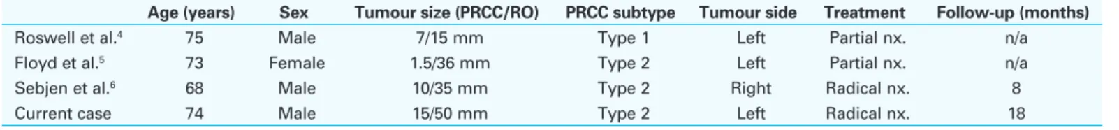
inciden-tally during imaging studies for other reasons and none of these were larger than 4 cm. Two of these cases were treated with partial nephrectomy and one with radical nephrectomy. In our case, we performed radical nephrectomy because the 5 cm in diameter tumour was close to the renal hilum. This case, which has the largest renal oncocytoma and papillary RCC diameter also had the longest follow-up period among the cases reported so far (Table 1).
Conclusion
Sometimes chromophobe RCC arises within a renal onco-cytoma. However, the combination of papillary RCC and
oncocytoma is extremely rare because these tumours are developed from different origins. This case shows us once again that although very rare, renal oncocytoma may con-tain malignant tumour, such as RCC. Therefore, even if the radiological screening strongly suggests that the tumour is oncocytoma, this rare entity should also be kept in mind.
Competing interests: Authors declare no competing financial or personal interests. This paper has been peer-reviewed.
References
1. Lopez-Beltran A, Scarpelli M, Montironi R, et al. 2004 WHO classification of the renal tumors of the adults. Eur Urol 2006;49:798-805.
2. Benatiya MA, Rais G, Tahri M, et al. Renal oncocytoma: Experience of Clinical Urology A, Urology Department, CHU Ibn Sina, Rabat, Morocco and literature review. Pan Afr Med J 2012;12:84. 3. Burger M, Denzinger S, Filbeck T, et al. A metachronous, atypical, multifocal renal oncocytoma with a
concomitant renal cell carcinoma of the contralateral side and bilateral multifocal oncocytomas: Two case reports and review of literature. ScientificWorldJournal 2005;5:545-9. http://dx.doi.org/10.1100/ tsw.2005.73
Fig. 1. Immunohistochemical staining of oncocytoma areas (A) Positive
E-cadherin expression (×400). (B) Positive Pax2 (×200). (C) Weakly positive CD117 expression (×400).
Fig. 2. (A) On hematoxylin-eosin (H&E) staining (×40), papillary renal cell
carcinoma (RCC) (arrow) within the renal oncocytoma (arrowhead). (B) Oncocytoma (arrowhead) and papillary RCC (arrow) on H&E staining (×100). (C) Positive staining with vimentin (×200) in papillary RCC (arrow), negative staining in oncocytoma (arrowhead). (D) Positive staining with cytokeratin 7 (×200) in papillary RCC (arrow), negative staining in oncocytoma (arrowhead).
Table 1. Summary of the cases reported so far and current case
Age (years) Sex Tumour size (PRCC/RO) PRCC subtype Tumour side Treatment Follow-up (months)
Roswell et al.4 75 Male 7/15 mm Type 1 Left Partial nx. n/a
Floyd et al.5 73 Female 1.5/36 mm Type 2 Left Partial nx. n/a
Sebjen et al.6 68 Male 10/35 mm Type 2 Right Radical nx. 8
Current case 74 Male 15/50 mm Type 2 Left Radical nx. 18
CUAJ • November-December 2014 • Volume 8, Issues 11-12
E930
Özer et al.
4. Rowsell C, Fleshner N, Marrano P, et al. Papillary renal cell carcinoma within a renal oncocytoma: Case report of an incidental finding of a tumour within a tumour. J Clin Pathol 2007;60:426-8. http://dx.doi. org/10.1136/jcp.2006.041129
5. Floyd MS Jr, Javed S, Pradeep KE, et al. Composite oncocytoma and papillary renal cell carcinoma of the kidney treated by partial nephrectomy: A case report. ScientificWorldJournal 2011;11:1173-7. http:// dx.doi.org/10.1100/tsw.2011.120
6. Sejben I, Szabó Z, Lukács N, et al. Papillary renal cell carcinoma embedded in an oncocytoma: Case report of a rare combined tumour of the kidney. Can Urol Assoc J 2013;7:E513-6. http://dx.doi. org/10.5489/cuaj.414
7. Theodosopoulos T, Yiallourou A, Kyriazi M, et al. Unilateral simultaneous renal oncocytoma and angi-omyolipoma: Case report. Cases J 2009;2:9093. http://dx.doi.org/10.1186/1757-1626-2-9093
8. Ahmad S, Manecksha R, Hayes BD, et al. Case report of a symptomatic giant renal oncocytoma. Int J
Surg Case Rep 2011;2:83-5. http://dx.doi.org/10.1016/j.ijscr.2010.11.006
9. Anastasiadis A, Dimitriadis G, Radopoulos D. Incidental giant renal oncocytoma: A case report. J Med Case
Rep 2010;4:358. http://dx.doi.org/10.1186/1752-1947-4-358
10. Yusenko MV. Molecular pathology of renal oncocytoma: A review. Int J Urol 2010;17:602-12. http:// dx.doi.org/10.1111/j.1442-2042.2010.02574.x
11. Kuroda N, Tanaka A. Recent classification of renal epithelial tumors. Med Mol Morphol 2014;47:68-75. Epub 2013 Mar 26. http://dx.doi.org/10.1007/s00795-013-0033-0
Correspondence: Dr. Cevahir Özer, Baskent University Adana Medical and Research Center, Department of Urology, Adana, Turkey; [email protected]