Примљено • Received: March 01, 2016 Ревизија • Revised: October 18, 2016 Прихваћено • Accepted: Decembar 8, 2016 Online fi rst: February 24, 2017 Correspondence to: Murat SADIC
Clinic of Nuclear Medicine Ankara Training and Research Hospital
Ankara, TURKEY
[email protected] SUMMARY
Introduction/Objective Medical protection of kidneys against ischemia reperfusion injury is very im-portant. Many agents have been used for the protection of ischemia reperfusion renal tissue injury. We aimed to evaluate the radioprotective effect of lycopene on kidneys in ischemia reperfusion injury with histopathological, biochemical, and scintigraphic parameters.
Methods Twenty-one Wistar male albino rats were divided into the following three groups: lycopene, control, and sham group. In the lycopene group, lycopene was started three days before right renal isch-emia reperfusion injury and continued for 15 days. In the control group, right renal ischisch-emia reperfusion injury was applied with no medication. In the sham group, neither right renal ischemia reperfusion injury nor medication were applied. On the 15th day, all rats were sacrificed after 99mTc-dimercaptosuccinic acid (DMSA) scintigraphies were taken. Histopathological, biochemical, and scintigraphic evaluations were made.
Results The histopathological score was lower in the lycopene group. In biochemical analysis, myeloper-oxidase levels were lower in the lycopene group than in the control group, but not statistically significant. Malondialdehyde and nitrite levels were lower in the lycopene group than in the control group. The postoperative mean 99mTc-DMSA uptake values were 44.82 ± 1.84 in the lycopene group, 38.92 ± 1.17 in the control group, and 50.21 ± 1.35 in the sham group. DMSA uptake values were higher in the lycopene group than in the control group.
Conclusion Lycopene seems to be an effective agent for protection of kidneys in ischemia reperfusion injury as demonstrated by the histopathological, biochemical, and scintigraphic parameters.
Keywords: renal ischemia/reperfusion; kidney; lycopene
INTRODUCTION
Acute renal failure from ischemic damage to the kidney cause morbidity and mortality. Tu-bular epithelial cell death is the most common cause of this renal failure [1]. Even after reper-fusion of extended ischemia of kidney, injury occurs [2]. Although reperfusion is essential for the survival of ischemic renal tissue, it may cause additional damage to kidney [3]. Renal damage caused by ischemia reperfusion injury (IRI) occurs in surgical procedures in which the kidney remains without blood supply for some time. IRI is the most common cause of acute renal failure after renal transplantation, major abdominal and vascular surgery, coro-nary bypass graft surgery, trauma, and sepsis [4]. IRI seen in kidney transplantation effects the short- and long-term graft survival [5]. IRI results in persistent intrarenal vasoconstriction, injury of microvascular endothelial and tubu-lar epithelial cells [6]. Reperfusion of ischemia
causes a rapid burst of free radicals, which is responsible for endothelial injury and edema. Reperfusion damages endoplasmic reticulum, leading to autophagosome formation and cel-lular disintegration [7].
Medical protection of the kidney against IRI is very important for nephron sparing surgery and renal transplantation, so many studies con-tinue in this area. Many agents have been used for the protection of ischemia reperfusion renal tissue injury, such as recombinant human man-ganese superoxide dismutase, n-acetylcysteine and desferrioxamine, telmisartan, exendin-4, and sitagliptin [8, 9, 10].
In this study we aimed to evaluate the radio-protective effect of lycopene on the kidney after unilateral ischemia due to renal artery tempo-rary clamping through the use of histopatho-logical evaluation, malondialdehyde (MDA), myeloperoxidase (MPO) and total nitrite level analysis and 99mTc-dimercaptosuccinic acid
(DMSA) scintigraphy uptake.
ORIGINAL ARTICLE / ОРИГИНАЛНИ РАД
Scintigraphic, histopathologic, and biochemical
evaluation of lycopene effects on renal ischemia
reperfusion injury in rats
Murat Sadıc1, Hasan Ikbal Atılgan2, Arif Aydın3, Gökhan Koca1, Meliha Korkmaz1, Tolga Karakan4, Hatice Surer5, Pelin Borcek6
1Ankara Training and Research Hospital, Clinic of Nuclear Medicine, Ankara, Turkey;
2Kahramanmaras Necip Fazil City Hospital, Department of Nuclear Medicine, Kahramanmaras, Turkey; 3Sanliurfa Mehmet Akif Inan Training and Research Hospital, Clinic of Urology, Sanliurfa, Turkey; 4Ankara Training and Research Hospital, Clinic of Urology, Ankara, Turkey;
5Ankara Training and Research Hospital, Department of Biochemistry, Ankara, Turkey; 6Baskent University, Department of Pathology, Ankara, Turkey
154
METHODS
This experimental study was approved by the Ankara Training and Research Hospital Local Ethics Committee of Animal Experiments and conducted in Hüsnü Sakal Experimental and Clinical Practice Center at the same hospital. The study group consists of total of 21 Wistar male albino rats (260 ± 45 grams, three to five months old). The rats were acclimated for at least one week prior to the study and housed under standard laboratory conditions [constant temperature (21°C ± 2°C) with relative humidity of 50–60%, with 12-hour light and dark cycles). During the study, the animals had ad libitum access to water and standard food.
Three groups of rats were formed and evaluated. Group 1 (n = 9) was administered lycopene [5 mg per kg of body weight per day (LYC-O-MATO®, GNC Holdings Inc., Pitts-burgh, PA, USA)] starting three days before right renal ischemia reperfusion injury and this was continued for 15 days. Group 2 was the control group (n = 9), to which right renal ischemia reperfusion injury was applied without any medication. Group 3 (n = 3) was the sham group, to which neither right renal ischemia reperfusion injury nor medication were applied.
For static renal scintigraphic examination, 99mTc-DMSA
images were obtained preoperatively and on postoperative day 15 for each group. Surgery and 99mTc-DMSA
scintigra-phy were applied under anesthesia of 40 mg/kg ketamine hydrochloride (Ketalar, Parke-Davis/Eczacıbaşı, Istanbul, Turkey) and 5 mg/kg xylazine (Rompun, Bayer, Istanbul, Turkey) applied intramuscularly. The subjects were sacri-ficed by decapitation under 50 mg/kg, intraperitoneally, with propofol anesthesia (Abbott Laboratory Corporation, Istanbul, Turkey). After the sacrification for each group, left and right kidneys were removed surgically for histo-pathological examination.
Surgical method
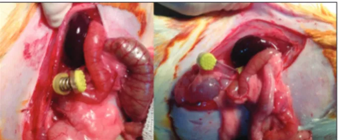
DMSA renal scintigraphy was performed in all rats be-fore the surgical procedure. General anesthesia was ad-ministered intramuscularly as a combination of 40 mg/ kg ketamine hydrochloride (HCl) (Ketalar, Parke-Davis/ Eczacıbaşı) and 10 mg/kg xylazine HCl (Rompun, Bayer). The incision area was shaved and the surgical site was cleaned preoperatively with soap and povidone-iodine. A sterile environment was prepared. By laparotomy via mid-line incision, the right kidney was reached and the right renal pedicle was isolated. The right pedicle was occluded for 45 minutes to induce ischemia and then subjected to reperfusion for six hours (I/R groups). After the reper-fusion, DMSA renal scintigraphy was performed (Figure 1). Before the sacrification, blood samples had been taken from all the rats, and the values of urea and creatinine (mg/dL) parameters were determined in plasma to assess the renal activity. We performed the right nephrectomy 24 hours after all the rats were sacrificed. Surgically ex-cised right kidney tissues obtained at 24 hours were placed into aseptic containers for biochemical examination and
histopathological analysis (placed in liquid nitrogen for immediate freezing and protein denaturation) and were stored at -80°C in special containers and racks in freezer. The right kidney was evaluated histopathologically and biochemically for oxidative stress markers in the tissue, including the level of MDA, which are indicators of lipid peroxidation, and MPO and total nitrite.
Histopathological evaluation
Kidney tissues obtained after sacrificing the rats were fixed immediately in 10% formaldehyde and were processed in paraffin tissue blocks and macroscopic sections were taken to include the renal cortex and pelvis. Sections of 5 μm thickness cut from formalin fixed paraffin-embedded blocks were stained with hematoxylin and eosin. Histo-pathological examination was performed in 40–100–200– 400 × original magnification with light microscopy (Figure 2). For the histopathological score the following criteria were used:
• Normal histology: 0 points;
• Swelling of tubule cells, loss of brush border and nu-clear condensation of ≤ 1/3: 1 point;
• In addition to the changes in 1 point, 1/3–2/3 tubule changes: 2 points;
• Tubular changes more than 2/3: 3 points.
All kidneys were examined in 100 areas with a maxi-mum score of 300.
Figure 1. Placing atraumatic microvascular clamps to the renal pedicle
for renal ischemia
Figure 2. Areas of unaffected tubules displaying normal nuclei with
open chromatin, retained brush borders, and cytoplasmic integrity (H&E, × 400)
Biochemical Evaluation
Tissue samples were stored at -80°C until analysis. Kidney samples were homogenized with 0.15M KCl at a rate of 1/10 (weight per volume). MDA, MPO, and total nitrite levels were measured in the tissue samples.
Determination of MPO level
MPO activity was obtained spectrophotometrically by de-termining the decomposition of hydrogen peroxide using o-dianisidine as the hydrogen donor. Tissue samples of ap-proximately 50 mg were taken, weighed and homogenized three times for 30 seconds at 4°C in 1 ml of ice-cold 0.5% hexadecyltrimethylammonium bromide in 50 mmol/L phosphate buffer (pH 6). The homogenate was subjected to three freeze/thaw cycles and centrifuged for 15 minutes at 40,000 × g. MPO activity was determined by the addi-tion of 0.1 ml of the supernatant to 2.9 ml of 50 mmol/L phosphate buffer containing 0.167 mg/mL o-dianisidine dihydrochloride and 0.0005% hydrogen peroxide. The change in absorbance at 460 nm over a five-minute pe-riod was measured at 25°C. The data are expressed as the change in absorbance per minute per gram of tissue (Δabs/ min/g of tissue) [11].
Determination of MDA level
MDA levels were calculated by the fluorometric method, developed by Wasowicz et al. [12]. After the reaction of thiobarbituric acid with MDA, the reaction product was extracted in butanol and was measured spectrofluoromet-rically at wavelengths of 525 nm for excitation and 547 nm for emission. As standard, 0–5 μmol/L 1,1’,3,3’-tetraeth-oxypropane solutions were used. For the measurement of tissue MDA, 50 μL of homogenate was added and intro-duced into 10 mL glass tubes containing 1 ml of distilled water. After the addition of 1 ml of the solution containing 29 mmol/L thiobarbituric acid in acetic acid and mixing, the samples were placed in a water bath and heated for one hour at 95–100°C. After the samples cooled, 25 μL of 5 mol/L HCL was added and the reaction mixture was ex-tracted by agitation for five minutes with 3.5 mL n-butanol. After separation of the butanol phase by centrifugation at 1,500 × g for 10 minutes, the fluorescence of the butanol extract was measured with F-2500 fluorometer (Hitachi Ltd., Chiyoda, Tokyo, Japan) at wavelengths of 525 nm for excitation and 547 nm for emission. As standard, 0–5 μmol/L 1,1’,3,3’-tetraethoxypropane solutions were used. MDA levels are given as μmol per gram of wet tissue.
Determination of total nitrite level
A total of 300 μL of homogenate was added to 300 μLl of KH2PO4/K2HPO4 buffer (pH 7.5), 50 μl of 2 mmol/L NADPH, 50 μl of 50 μmol/L FAD, and 50 μl of 1 unit/ mL aspergillus nitrate reductase. This was incubated at room temperature for one hour followed by the addition of 500 μl of 0.2 mol/L ZnSO4 and 70 μL 2 mol/L NaOH to
deproteinate the sample. After centrifugation, 0.75 mL of the supernatant was added to 1 mL of 1% sulphanilic acid (in 4 mol/L HCl). After 10 minutes at room temperature, 0.75 mL of freshly prepared 1% N-(1-Naphthyl)ethylene-diamine was also added. The resultant color change was measured at 548 nm using a spectrophotometer. Nitrite concentration was calculated from 5, 12.5, 25, 50 μmol/L sodium nitrite standards [13]. Total nitrite levels are given as nmol per liter per gram of wet tissue.
99mTc-DMSA scintigraphy evaluation
Preoperatively and on postoperative 15th day of the renal injury, adequate hydration with sterile saline and induc-tion of anesthesia were carried out before the scintigraph-ic study. A commercially available kit of DMSA (MON. DMSA KIT, Monrol Eczacıbaşı, Istanbul, Turkey) was labeled with 99mTc in accordance with the manufacturer’s
leaflet for use. A 37 MBq (1 mCi) dose of 99mTc-DMSA in a
0.1 ml volume was administered to the tail vein. Two hours after injection, static renal images were taken under anes-thesia. Renal static scintigraphies were performed using a single-head e.Cam gamma camera (Siemens Healthcare, Erlangen, Germany) equipped with pinhole collimator, energy peak adjusted to 140 keV ± 20% with 256 × 256 matrix and 2.67 zoom factor for five minutes in posterior position. ROIs (regions of interest) were drawn on the anterior and posterior images and the geometric mean of these values was accepted as the relative uptake percentage of the kidneys.
Statistical analysis
Statistical analysis was performed with SPSS Statistics for Windows, Version 20.0 (IBM Corp., Armonk, NY, USA). Continuous and categorical variables are expressed as the mean value ± standard deviation and frequency (%) of numbers. Group comparisons for each parameter were done using the nonparametric Mann–Whitney U-test and the Kruskal–Wallis tests. A p–value less than 0.05 was con-sidered to be a statistically significant difference.
RESULTS
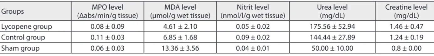
The histopathological score was 45.00 ± 16.58 in the lycopene group, 95.56 ± 40.34 in the control group, and 30.00 ± 5.00 in the sham group (Table 1). The histopathological score was lower in the lycopene group than in the control group (p < 0.001) and higher in the control group than in the sham group (p = 0.012). The histopathological score of the
Table 1. Histopathological scores of the groups; values are given as mean ± standard deviation (SD)
Minimum score Maximum score Mean score ± SD Lycopene group 10 60 45 ± 16.58 Control group 75 200 95.56 ± 40.35 Sham group 25 35 30 ± 5
156
lycopene group was higher than that of the sham group, but it was not statistically significant (p = 0.093) (Table 2).
In biochemical analysis, the mean value of MPO level (Δabs/min/g of tissue) was 0.08 ± 0.09 in the lycopene group, 0.11 ± 0.03 in the control group, and 0.06 ± 0.03 in the sham group. The MPO level was lower in the lycopene group than in the control group, and higher in the control group than in the sham group, but not statistically signifi-cant. The mean value of the MDA level (µmol/g of wet tis-sue) was 4.61 ± 2.10 in the lycopene group, 6.85 ± 1.68 in the control group and 13.36 ± 3.56 in the sham group. The MDA level was lower in the lycopene group than in the control group (p = 0.009) and the sham group (p = 0.013), and lower in the control group than in the sham group (p = 0.021). The mean value of the nitrite level (nmol/l/gm wet tissue) was 0.05 ± 0.02 in the lycopene group, 0.09 ± 0.02 in the control group, and 0.04 ± 0.01 in the sham group. The nitrite level was lower in the lycopene group than in the control group (p = 0.003) and higher in the control group than in the sham group (p = 0.013). The mean value of urea level (mg/dL) was 175.56 ± 52.94 in the lycopene group, 144.44 ± 27.89 in the control group and 50 ± 10 in the sham group. The urea level was higher in the lycopene group than in the control group but not statistically significant, and higher in the control group than in the sham group (p = 0.012). The mean value of creatine level (mg/dL) was 1.46 ± 0.47 in the lycopene group, 1.24 ± 0.19 in the control group and 0.8 ± 0 in the sham group. The creatine level was higher in the lycopene group than in the control group but not statistically significant, and higher in the control group than in the sham group (p = 0.011) (Tables 2 and 3).
In the scintigraphic analysis, the preoperative mean uptake values were 50.16 ± 1.49 in the lycopene group, 50.56 ± 1.38 in the control group and 50.18 ± 0.34 in the sham group. Preoperative 99mTc-DMSA uptake values were
not statistically significant between the groups (p = 0.767). The postoperative mean uptake values were 44.82 ± 1.84 in the lycopene group, 38.92 ± 1.17 in the control group and 50.21 ± 1.35 in the sham group. There was statistically sig-nificant difference between the lycopene and control groups, higher in the lycopene group (p < 0.001). The uptake values of the lycopene and control groups were lower than those of the sham group, as expected (p = 0.013, both) (Figure 3).
DISCUSSION
In our study, lycopene showed histopathological protective effect in IRI of the kidney. In the histopathological evalua-tion, the histopathological score was lower in the lycopene group than in the control group, and histopathological score of the control group was higher than that in the sham group. MPO level in the lycopene group was lower than in the control group, but not statistically significant. MDA and nitrite levels in the lycopene group were lower than in the control group. These biochemical parameters indicate that lycopene has protective effect against IRI. 99mTc-DMSA
uptake of the effected kidneys were higher in the lycopene group than in the control group, which indicates lycopene has protective effect on renal parenchyma.
In animal models, reperfusion of the ischemic kidney is followed by tissue destruction and many morbidities ensue. Neutrophils accumulate in the area of ischemic tissues after reperfusion and tissue damage is mediated through neutrophil-mediated oxidative stress. Reactive oxygen species are produced at the sites of inflammation by neutrophils and cause formation of lipid peroxides, damage
Table 2. Statistical analysis (p-values) of biochemical parameters and histopathological scores between the groups
Groups MPO levellevel MDA level Nitrite level Urea (mg/dL) level Creatine levellevel Microscopicscore
Lycopene group vs. control group 0.07 0.009* 0.003* 0.306 0.424 0.000*
Lycopene group vs. sham group 0.782 0.013* 0.926 0.013* 0.012 0.093
Control group vs. sham group 0.079 0.021* 0.013* 0.012* 0.011* 0.012*
MPO – myeloperoxidase; MDA – malondialdehyde
Table 3. Biochemical parameters of the groups; values are given as mean ± standard deviation (SD)
Groups (Δabs/min/g tissue)MPO level (µmol/g wet tissue)MDA level (nmol/l/g wet tissue)Nitrit level Urea level(mg/dL) Creatine level (mg/dL)
Lycopene group 0.08 ± 0.09 4.61 ± 2.10 0.05 ± 0.02 175.56 ± 52.94 1.46 ± 0.47
Control group 0.11 ± 0.03 6.85 ± 1.68 0.09 ± 0.02 144.44 ± 27.89 1.24 ± 0.19
Sham group 0.06 ± 0.03 13.36 ± 3.56 0.04 ± 0.01 50.00 ± 10.00 0.8 ± 0.00
MPO – myeloperoxidase; MDA – malondialdehyde
Figure 3. Preoperative (a) and postoperative (b) DMSA scintigraphy in
posterior image of a rat in the control group (uptake value of the right kidney; preoperative = 37.45%, postoperative = 48.89%), preoperative (c) and postoperative (d) DMSA scintigraphy in posterior image of a rat in the lycopene group (uptake value of the right kidney; preopera- tive = 47.56%, postoperative = 50.63%)
of cell membrane and destruction of antioxidative defense mechanism. Reperfusion may increase the damage after ischemia in tissues. After reperfusion of ischemic tissue, inflammatory and metabolic damage occurs as a result of disruption of cellular integrity. When the cascade begins, various distant organs, such as lungs, liver and heart, are af-fected by many activated system and toxic mediators [14].
Effects of IRI on kidneys have been studied by various methods such as biochemical assays, histopathological ex-amination, and scintigraphic imaging, but there is no con-sensus for the best method for the evaluation of impaired renal functions [15]. After reperfusion of ischemic kidney, MDA and MPO levels increase and injure the renal tissue to a greater extent. Reactive oxygen species released by the neutrophils increase the tissue damage further during reperfusion [14, 16]. MDA,which is a marker of lipid per-oxidation, increases during reperfusion after an ischemic renal episode [17]. MPO activity increases if neutrophil infiltration into the tissues occurs as in IRI, and total nitrite level is the marker of total lipid membrane damage [18].
Lycopene is the most common studied compound on the preventive effects of dietary intake of tomato products [19, 20]. Lycopene is the most potent antioxidant among vari-ous carotenoids. Lycopene has the best relative radical scav-enging abilities among the carotenes. Carotenoids prevent damage to DNA, proteins, cell membranes, lipids, and other structures, which occurs after oxidative injury. Lycopene has higher singlet oxygen quenching ability than β-carotene and α-tocopherol by its high number of conjugated double bonds [21]. Lycopene has antiproliferative properties to protect the development of prostate cancer and inhibits cholesterol synthesis and enhances low density lipoprotein degradation [22]. Lycopene has also many other functions in the immune system, metabolic pathways, and cell–cell communication. Lycopene normalizes the change of intrathymic T-cell dif-ferentiation seen in tumorigenesis [23].
Due to antioxidative properties, lycopene mediates free radical scavenging activity and results in the reduction of infarct volume in ischemia reperfusion brain injury [22]. Lycopene ameliorated the ischemia reperfusion induced tissue damage and was found to protect the germ cells after testicular torsion [24]. However, in another study,
lycopene was not effective for testicular torsion in the long term, there was no improvement in the groups treated with lycopene for therapeutic purposes [25]. Liu et al. [26] identified the pro-regenerative, anti-apoptotic and anti-oxidant properties of mesenchymal stromal cells in IRI. Lycopene ameliorated lysosomal membrane damage as well as alterations in cardiac enzymes, lipid profile, and oxidative stress markers. Yue et al. [27] thought that lyco-pene protects myocardium against hypoxia reoxygenara-tion induced apoptosis by maintaining the mitochondrial function. Lycopene was also found effective in pancreatitis by inhibition of neutrophil infiltration and lipid peroxida-tion [20]. Bayramoglu et al. [21] used lycopene in IRI of liver in different doses of 2.5 and 5 mg/kg body weight. Improvements of alanine aminotransferase, aspartate ami-notransferase, lactate dehydrogenase, and MDA levels were partial and dose dependent. Lycopene showed the protec-tive effect as a decrease in MDA level, nitrite level, and histopathological score, and an increase in DMSA uptake of the kidney.
Pektaş et al. [17] evaluated the effectiveness of lycopene in IRI with biochemical and histopathological parameters and found that lycopene may have a protective effect on IRI. Kaya et al. [28] also used high dose of lycopene (100 mg/kg) in a single dose in IRI of the kidney. They also used bio-chemical and histopathological parameters and mentioned that lycopene administered prior to renal IRI prevented renal damage. A few studies used 99mTc-DMSA scintigraphy
for the evaluation of IRI of the kidney. DMSA scintigraphy was found to be an effective non-invasive method in the evaluation of kidney restoration after IRI injury [29]. In our study, we also used DMSA scintigraphy in addition to biochemical and histopathological parameters.
CONCLUSION
In conclusion, lycopene seems to be an effective agent for protection of the kidney in reperfusion injury after renal ischemia, as demonstrated by the histopathological, bio-chemical, and scintigraphic parameters. However, further larger studies are necessary for clinical use.
REFERENCES
1. Hamar P, Song E, Kökény G, Chen A, Ouyang N, Lieberman J. Small interfering RNA targeting Fas protects mice against renal ischemia-reperfusion injury. Proc Natl Acad Sci U S A. 2004; 101(41):14883–8.
2. Zhou W, Farrar CA, Abe K, Pratt JR, Marsh JE, Wang Y, et al. Predominant role for C5b-9 in renal ischemia/reperfusion injury. J Clin Invest. 2000; 105(10):1363–71.
3. Chatterjee PK, Patel NS, Kvale EO, Cuzzocrea S, Brown PA, Stewart KN, et al. Inhibition of inducible nitric oxide synthase reduces renal ischemia/reperfusion injury. Kidney Int. 2002; 61(3):862–71. 4. de Vries B, Köhl J, Leclercq WK, Wolfs TG, van Bijnen AA, Heeringa
P, et al. Complement factor C5a mediates renal
ischemia-reperfusion injury independent from neutrophils. J Immunol. 2003; 170(7):3883–9.
5. Wu H, Chen G, Wyburn KR, Yin J, Bertolino P, Eris JM, et al. TLR4 activation mediates kidney ischemia/reperfusion injury. J Clin Invest. 2007; 117(10):2847–59.
6. Neto JS, Nakao A, Kimizuka K, Romanosky AJ, Stolz DB, Uchiyama T, et al. Protection of transplant-induced renal ischemia-reperfusion injury with carbon monoxide. Am J Physiol Renal Physiol. 2004; 287(5):F979–89.
7. Cusumano G, Romagnoli J, Liuzzo G, Ciavarella LP, Severino A, Copponi G, et al. N-Acetylcysteine and High-Dose Atorvastatin Reduce Oxidative Stress in an Ischemia-Reperfusion Model in the Rat Kidney. Transplant Proc. 2015; 47(9):2757–62.
8. Bernardi RM, Constantino L, Machado RA, Vuolo F, Budni P, Ritter C, et al. N-acetylcysteine and deferrioxamine protects against acute renal failure induced by ischemia/reperfusion in rats. Rev Bras Ter Intensiva. 2012; 24(3):219–23.
9. Tawfik MK. Renoprotective activity of telmisartan versus pioglitazone on ischemia/reperfusion induced renal damage in diabetic rats. Eur Rev Med Pharmacol Sci. 2012; 16(5):600–9. 10. Chen YT, Tsai TH, Yang CC, Sun CK, Chang LT, Chen HH, et al.
158
injury through suppressing oxidative stress and inflammatory reaction. J Transl Med. 2013; 11:270.
11. Bradley PP, Priebat DA, Christensen RD, Rothstein G. Measurement of cutaneous inflammation: estimation of neutrophil content with an enzyme marker. J Invest Dermatol. 1982; 78(3):206–9. 12. Wasowicz W, Neve J, Peretz A. Optimized steps in fluorometric
determination of thiobarbituric acid-reactive substance in serum: Importance of extraction pH and influence of sample preservation and storage. Clin Chem. 1993; 39:2522–6.
13. Smarason AK, Allman KG, Young D, Redman CW. Elevated levels of serum nitrate, a stable end product of nitric oxide, in women with pre-eclampsia. Br J Obstet Gynaecol. 1997; 104(5):538–43. 14. Serteser M, Koken T, Kahraman A, Yilmaz K, Akbulut G, Dilek ON.
Changes in hepatic TNF-alpha levels, antioxidant status, and oxidation products after renal ischemia/reperfusion injury in mice. J Surg Res. 2002; 107(2):234–40.
15. Gültekin SS, Odabaş Ö, Giniş Z, Gökçe A, Yiğman M, Doğan S, et al. Scintigraphic comparison of renal ischemia-reperfusion injury models in rats: correlations with biochemical and histopathological findings. Ann Nucl Med. 2013; 27(6):564–71.
16. Suleyman Z, Sener E, Kurt N, Comez M, Yapanoglu T. The effect of nimesulide on oxidative damage inflicted by ischemia-reperfusion on the rat renal tissue. Ren Fail. 2015; 37(2):323–31.
17. Pektaş A, Gemalmaz H, Balkaya M, Ünsal C, Yenisey Ç, Kılıçarslan N, et al. The short-term protective effects of lycopene on renal ischemia-reperfusion injury in rats. Turk J Urol. 2014; 40(1):46–51. 18. Lipińska J, Lipińska S, Stańczyk J, Sarniak A, Przymińska vel Prymont
A, Kasielski M, et al. Reactive oxygen species and serum antioxidant defense in juvenile idiopathic arthritis. Clin Rheumatol. 2015; 34(3):451–6.
19. Lavelli V, Peri C, Rizzolo A. Antioxidant activity of tomato products as studied by model reactions using xanthine oxidase, myeloperoxidase, and copper-induced lipid peroxidation. J Agric Food Chem. 2000; 48(5):1442–8.
20. Ozkan E, Akyüz C, Dulundu E, Topaloğlu U, Sehirli AÖ, Ercan F, et al. Protective effects of lycopene on cerulein-induced experimental acute pancreatitis in rats. J Surg Res. 2012; 176(1):232–8. 21. Bayramoglu G, Bayramoglu A, Altuner Y, Uyanoglu M, Colak S. The
effects of lycopene on hepatic ischemia/reperfusion injury in rats. Cytotechnology. 2015; 67(3):487–91.
22. Hsiao G, Fong TH, Tzu NH, Lin KH, Chou DS, Sheu JR. A potent antioxidant, lycopene, affords neuroprotection against microglia activation and focal cerebral ischemia in rats. In Vivo. 2004; 18(3):351–6.
23. Kobayashi T, Iijima K, Mitamura T, Toriizuka K, Cyong JC, Nagasawa H. Effects of lycopene, a carotenoid, on intrathymic T cell differentiation and peripheral CD4/CD8 ratio in a high mammary tumor strain of SHN retired mice. Anticancer Drugs. 1996; 7(2):195–8.
24. Hekimoglu A, Kurcer Z, Aral F, Baba F, Sahna E, Atessahin A. Lycopene, an antioxidant carotenoid, attenuates testicular injury caused by ischemia/reperfusion in rats. Tohoku J Exp Med. 2009; 218(2):141–7.
25. Güzel M, Sönmez MF, Baştuğ O, Aras NF, Öztürk AB, Küçükaydın M, et al. Effectiveness of lycopene on experimental testicular torsion. J Pediatr Surg. 2016; 51(7):1187–91.
26. Liu H, McTaggart SJ, Johnson DW, Gobe GC. Original article anti-oxidant pathways are stimulated by mesenchymal stromal cells in renal repair after ischemic injury. Cytotherapy. 2012; 14(2):162–72. 27. Yue R, Hu H, Yiu KH, Luo T, Zhou Z, Xu L, et al. Lycopene
protects against hypoxia/reoxygenation-induced apoptosis by preventing mitochondrial dysfunction in primary neonatal mouse cardiomyocytes. PLoS One. 2012; 7(11):e50778.
28. Kaya C, Karabulut R, Turkyilmaz Z, Sonmez K, Kulduk G, Gülbahar Ö, et al. Lycopene has reduced renal damage histopathologically and biochemically in experimental renal ischemia-reperfusion injury. Ren Fail. 2015; 37(8):1390–5.
29. Kwak W, Jang HS, Belay T, Kim J, Ha YS, Lee SW, et al. Evaluation of kidney repair capacity using 99mTc-DMSA in ischemia/reperfusion injury models. Biochem Biophys Res Commun. 2011; 406(1):7–12.
САЖЕТАК Увод/Циљ Заштита бубрега од реперфузионих оштећења после исхемије је врло значајна. За ову заштиту су коришће-на многа средства. Циљ рада је процена радиопротективног ефекта ликопена на реперфузиона оштећења бубрега после исхемије. Методе Група од 21 мужјака вистар албино пацова подеље-на је у следеће три групе: ликопенску, контролну и псеудо-групу. Пацовима у ликопенској групи ликопен је даван три дана пре и 15 дана после реперфузионог оштећења бубрега, контролној групи није даван ликопен после оштећења, а пацови псеудогрупе нису имали исхемију бубрега и није им даван ликопен. После 15 дана урађена је сцинтиграфија са 99mTc-ДМСК (димеркаптосукцинска киселина), а потом су пацови жртвовани и урађена су патохистолошка и биохе-мијска истраживања. Резултати Патохистолошки скор је био нижи у ликопенској групи. Биохемијска анализа је показала да је ниво мијелопе-роксидазе био нижи у ликопенској групи него у контролној, али не статистички значајно. Нивои малондиалдехида и ни-трата су били нижи у ликопенској него у контролној групи. Прихват 99mTc-ДМСК у ликопенској групи био је 44,82 ± 1,84, у контролној групи 38,92 ± 1,17 и 50,21 ± 1,35 у псеудогрупи. Закључак Ликопен би могао бити ефикасно средство за заштиту бубрега од реперфузионог оштећења после исхе-мије, што је показано патохистолошким, биохемијским и сцинтиграфским параметрима. Кључне речи: бубрежна исхемија / реперфузија, бубрег, ликопен