A rare case of a gastric adenomyoma mimicking a gastric
duplication cyst
Esra Elif Arslan1 , Tuğba Acer Demir1 , Lütfi Hakan Güney1 , Merih Tepeoğlu2 , Müge Sağnak Akıllı1 , Akgün Hiçsönmez1 1Department of Pediatric Surgery, Başkent University School of Medicine, Ankara, Turkey
2Department of Pathology, Başkent University School of Medicine, Ankara, Turkey
Dear Editor,
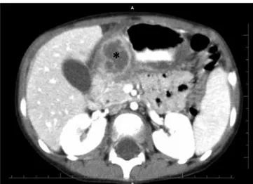
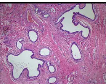
A 5-year-old girl was admitted to our facility with the complaints of abdominal pain, poor oral intake, and fe-ver. The patient had suffered from abdominal pain for 2 years. A physical examination revealed tenderness in the epigastric area. Her serum C-reactive protein level was 84.4 mg/dL (normal: 0-5 mg/dL). Abdominal ultra-sonography (USG) was performed, and an intramural hy-poechoic heterogeneous nodular mass measuring 33×25 mm was detected in the pyloric region of the stomach. Computed tomography (CT) revealed a cystic lesion measuring 30×28 mm in the gastric and pyloric antrum, which was thought to be formed by inflammation, ab-scess, or an infected cyst (Figure 1). The patient was hos-pitalized and treated with antibiotics and antacids. CT for control examination revealed that the cystic lesion had disappeared. The patient was readmitted to our facility after 8 months with abdominal pain and poor oral in-take. Abdominal USG reported a cystic lesion measuring 32×26×20 mm in size and interpreted it as a duplication cyst (Figure 2). Abdominal CT revealed soft tissue thick-ening at the level of the gastric antrum extending to the pylorus (Figure 3). The patient underwent exploratory lap-arotomy; a 3.5×3-cm mass present at the anterior-inferi-or wall of the pre-pylanterior-inferi-oric region was completely excised, and double-layer transverse anastomosis was performed. Pathological examination revealed gastric adenomyoma, which was composed of cysts and glandular structures lined by cuboidal to columnar epithelium surrounded by hypertrophic smooth muscle bundles. Foreign-body gi-ant cells and xgi-anthogranulomatous inflammation were
also detected on the serosal surface (Figure 4). Three months later, control radiological examinations revealed normal gastric anatomy.
Approximately 60% of gastrointestinal adenomyomas are identified in the stomach, and gastric adenomyoma is most frequently found in the gastric antrum (85%) and pyloric region (15%) (1-3). The ages of reported cases ranged from 1 week to 81 years, although most were be-tween the fourth and sixth decades (1,2,4).
Previously, five pediatric cases of gastric adenomy-oma have been reported in the literature. The major complaint of the patients was vomiting (1-5). Among
613 Cite this article as: Arslan EE, Acer Demir T, Güney LH, Tepeoğlu M, Sağnak Akıllı M, Hiçsönmez A. A rare case of a gastric adenomyoma mimicking a gastric duplication cyst. Turk J Gastroenterol 2018; 29: 613-5.
ORCID IDs of the authors: E.E.A. 4528-4671; T.A.D. 0000-0001-5391-9094; L.H.G. 2500-5401; M.T. 0000-0002-9894-8005; M.S.A. 0000-0003-2573-6172; A.H. 0000-0003-2893-393X.
This study was presented at the 16th European Congress of Paediatric Surgery (European Paediatric Surgeons’ Association), June 2015,
Ljubljana, Slovenia.
Corresponding Author: Tuğba Acer Demir; [email protected]
Received: July 28, 2018 Accepted: March 6, 2018 Available online date: August 9, 2018
© Copyright 2018 by The Turkish Society of Gastroenterology • Available online at www.turkjgastroenterol.org DOI: 10.5152/tjg.2018.17470
LETTER TO THE EDITOR
Figure 1. The initial computed tomography image: A cystic lesion (asterisk) measuring 30×28 mm at the gastric antrum
these patients, 4 patients [1-week-old boy (4), 13-day-old boy (5), 1-month-13-day-old girl (1), and 4-month-13-day-old girl (3)] were misdiagnosed as having hypertrophic pyloric stenosis. The presence of gastric duplication cyst was considered in differential diagnosis of a 5-year-old girl whose clinical and radiological findings were similar to our case (2). These pathologies are more frequent at etiology of vomiting in the pediatric age group (1-5). A misdiagnosis may lead to a delay in the excision of the mass and pathological diagnosis. USG or CT of all the reported cases showed cystic or heterogenous
in-tramural masses, and surgical exploration and excision of the mass led to a definitive pathological diagnosis of gastric adenomyoma (1-5).
Gastric adenomyoma should be considered a differential diagnosis of hypertrophic pyloric stenosis and gastric du-plication in children admitted with vomiting. As it resem-bles abscess formation, a differential diagnosis of gastric adenomyoma should also be considered when a cystic mass is found in the stomach.
Peer-review: Externally peer-reviewed.
Author Contributions: Concept - T.A.D.; Supervision - E.E.A., T.A.D., L.H.G., M.T., M.S.A., A.H.; Data Collection and/or Interpretation - E.E.A., T.A.D., L.H.G., M.T., M.S.A., A.H.; Analysis and/or Interpretation - E.E.A., T.A.D., L.H.G., M.T., M.S.A., A.H.; Literature Search - E.E.A., T.A.D., L.H.G., M.T., M.S.A., A.H.; Writing Manuscript - E.E.A., T.A.D., L.H.G., M.T., M.S.A., A.H.; Critical Review - E.E.A., T.A.D., L.H.G., M.T., M.S.A., A.H.
Acknowledgements: We would like to thank Editage (editage.com) for English language editing.
Conflict of Interest: The authors have no conflict of interest to de-clare.
Financial Disclosure: The authors declared that this study has re-ceived no financial support.
REFERENCES
1. Takeyama J, Sato T, Tanaka H, Nyo M. Adenomyoma of the stom-ach mimicking infantile hypertrophic pyloric stenosis. J Pediatr Surg 2007; 42: E11-2. [CrossRef]
614
Arslan et al. Gastric adenomyoma mimicking a duplication cyst Turk J Gastroenterol 2018; 29(5): 613-5
Figure 2. Abdominal ultrasonography showed a thickened wall cystic lesion measuring 32×26×20 mm on the anterior wall of the stomach
in the pyloric region
Figure 3. Preoperative computed tomography: Soft tissue thicken-ing at the anterior wall of stomach and pylorus (asterisk); the widest
segment was 19×12 mm
Figure 4. Pathological section from adenomyoma (glandular struc-tures in the muscularis propria)
2. Min SH, Kim HY, Kim SH, et al. Gastric adenomyoma mimicking gastric duplication cyst in a 5-year-old girl. J Pediatr Surg 2012; 47: 1019-22. [CrossRef]
3. Castain C, Rullier A. Pyloric adenomyoma: a rare cause of gastric outlet obstruction in childhood. Diagnostic Histopathology 2012; 18: 511-3. [CrossRef]
4. Rhim JH, Kim WS, Choi YH, Cheon JE, Park SH. Radiological find-ings of gastric adenomyoma in a neonate presenting with gastric outlet obstruction. Pediatr Radiol 2013; 43: 628-30. [CrossRef]
5. Aljahdali A, Oviedo A, Blair GK. Gastric hamartoma of the pylorus in an infant. J Pediatr Surg 2012; 47: E29-31. [CrossRef]
615 Turk J Gastroenterol 2018; 29(5): 613-5 Arslan et al. Gastric adenomyoma mimicking a duplication cyst