Abstract
I
ntroductIonLymphocysts generally occur after pelvic and paraaortic lymph node dissection (LND) in patients with gynecologic cancer. Lymphocyst is a localized collection of lymphatic fluid, which does not have an epithelial wall, and has been reported to develop in approximately 30% of patients who
have undergone to lymphadenectomy during pelvic surgery.[1]
The LND is important in the surgical treatment of gynecologic cancers and in the staging and treatment of epithelial ovarian cancers, endometrial cancers, cervical cancers, and
Objectives: Here, we compare the success of percutaneous transcatheter sclerosant alcohol therapy (PTSAT) for the postoperative treatment of benign pelvic cysts that occurred after gynecologic surgery.
Materials and Methods: The study is a retrospective case–control trial. Gynecological patients who had symptoms due to postoperative pelvic cysts and received PTSAT after gynecologic surgery, between October 2008 and January 2018, were examined in a single training and research hospital in Turkey. Some factors were investigated for associations with postoperative pelvic cyst formation in patients who underwent gynecologic operations for malignancies or benign conditions. Statistical analysis used: The association between two independent and nonnormally distributed continuous variables was analyzed with the Mann–Whitney U-test. Spearman’s rho correlation analysis was conducted to determine the correlation of two nonnormally distributed variables. Chi-square (or Fisher’s exact test, when more suitable) was used to examine the correlation between categorical variables.
Results: Statistically significant differences were found in terms of the average age was higher in patients with malignancies, and the average postoperative pelvic cyst detection time was higher in patients with benign pelvic cysts. While all patients were treated with PTSAT, repetitive PTSAT was required for seven benign and ten malign cases.
Conclusion: Patients with pelvic cysts that occurred after gynecologic surgery for malignant conditions, large volume pelvic cysts and patients with benign cysts who underwent more than one surgery required recurrent PTSAT.
Keywords: Gynecologic, gynecological; sclerosing solutions; neoplasms, surgery
Address for correspondence: Dr. Cihan Comba,
Department of Gynecology and Obstetrics, Division of Gynecologic Oncology, University of Health Sciences, Sultangazi Haseki Training and Research Hospital, Istanbul, Turkey. E‑mail: [email protected]
Access this article online
Quick Response Code:
Website:
www.e-gmit.com
DOI:
10.4103/GMIT.GMIT_107_18
This is an open access journal, and articles are distributed under the terms of the Creative Commons Attribution‑NonCommercial‑ShareAlike 4.0 License, which allows others to remix, tweak, and build upon the work non‑commercially, as long as appropriate credit is given and the new creations are licensed under the identical terms.
For reprints contact: [email protected]
How to cite this article: Comba C, Salik AE, Demirayak G, Erdogan SV,
Sacan F, Ozdemir IA. Comparison of postoperative benign pelvic cysts occurred after gynecologic or gyne-oncologic surgery treated with percutaneous transcatheteric sclerosant alcohol therapy. Gynecol Minim Invasive Ther 2020;9:198-203.
Comparison of Postoperative Benign Pelvic Cysts Occurred
after Gynecologic or Gyne‑oncologic Surgery Treated with
Percutaneous Transcatheteric Sclerosant Alcohol Therapy
Cihan Comba1*, Aysun Erbahceci Salik2, Gokhan Demirayak3, Sakir Volkan Erdogan4, Filiz Sacan5, Isa Aykut Ozdemir6
1Department of Gynecology and Obstetrics, Division of Gynecologic Oncology, University of Health Sciences, Sultangazi Haseki Training and Research Hospital, 2Department of Interventional Radiology, University of Health Sciences, Bakirkoy Dr. Sadi Konuk Training and Research Hospital, 3Department of Gynecology and Obstetrics, Division of Gynecologic Oncology, University of Health Sciences, Bakirkoy Dr. Sadi Konuk Training and Research Hospital, 4Department of Gynecology and Obstetrics, University of Health Sciences, Bagcilar Training and Research Hospital, 5Department of Interventional Radiology, Acıbadem Altunizade Hospital,
6Department of Gynecology and Obstetrics, Division of Gynecologic Oncology, Medipol Mega University Hospital, Istanbul, Turkey
Article History:
Submitted: 6 November 2018 Revised: 16 April 2019 Accepted: 15 June 2020 Published: 15 October 2020
vulvar cancers.[2] Although the LND provides important
information, it is associated with various complications, such as hemorrhages, infections, and lymphedemas.[3]
Lymphocysts may also occur following renal transplantation and urological malignancies.[4] Less frequent causes of
lymphocysts are extensive vascular or spinal surgeries.[5]
Injuries to the lymphatic vessels are associated with continued lymphatic leakage because the lymphatic fluid has no platelets and very low concentration of clotting factors. The predisposing factors for lymphocyst formation are elevated body mass index, excessive number of removed lymph nodes, the presence of nodal metastasis, inadequate intraoperative lymphostasis, chemotherapy, previous radiation in the operation field, and the use of anticoagulants and diuretics.[6] However, tumor stage or histology has not been
found to associate with lymphocyst formation.[7] Lymphocysts
usually appear 3–8 weeks after surgery.[8] Although a large
number of postsurgical lymphocysts remain asymptomatic, some may be associated with serious complications. Larger lymphocysts may cause symptoms related to compression of closed structures, leading to symptoms of constipation, urinary frequency, lower abdominal pain, abdominal fullness, and edema of the genital organs or lower extremities. Serious sequelaes include infection of the lymphocysts, obstruction and infection of the urinary tract, hydronephrosis, intestinal obstruction, venous thrombosis, pulmonary embolism, and lymphatic fistula formation.[9] In these conditions, needle
aspiration, percutaneous catheter drainage with or without sclerotherapy, and surgical marsupialization may be required.[6]
Pelvic inclusion cyst is usually occur after abdominopelvic surgery.[10] Complex pelvic inclusion cyst may resemble
malign ovarian conditions and causes laparotomy for maximal debulking surgery.[11]
Here, we compare the success of percutaneous transcatheteric sclerosant alcohol therapy (PTSAT) for the postoperative treatment of pelvic cysts that occurred after gynecologic surgery for malignant or benign conditions. The aim of this study was detected success of the procedure on postoperative pelvic cysts, especially in gynecologic cancers. Postoperative pelvic cysts are distinguished benign cysts such as lymphocysts, pelvic inclusion cyst (pseudocysts, seromas, hematomas), and malignant cysts such as recurrent serous malignancies through imaging and biopsies.
M
AterIAlsAndM
ethodsGynecology patients who were symptomatic (pelvic pain, pelvic mass sensation, bowel dysfunction, or symptoms of urinary dysfunction) because of postoperative pelvic cysts occurred after gynecologic surgery and received PTSAT between October 2008 and January 2018 were
retrospectively examined [Figure 1]. Informed consent was obtained from all individual participants included in the study. This study received approval from the ethical board numbered 2017/259 of identified using a computer program, and patients who received PTSAT were analyzed pelvic pain, pelvic mass sensation, bowel dysfunction, or symptoms of urinary dysfunction and postoperative pelvic cysts. All PTSAT procedures were performed by the same interventional radiologist. The Seldinger technique was utilized for catheterization of all postoperative pelvic cysts. Before performing alcohol sclerotherapy, all postoperative pelvic cysts were catheterized. After the selection of a safe entry site to the pelvic cysts, an 18G needle was employed for the puncture of the cyst under US guidance after local anesthesia. Approximately 20% of the volume of the cyst aspirated and a cavitography was performed after the introduction of the needle. After excluding the probability of leakage of the cyst content, again under US guidance, a stiff guide-wire was advanced into the cavity. An 8–10 french catheter was placed into the cavity according to the size of the cyst. The first session of sclerotherapy was performed at the time of the catheterization procedure. Ethanol (95% ethanol at 30%–50% of the aspirated cavity volume) was instilled and left in the cavity for 15 min. After 15 min, all of the cyst content, including the ethanol, was reaspirated through the catheter. The catheter was then fixed to the skin with a single suture and allowed to drain by gravity. When the daily drainage volume from the cavity was <10 mL, a cystogram under fluoroscopic guidance was obtained to assess possible leakage. After the exclusion of the leakage, another session of ethanol sclerotherapy was performed, and the catheter was withdrawn. If the daily drainage volume was >10 mL, for the large pelvic cysts, multiple sessions of ethanol sclerotherapy were performed with 2–3 days interval until the daily drainage dropped <10 mL. During the first catheterization procedure, a sample of aspirated fluid was obtained and sent for microbiologic and cytological analyses. Among patients received operations for pelvic cysts occurred after malignant gynecologic surgery, we compared patient age, gravid, parity, cyst detection time, preoperative and postoperative hematocrit, number of days hospitalized, average follow-up time, measured cyst volume, first operations with drainage volume, complications, number of removed pelvic and/or paraaortic lymph nodes. The efficiencies of PTSAT procedures were also examined. Descriptive statistics were used to define continuous variables (averages, standard deviations, minimums, medians, and maximums). The association between two independent and non-normally distributed continuous variables was analyzed with the Mann–Whitney U test. Spearman’s rho correlation analysis was conducted to determine the correlation of two non-normally distributed
variables. Chi-square (or Fisher’s exact test, when more suitable) was used to examine the correlation between categorical variables. Values of P < 0.05 were considered statistically significant. Analyses were conducted using the MedCalc Statistical Software, version 12.7.7 (MedCalc Software bvba, Ostend, Belgium; http://www.medcalc.org; 2013).
r
esultsIn this study, the results of 28 patients with pelvic pain who had received percutaneous catheter drainage for pelvic cysts occurred after gynecologic surgery for malignant or benign conditions, in line with the criteria of our study, were examined. Of these patients, 11 patients had gynecologic malignancies, whereas 17 received operations for benign conditions. Of the patients with malignancies, four had endometrial cancer, four had ovarian cancer, and three had cervical cancer. Two of these patients received operations for endometrial cancer were diagnosed with endometrioid type grade 1 carcinoma, one of whom had stage 1A, while the other two had stage 3 C2. One of the four patients with endometrial cancer had endometrioid type stage 1A, grade 3, and the remaining patient had stage 2 serous-type. Two of the four patients with ovarian cancer exhibited serous histology, one of whom was stage 1B, whereas the other was stage 3C. Of the other two patients, one had seromucinous histology, stage 1 C1, whereas the other had clear cell histology, stage 1 A. Of the patients with cervical carcinoma, two had adenocarcinoma, one had stage 4B peritoneal carcinomatous (was performed palliative ileostomy), one had stage 1B1, and one had stage 1B1 with adenosquamous histology.
Two of the patients who underwent operations for benign conditions had received more than one operation (one patient received a cystectomy and hysterectomy, and the other received a myomectomy) [Table 1]. Six patients had various postoperative complications; two vesicovaginal fistulas, two wound infections, one pseudocyst, one pelvic abscess. Cysts recurred in two patients without any symptoms after final treatments. Both of these patients had undergone operations for benign conditions and received PTSAT (one received PTSAT twice, whereas the other received PTSAT three times). In one of these patients, the cyst had ruptured, and no problems were encountered during the follow-up. When patients who had percutaneous transcatheter ethanol sclerotherapies were grouped into pelvic cysts that occurred after gynecologic surgery for malignant and benign conditions cohorts, the ages of patients with malignancies were found to be statistically higher with shorter pelvic cyst detection times [Table 2]. No differences were found between the cyst
Table 1: Patients’ operation list
Operations n (%)
Debulking surgery 3 (10.71)
Hysterectomy 3 (10.71)
Laparoscopic cystectomy 1 (3.57)
Cystectomy (4 times), hysterectomy 1 (3.57)
Laparotomic Bso+omentectomy 1 (3.57)
Laparotomic abscess drainage 1 (3.57)
Laparotomic cystectomy 3 (10.71)
Laparotomic Uso 1 (3.57)
Laparoscopic Uso 1 (3.57)
Laparoscopic hysterectomy 1 (3.57)
Myomectomy (2 times) appendectomy 1 (3.57)
Ovarian cyst aspiration 2 (7.14)
Diagnostic laparoscopy for ovarian cyst rupture 1 (3.57)
Salpingostomy 1 (3.57)
Cesarean section 3 (10.71)
Tah + Bso 1 (3.57)
Tah Bso + Pplnd 1 (3.57)
Type C2 hysterectomy 2 (7.14)
Bso: Bilateral salpingoophorectomy, Uso: Unilateral
salpingoophorectomy, Tah: Total abdominal hysterectomy, Pplnd: Pelvic and paraaortic lymph node dissection
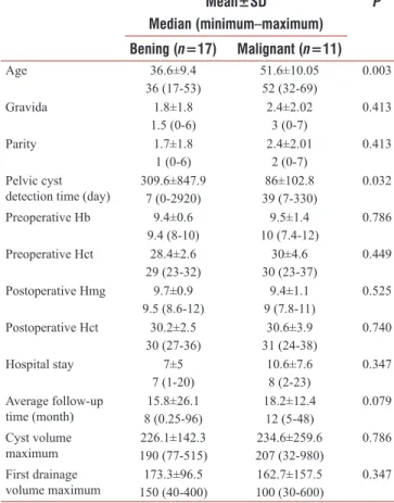
Table 2: Comparison of performed percutaneous transcatheter sclerosant alcohol therapy in patient with gynecologic surgery for benign and malignant conditions Mean±SD Median (minimum–maximum) P Bening (n=17) Malignant (n=11) Age 36.6±9.4 36 (17-53) 51.6±10.0552 (32-69) 0.003 Gravida 1.8±1.8 1.5 (0-6) 2.4±2.023 (0-7) 0.413 Parity 1.7±1.8 1 (0-6) 2.4±2.01 2 (0-7) 0.413 Pelvic cyst
detection time (day) 309.6±847.97 (0-2920)
86±102.8 39 (7-330) 0.032 Preoperative Hb 9.4±0.6 9.4 (8-10) 9.5±1.4 10 (7.4-12) 0.786 Preoperative Hct 28.4±2.6 29 (23-32) 30 (23-37)30±4.6 0.449 Postoperative Hmg 9.7±0.9 9.5 (8.6-12) 9 (7.8-11)9.4±1.1 0.525 Postoperative Hct 30.2±2.5 30 (27-36) 30.6±3.9 31 (24-38) 0.740 Hospital stay 7±5 7 (1-20) 10.6±7.6 8 (2-23) 0.347 Average follow-up time (month) 8 (0.25-96)15.8±26.1 18.2±12.412 (5-48) 0.079 Cyst volume maximum 190 (77-515)226.1±142.3 207 (32-980)234.6±259.6 0.786 First drainage volume maximum 150 (40-400)173.3±96.5 162.7±157.5 100 (30-600) 0.347 Hb: Hemoglobine, Hct: Hematocrit, SD: Standard deviation
and first drainage volumes. Both of the patients with pelvic cysts that occurred after surgery for benign conditions who had three interventions had received more than one operation. In addition, the patient who received four interventions also had the largest cyst volume. The patient who received three interventions because of benign conditions also had a greater cyst volume than those who received fewer interventions [Table 3].
The average pelvic lymph node number of the nine patients who received pelvic lymph node dissections was
31.8, whereas the average paraaortic lymph node number of the eight patients who received paraaortic LND was 14.3 [Table 4]. The effect of lymph node number on maximum cyst volume was not found to be statistically significant; however, the lymph node number was found to influence the success of the procedure when compared to the not performed lymph, not dissection.
d
IscussIonTreatment options for symptomatic postoperative lymphocysts include aspiration, image-guided percutaneous drainage with or without the sclerotherapy, and surgery. Surgery has been the most popular treatment in the past, with success rates of 50%–70% for external drainage and 80%–90% for internal marsupialization.[12] Relatively long hospitalization
is a disadvantage of surgery. Percutaneous cyst aspiration is reserved for very small lymphocysts, as a result of its high recurrence (90%) and infection (50%) rates.[13] Instillation
of sclerosing agents into the lymphocyst cavity is the next treatment step when aspiration is unsuccessful. Several agents, such as alcohol,[2] povidone-iodine,[14] and doxycycline, have
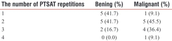
Table 3: Comparison number of percutaneous transcatheter sclerosant alcohol therapy repetitions between in patient with gynecologic surgery for benign and malignant conditions
The number of PTSAT repetitions Bening (%) Malignant (%)
1 5 (41.7) 1 (9.1)
2 5 (41.7) 5 (45.5)
3 2 (16.7) 4 (36.4)
4 0 (0.0) 1 (9.1)
PTSAT: Percutaneous transcatheter sclerosant alcohol therapy
Table 4: Comparison the number of percutaneous transcatheter sclerosant alcohol therapy repetitions
The number of PTSAT repetitions Mean±SD
Median (minimum‑maximum) P 1 2 3 Age 39±10 40 (25-53) 41.6±11.5 41 (17-55) 50.8±14.4 52 (33-69) 0.314 Gravida 2.5±2.1 2.5 (0-6) 1.2±1.2 1 (0-3) 2.5±1.4 2.5 (1-4) 0.161 Parity 2.3±2.2 2 (0-6) 1.1±1.11 (0-3) 2.5 (1-4)2.5±1.4 0.145
Pelvic cyst detection time (day) 8.3±5.4
9.5 (0-16) 408.7±910.230 (0-2920) 32.3±32.230 (0-90) 0.267 Preoperative Hb 9.7±0.7 9.7 (9-11) 9.5±1.2 9.4 (7.8-12) 8.9±0.9 9.2 (7.4-10) 0.323 Preoperative Hct 28.7±3.3 29.5 (23-32) 29.2±3.8 29 (23-36) 28.8±4.3 28 (25-37) 0.814 Postoperative Hb 9.8±1.2 9.7 (8.6-12) 9.7±1.05 9.5 (7.8-11) 9.1±0.7 9.2 (8-10) 0.400 Postoperative Hct 30.3±3.3 30.5 (27-36) 30 (24-38)30.7±3.9 29.5 (27-33)29.5±2.1 0.721 Hospital stay 9.5±7.4 7 (1-20) 7.5 (2-21)8.5±5.9 8 (1-23)9.5±7.6 0.954 Average follow-up time (month) 7±5.1
7 (2-12) 24.4±28.6 15 (0.25-96) 13.3±7.7 13.5 (5-24) 0.168
Cyst volume maximum 122.8±78.3
96 (32-234) 180.1±125.6 148 (80-515) 296±91.5 261.5 (210-417) 0.012 First drainage volume maximum 93.3±57.1
90 (30-160) 140 (50-250)145±62.4 200 (60-400)210±113.1 0.086
Post hoc dual comparison (P*) 1 versus 2 1 versus 3 2 versus 3
Cyst volume maximum 0.368 0.015 0.007
The patient who had four times sclerosing alcohol therapy did not participate in the evaluation because there was a patient. *Kruskal-Wallis, Mann-Whitney U test. Hb: Hemoglobin, Hct: Hematocrit, SD: Standard deviation, PTSAT: Percutaneous transcatheter sclerosant alcohol therapy
been used for sclerosing cysts.[1] The reported success rate of
sclerotherapy is 88%.[2] When sclerotherapy is unsuccessful
or undesirable, other alternatives, such as laparoscopic or open lymphocyst marsupializations, may be used.[15]
Postsurgical pelvic adhesions usually cause pelvic inclusion cysts, also known as pelvic inflammatory cysts.[10] Adnexa and
peritoneal fluid are caught in adhesions and form pseudocysts, which have complex appearances and lack true cystic walls. Large, complex pelvic inflammatory cysts can mimic ovarian tumors.[11] Ultrasound-guided aspiration can be used as the
treatment of choice for the minimally invasive management of pseudocysts; however, recurrence is common, and repeated aspirations are inescapable and cause considerable morbidity.[16] Laparotomy and laparoscopy can certainly
resolve the problem of recurrence, and laparoscopic surgeries have yielded good results for peritoneal and intra-abdominal adhesions.[17]
Intracystic ethanol injection may cause sclerosis and prevent a recurrence. Ethanol is a toxic agent, and when ethanol contacts with the cyst epithelial cells, it renders them nonviable. Simultaneously, ethanol fixes the nucleus and cytoplasm, and as such future cytological examination remains possible.[18]
Bean reported the successful sclerosis of symptomatic renal cysts by alcohol injection for the first time in 1981.[19] The
success of this technique has also been reported for ovarian cysts in postmenopausal women. Nonetheless, this procedure remains controversial, mainly due to concerns regarding the possibility of malignancy.[18] Alcohol sclerosis is easy, well
tolerated, readily available, and yields wonderful results. Thus, alcohol is the most widely used sclerosing agent for simple cysts of the abdomen and pelvis. Various agents, such as tetracycline, bleomycin, glucose, urea chlorhydrolactate, n-butyl cyanoacrylate with iodized oil, and phenol are also used for the sclerosis of cysts.[20] In the present study, we used
alcohol sclerosis for all cases.
Ditto et al. studied 111 patients with early-stage ovarian cancer in 2012. Overall postoperative lymphadenectomy-related complications (lymphocysts and lymphorrhea) were observed in 16 patients (14.4%). Ultrasound-guided transabdominal drainage was required in five patients with lymphocysts.[21] Cystic lesions in the pelvis may be
inaccessible to percutaneous biopsy and drainage. The bowel, urinary bladder, and subcutaneous fatty tissue may make it impossible to use an anterior approach, whereas the bony pelvis limits lateral and posterior access. Thus, a transvaginal route may be used, depending on the size, location, sonographic characteristics, and clinical presentation of the lesion.[22] However, we did not use a transvaginal route, and
all procedures were performed transabdominally.
A recent study reported that patients with pelvic cysts had increased incidences of lymphedema, lymphangitis, and deep-vein thrombosis. Furthermore, persistent lymphocysts were found to be an independent risk factor for lymphedema; and therefore, patients with persistent lymphocysts must be efficiently treated.[23]
In 2015, Hiramatsu et al. investigated 282 patients with lymphocysts, and found that infections occurred in 33 patients (12%), among whom two experienced an infection recurrence. Thus, 35-infected lymphocysts were analyzed in this study. The incidence of lymphocyst infection was not associated with the type of hysterectomy, the region of lymphadenectomy, or tumor origin.[7] The study included one
patient who had an abscess after PTSAT.
Six patients with clinical and ultrasonic benign characteristics of postsurgical pelvic peritoneal cysts underwent transvaginal ultrasound-guided drainage and instillation of 20–30 mL of ethanol into the cyst cavity. These treatments represent the first successful report in the English literature. In only one case, a 15 cm peritoneal cyst recurred 3 months after ethanol instillation, and for over 1 year, the procedure was reiterated with no subsequent recurrence. In the other five patients, no recurrences were observed.[24]
Conclusion and Limitation
Our study only included a few patients with pelvic cysts occurred after gynecologic surgery for malignant conditions, and as such, we do not know how to treat patients who are asymptomatic. In future, we need to determine the scenarios where percutaneous drainage with or without sclerosing agent or operation should be chosen, respectively.
PTSAT is minimally invasive and effective treatment for postoperative intrapelvic cysts in gynecologic patients. Especially symptomatic cysts after surgery should be treated with PTSAT. Since it is an effective, easily usable, and less complicated procedure from surgery. However, patients All postoperative pelvic cysts (n=77)
Asymptomatic (n=49)
(excluded) Symptomatic (n=28)(included)
Gynecologic surgery for malignant conditions (n=11) Gynecologic surgery for benign conditions (n=17)
who required recurrent PTSAT likely had pelvic cysts that occurred after gynecologic surgery for malignant conditions with large volume or had pelvic cysts that occurred after gynecologic surgery for benign conditions and underwent more than one surgery. Surgeons should treat postoperative pelvic cysts through percutaneous catheter drainage with or without sclerotherapy primarily and discuss the possibility of repeated PTSAT with patients, occurred after gynecologic surgery for malignant or benign conditions.
Acknowledgment
We would like to thank to www.editage.com for editing this paper.
Financial support and sponsorship
Nil.
Conflicts of interest
There are no conflicts of interest.
r
eferences1. Caliendo MV, Lee DE, Queiroz R, Waldman DL. Sclerotherapy with use of doxycycline after percutaneous drainage of postoperative lymphoceles. J Vasc Interv Radiol 2001;12:73-7.
2. Akhan O, Karcaaltincaba M, Ozmen MN, Akinci D, Karcaaltincaba D, Ayhan A. Percutaneous transcatheter ethanol sclerotherapy and catheter drainage of postoperative pelvic lymphoceles. Cardiovasc Intervent Radiol 2007;30:237-40.
3. Achouri A, Huchon C, Bats AS, Bensaid C, Nos C, Lécuru F. Complications of lymphadenectomy for gynecologic cancer. Eur J Surg Oncol 2013;39:81-6.
4. Braun WE, Banowsky LH, Straffon RA, Nakamoto S, Kiser WS, Popowniak KL, et al. Lymphocysts associated with renal transplantation. Report of 15 cases and review of the literature. Am J Med 1974;57:714-29.
5. Jensen SR, Voegeli DR, McDermott JC, Crummy AB, Turnipseed WD. Lymphatic disruption following abdominal aortic surgery. Cardiovasc Intervent Radiol 1986;9:199-201.
6. Kim HY, Kim JW, Kim SH, Kim YT, Kim JH. An analysis of the risk factors and management of lymphocele after pelvic lymphadenectomy in patients with gynecologic malignancies. Cancer Res Treat 2004;36:377-83.
7. Hiramatsu K, Kobayashi E, Ueda Y, Egawa-Takata T, Matsuzaki S, Kimura T, et al. Optimal timing for drainage of infected lymphocysts after lymphadenectomy for gynecologic cancer. Int J Gynecol Cancer 2015;25:337-41.
8. Suzuki M, Ohwada M, Sato I. Pelvic lymphocysts following
retroperitoneal lymphadenectomy: Retroperitoneal partial ‘‘no-closure’’ for ovarian and endometrial cancers. J Surg Oncol 1998;68:149-52. 9. Metcalf KS, Peel KR. Lymphocele. Ann R Coll Surg Engl
1993;75:387-92.
10. McFadden DE, Clement PB. Peritoneal inclusion cysts with mural mesothelial proliferation. A clinicopathological analysis of six cases. Am J Surg Pathol 1986;10:844-54.
11. Huang YH, Chao A, Chao AS, Wang CJ. Laparoscopic adhesiolysis and marsupialization of a rapidly progressing pelvic pseudocyst. Taiwan J Obstet Gynecol 2012;51:455-7.
12. Hsu TH, Gill IS, Grune MT, Andersen R, Eckhoff D, Goldfarb DA, et al. Laparoscopic lymphocelectomy: A multiinstitutional analysis. J Urol 2000;163:1096-8.
13. White M, Mueller PR, Ferrucci JT Jr, Butch RJ, Simeone JF, Neff CC,
et al. Percutaneous drainage of postoperative abdominal and pelvic
lymphoceles. AJR Am J Roentgenol 1985;145:1065-9.
14. Cohan RH, Saeed M, Schwab SJ, Perlmutt LM, Dunnick NR. Povidone-iodine sclerosis of pelvic lymphoceles: A prospective study. Urol Radiol 1988;10:203-6.
15. Zietek Z, Sulikowski T, Tejchman K, Sieńko J, Janeczek M, Iwan-Zietek I, et al. Lymphocele after kidney transplantation. Transplant Proc 2007;39:2744-7.
16. Chopra R, McVay C, Phillips E, Khalili TM. Laparoscopic lysis of adhesions. Am Surg 2003;69:966-8.
17. Swank DJ, Van Erp WF, Repelaer Van Driel OJ, Hop WC, Bonjer HJ, Jeekel H. A prospective analysis of predictive factors on the results of laparoscopic adhesiolysis in patients with chronic abdominal pain. Surg Laparosc Endosc Percutan Tech 2003;13:88-94.
18. Bret PM, Atri M, Guibaud L, Gillett P, Seymour RJ, Senterman MK. Ovarian cysts in postmenopausal women: Preliminary results with transvaginal alcohol sclerosis. Work in progress. Radiology 1992;184:661-3.
19. Bean WJ. Renal cysts: Treatment with alcohol. Radiology 1981;138:329-31.
20. Kim SH, Moon MW, Lee HJ, Sim JS, Kim SH, Ahn C. Renal cyst ablation with n-butyl cyanoacrylate and iodized oil in symptomatic patients with autosomal dominant polycystic kidney disease: Preliminary report. Radiology 2003;226:573-6.
21. Ditto A, Martinelli F, Reato C, Kusamura S, Solima E, Fontanelli R,
et al. Systematic para-aortic and pelvic lymphadenectomy in early
stage epithelial ovarian cancer: A prospective study. Ann Surg Oncol 2012;19:3849-55.
22. Lucey BC, Kuligowska E. Radiologic management of cysts in the abdomen and pelvis. AJR Am J Roentgenol 2006;186:562-73. 23. Kondo E, Tabata T, Shiozaki T, Motohashi T, Tanida K, Okugawa T,
et al. Large or persistent lymphocyst increases the risk of lymphedema,
lymphangitis, and deep vein thrombosis after retroperitoneal lymphadenectomy for gynecologic malignancy. Arch Gynecol Obstet 2013;288:587-93.
24. Lipitz S, Seidman DS, Schiff E, Achiron R, Menczer J. Treatment of pelvic peritoneal cysts by drainage and ethanol instillation. Obstet Gynecol 1995;86:297-9.