Objective: To demonstrate the diffusion-weighted imaging (DWI) findings of ectopic pregnancy (EP) and introduce the “ring of restriction” sign by discussing possible causes.
Methods: Between January 2014 and January 2017, patients with EP and examined by MRI on a 3T scanner were retrospectively evaluated. MRI and DWI findings were recorded.
Results: A total of 40 patients were diagnosed with EP at our university hospital, 8 of whom (20%) were evaluated by MRI and DWI. All of them were haemod-ynamically and clinically stable and could be imaged adequately. Locations were ovary (n = 3, 37.5%), tuba
(n = 2, 25%), Caesarean section scar (n = 2, 25%) and parauterine (n = 1, 12.5%). In all eight EPs, the gestational sac diameter was compatible with 7–9 weeks. With DWI, the gestational sac was seen as a thick-walled cyst-like structure. The thick wall showed diffusion restrictions in all patients (ring of restriction sign).
Conclusions: DWI through its cellular and molecular evidence may contribute diagnosis of EP.
Advances in knowledge: Being aware of the EP wall shows diffusion restriction (ring of restriction) avoids interpretation errors especially in appropriate clinical setting with no need for contrast material.
Cite this article as:
Durur-Karakaya A, Seker M, Durur-Subasi İ. Diffusion-weighted imaging in ectopic pregnancy: ring of restriction sign. Br J Radiol 2018; 91: 20170528.
ShORt COMMuniCAtiOn
Diffusion-weighted imaging in ectopic pregnancy: ring
of restriction sign
1AfAk DuRuR-kARAkAyA, MD, 1MehMet SekeR, MD and 2İRMAk DuRuR-SubASi, MD, PhD
1Department of Radiology, Faculty of Medicine, Istanbul Medipol University, Istanbul, Turkey
2University of Health Sciences, Diskapi Yildirim Beyazit Training and Research Hospital, Clinic of Radiology, Ankara, Turkey
Address correspondence to: Dr Afak Durur-Karakaya E-mail: afakdurur@ yahoo. com
intRODuCtiOn
Ectopic pregnancy (EP) is the implantation of a devel-oping blastocyst at a site other than the uterine endo-metrium. The classic triad of EP symptoms includes abdominal or pelvic pain, vaginal bleeding and a tender adnexal mass that classically emerges 6 to 8 weeks after the last menstruation.1 The rapid and correct diagnosis of this life-threatening condition is of great importance. EP’s main risk factors are a prior history of EP, pelvic inflammatory disease and gynaecologic and other abdom-inal surgeries, history of infertility, placenta previa, intra-uterine device, in vitro fertilization, congenital uterine anomalies, endometriosis, smoking, exposure to diethylstilboestrol.2 Its incidence is expected to be rising currently.
The majority of EP cases (95%) are tubal in location, occur-ring most commonly in the ampullary portion.2 The finding of an adnexal mass that is distinct from the ovary is a typical finding of a tubal EP with all imaging modalities.3 Other cases in which EP is encountered are the abdominal cavity, uterine cervix, ovaries, interstitium, intraligamentous,
heterotopic pregnancy, Caesarean section scar, pregnancy after supracervical hysterectomy.
Sensitive human chorionic gonadotropin measurement devices and the common use of ultrasonography (US) and laparoscopy have led to early and accurate diagnoses. US features in a classical tubal EP encompass a live extra-uterine pregnancy, tubal ring, heterogeneous adnexal mass, haema-tosalpinx, and a “ring of fire” on a colour Doppler.4 The ring of fire is accepted as a measure of the hyper-vascularity of the ectopic gestational sac; its primary value is in discovering an obscure EP, especially one covered by a haematoma.4 It has been suggested that the enhancing ring in adnexa on CT images was correlated with the “ring of fire” sign on US of tubal pregnancy, as the endometrium may contain a thin-walled decidual cyst or a pseudogestational sac.2,5 MRI find-ings of tubal EP include haematosalpinx, a haemorrhagic or heterogeneous mass, bloody ascites, tubal dilatation and wall enhancement.6 The wall of the gestational sac is characteris-tically iso-intense on both T1- and T2 weighted images.7 This thick wall is enhanced after the administration of contrast material.7,8 MRI offers the benefits of multiplanar imaging, a relatively unlimited field of view compared with US, lack of Received:
2 of 5 birpublications.org/bjr Br J Radiol;91:20170528 Table 1. Features of the patients
Patient number Age Predisposing factor Localization diameter (mm)Gestational sac Gestational age (week)
1 29 Smoking Right ovary 14 7
2 31 – Left tuba 15 7
3 36 Prior Caesarean section Caesarean section scar 18 7
4 32 Endometriosis, prior laparoscopy Right ovary 14 7
5 41 In vitro fertilization Left ovary 13 7
6 37 Prior Caesarean section, smoking Caesarean section scar 12 7
7 33 – Right tuba 22 8
8 30 Prior pelvic inflammatory disease Parauterine 29 9
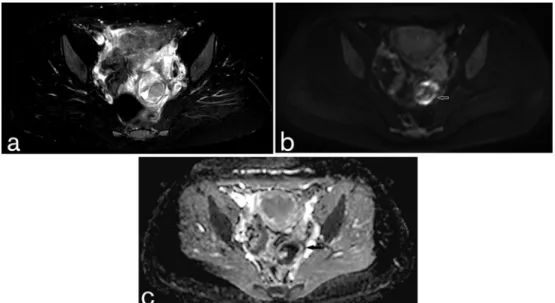
Figure 1. (a–c) EP at the site of Caesarean section scar (a) transverse fat-saturated T2 TSE, (b) DWI and (c) ADC map show a
thick-walled cyst with “ring of restriction” sign. ADC, apparent diffusion coefficient; DWI, diffusion-weighted imaging; EP, ectopic pregnancy; TSE, turbo spin echo.
ionizing radiation and greater soft-tissue contrast and tissue char-acterization options.9
In this paper, we present imaging findings of EP with special emphasis to “ring of restriction” sign on diffusion-weighted imaging (DWI) and discuss possible causes of “ring of restriction”.
MethODS AnD MAteRiAlS
After intuitional ethical committee approval gynaecology and radiology archives were evaluated retrospectively. Between January 2014 and January 2017, a total of 40 patients were diag-nosed with EP at our university hospital, 8 of whom (20%) were evaluated by both conventional MRI and DWI. Patients were examined by a 3-Tesla scanner (Achieva 3.0T, Philips Health-care, Cleveland, OH). The obtained sequences were coronal and transverse T2 turbo spin echo, transverse dual gradient echo, transverse single shot turbo spin echo with relatively short and
longer TE, transverse T2 spair, DWI. DWI parameters were TR 3400 ms, TE 60 ms, frequency and phase matrix 144 × 256, field of view 310–350 with rectangular FOV of 80%, EPI factor 109, sensitivity encoding factor (SENSE) 2, half scan factor 60%, b values of 50 and 800.
ReSultS
All of the 8 patients (all female, mean age 34; 29- to 41-year-old) were haemodynamically and clinically stable. Patients could not be imaged satisfactory by US due to limited field of view or non-specific imagind findings. MRI could reveal the patients adequately. Patients’ profile can be seen in Table 1. Locations of EP were ovary (n = 3, 37.5%), tubal (n = 2, 25%), Caesarean section scar (n = 2, 25%) and parauterine (n = 1, 12.5%). In all eight EPs, the gestational sac diameter was compatible with 7–9 weeks (mean 17 ± 5 mm). With DWI, the gestational sac was seen as a thick-walled cyst-like structure. The thick wall showed diffusion restrictions in all patients (Figures 1–3). We named this
Figure 2. (a–c) EP at parauterin region (a) transverse fat-saturated T2 TSE, (b) DWI and (c) ADC map show an enhancing
thick-walled cyst with “ring of restriction”. ADC, apparent diffusion coefficient; DWI, diffusion-weighted imaging; EP, ectopic preg-nancy; TSE, turbo spin echo.
Figure 3. (a–c) EP at the right tuba (a) coronal fat saturated T2 TSE, (b) DWI and (c) ADC map show a thick-walled cyst with “ring
of restriction”. ADC, apparent diffusion coefficient; DWI, diffusion-weighted imaging; EP, ectopic pregnancy; TSE, turbo spin echo.
relatively early finding as “ring of restriction” similar to “ring of fire” described by the other modalities.
Patients were treated conservatively. DiSCuSSiOn
DWI provides information at the molecular level of tissues and pathological processes. This method principally measures the diffusion of water molecules through the extracellular matrix of the tissue concerned. Dense cellular structures, aggressive tumours, high cellular proliferation rate, large nucleus size, intra-cellular macromolecules, high nucleus-cytoplasm ratio and the limited size of the extracellular matrix may all lead to diffusion restriction.10–12 Additionally, DWI requires no contrast agents. A study on conventional MRI and DWI findings of non-neoplastic
diseases of the Fallopian tube concluded that MRI is the method of choice for studying adnexal pelvic masses, while qualitative and quantitative functional imaging with DWI can be of help in the characterization of tubal diseases.13 However, this paper covers only tubal EP and gives entire diffusion restriction of the lesion. In our study group diffusion restriction has been encoun-tered at the wall of the EP. Additionally, in another study on find-ings and clinical utility of MRI for EP have mentioned ring or dote like hyperintensity on DWI similar to our results.14
The wall of the gestational sac contains trophoblasts. Higher cellularity of trophoblasts due to high ratio of cellular prolifera-tion, additional vascular-fibroblastic proliferaprolifera-tion, macromol-ecules within the cytoplasm and extracellular matrix may all contribute to the diffusion restriction of the lesion. The large,
4 of 5 birpublications.org/bjr Br J Radiol;91:20170528 Figure 4. Representative figure demonstrating ADC map findings of cystic adnexal masses. While EP shows “ring of restriction”, TOA displays diffusion restriction in their walls, septa and purulent content, CON in their cystic components and FOC shows no diffusion restriction. CON, cystic ovarian neoplasm; EP, ectopic pregnancy; FOC, functional ovarian cyst; TOA, tubo-ovarian abscess.
RefeRenCeS
1. Levine D. Ectopic pregnancy. Radiology 2007; 245: 385–97. doi: https:// doi. org/ 10. 1148/ radiol. 2452061031
2. Lin EP, Bhatt S, Dogra VS. Diagnostic clues to ectopic pregnancy. Radiographics 2008; 28: 1661–71. doi: https:// doi. org/ 10. 1148/ rg. 286085506
3. Parker RA, Yano M, Tai AW, Friedman M, Narra VR, Menias CO, et al. MR imaging findings of ectopic pregnancy: a pictorial review. Radiographics 2012; 32: 1445–60. doi: https:// doi. org/ 10. 1148/ rg. 325115153 4. Mausner Geffen E, Slywotzky C, Bennet G.
Pitfalls and tips in the diagnosis of ectopic pregnancy. Abdom Radiol 2017. doi: https:// doi. org/ 10. 1007/ s00261- 016- 1020-4 5. Pham H, Lin EC. Adnexal ring of ectopic
pregnancy detected by contrast-enhanced CT. Abdom Imaging 2007; 32: 56–8. doi: https:// doi. org/ 10. 1007/ s00261- 006- 9039-6
6. Kao LY, Scheinfeld MH, Chernyak V, Rozenblit AM, Oh S, Dym RJ. Beyond ultrasound: CT
and MRI of ectopic pregnancy. AJR Am J Roentgenol 2014; 202: 904–11. doi: https:// doi. org/ 10. 2214/ AJR. 13. 10644
7. Ascher SM, Edelman RR, Hesselink JR, Zlatkin MB. Benign conditions of the female pelvis. Clinical magnetic resonance imaging 2006; 3: 3036–43.
8. Ha HK, Jung JK, Kang SJ, Koong SE, Kim SJ, Kim JY, et al. MR imaging in the diagnosis of rare forms of ectopic pregnancy. AJR Am J Roentgenol 1993; 160: 1229–32. doi: https:// doi. org/ 10. 2214/ ajr. 160. 6. 8498223 9. Singh AK, Desai H, Novelline RA.
Emergency MRI of acute pelvic pain: MR protocol with no oral contrast. Emerg Radiol 2009; 16: 133–41. doi: https:// doi. org/ 10. 1007/ s10140- 008- 0748-8
10. Karaman A, Durur-Subasi I, Alper F, Durur-Karakaya A, Subasi M, Akgun M. Is it better to include necrosis in apparent diffusion coefficient (ADC) measurements? The necrosis/wall ADC ratio to differentiate
malignant and benign necrotic lung lesions: Preliminary results. J Magn Reson Imaging 2017; 46: 1001–6. doi: https:// doi. org/ 10. 1002/ jmri. 25649
11. Henzler T, Schmid-Bindert G, Schoenberg SO, Fink C. Diffusion and perfusion MRI of the lung and mediastinum. Eur J Radiol 2010; 76: 329–36. doi: https:// doi. org/ 10. 1016/ j. ejrad. 2010. 05. 005 12. Durur-Subasi I, Durur-Karakaya A, Karaman
A, Seker M, Demirci E, Alper F. Is the necrosis/ wall ADC ratio useful for the differentiation of benign and malignant breast lesions? Br J Radiol 2017; 90: 1073: 20160803: . doi: https:// doi. org/ 10. 1259/ bjr. 20160803
13. Foti PV, Ognibene N, Spadola S, Caltabiano R, Farina R, Palmucci S, et al. Non-neoplastic diseases of the fallopian tube: MR imaging with emphasis on diffusion-weighted imaging. Insights Imaging 2016; 7: 311–27. doi: https:// doi. org/ 10. 1007/ s13244- 016- 0484-7
round-to-oval-shaped nuclei were of different size, hyperchro-matic and featured prominent nuclear membranes. Glycos-aminoglycans are produced in cytotrophoblasts and released extracellularly.15 In addition, fibronectin is a high-molecular weight glycoprotein of the extracellular matrix of tropho-blastic activity.16 Fibronectin plays a major role in cell adhe-sion, growth, migration and differentiation.16 Another set of macromolecules, laminins, are high-molecular weight proteins of the extracellular matrix. Laminins affect cell differentiation, migration and adhesion.17
Up to the present study, all US, colour Doppler US, CT and MRI findings of EP have been primarily based on anatomical considerations and increased vascularity.1–6,14 However, DWI in our paper may show cellular, macromolecular and extracel-lular EP features, in addition to anatomical considerations and increased vascularity.
The differential diagnosis of an adnexal mass with peripheral enhancement includes tubo-ovarian abscess, cystic ovarian neoplasm and a functional ovarian cyst, including a corpus luteum cyst. Tubo-ovarian abscesses are expected to show diffusion restric-tion in their walls, septa and purulent content.13 Cystic ovarian neoplasms have been reported to have low ADC values in their cystic components.18 A functional ovarian cyst shows no diffu-sion restriction. Thus, DWI may serve as a discrimination tool (Figure 4).
As a conclusion, the EP gestational sac demonstrates diffusion restriction in its wall, which may be attributed to its cellular possessions, vascular-fibroblastic proliferation, macromol-ecules within the cytoplasm and the extracellular matrix. Studies conducted with larger population and supported by histopathology are required. DWI may be of value in helping to discriminate EP from other cystic lesions in this region.
14. Takahashi A, Takahama J, Marugami N, Takewa M, Itoh T, Kitano S, et al. Ectopic pregnancy: MRI findings and clinical utility. Abdom Imaging 2013; 38: 844–50. doi: https:// doi. org/ 10. 1007/ s00261- 012- 9969-0 15. Logothetou-Rella H, Kotoulas IG, Nesland JM, Kipiotis D, Abazis D. Early human trophoblast
cell cultures. A morphological and immunocytochemical study. Histol Histopathol 1989; 4: 367–74.
16. Pankov R, Yamada KM. Fibronectin at a glance. J Cell Sci 2002; 115(Pt 20): 3861–3. doi: https:// doi. org/ 10. 1242/ jcs. 00059 17. Timpl R, Rohde H, Robey PG, Rennard
SI, Foidart JM, Martin GR. Laminin--a
glycoprotein from basement membranes. J Biol Chem 1979; 254: 9933–7.
18. Namimoto T, Awai K, Nakaura T, Yanaga Y, Hirai T, Yamashita Y. Role of diffusion-weighted imaging in the diagnosis of gynecological diseases. Eur Radiol 2009; 19: 745–60. doi: https:// doi. org/ 10. 1007/ s00330- 008- 1185-5