Is ultrasonography useful in the diagnosis of the
polyneuropathy in diabetic patients?
Mehmet Agirman, MD1, 2)*, Ilker Yagci, MD2), Merve Akdeniz Leblebicier, MD2),
Demet Ozturk, MD2), Gulseren Derya Akyuz, MD2)
1) Department of Physical Medicine and Rehabilitation, Faculty of Medicine, Istanbul Medipol University: Medipol Universite Hastanesi, TEM Avrupa Otoyolu Göztepe Çıkışı, postal code: 34214, İstanbul, Turkey
2) Department of Physical Medicine and Rehabilitation, Faculty of Medicine, Marmara University, Turkey
Abstract. [Purpose] The aim of this study was to investigate the usefulness of ultrasonography for the diagnosis of polyneuropathy in diabetic patients by examination of the median and ulnar nerves. [Subjects and Methods] Sixty-three diabetic patients and fourteen controls were enrolled in the study. Nerve conduction studies were per-formed on both upper and lower limbs. Median and ulnar nerve cross-sectional areas were measured at the wrist and forearm levels in 140 hands by ultrasound. [Results] The median nerve cross-sectional area was increased at the hook of hamatum, pisiform bone, and radioulnar joint levels in patients with carpal tunnel syndrome. The ulnar nerve area at the medial epicondyle was significantly increased in the diabetic polyneuropathy (9.2 ± 1.6), diabetic polyneuropathy plus carpal tunnel syndrome (9.3 ± 1.4), and carpal tunnel syndrome (9.2 ± 1.9) groups compared with the control group (7.7 ± 1.1). In receiver operating characteristics analysis, the cutoff value of the ulnar nerve was 8.5 mm2 at ulnar epicondyle with 71.4% specificity and 70.4% sensitivity, corresponding to the highest
diag-nostic accuracy for diabetic polyneuropathy. [Conclusion] Ultrasonographic examination of the median and ulnar nerves can be an alternative or additional diagnostic modality for the evaluation of neuropathies in diabetic patients.
Key words: Polyneuropathy, Ultrasound, Diabetes mellitus
(This article was submitted Mar. 11, 2016, and was accepted May 31, 2016)
INTRODUCTION
Diabetic polyneuropathy (DPN) is the most common long-term complication in diabetes mellitus and affects more than 50% of patients1). DPN can affect any part of the nervous system (distal, proximal, large, small, motor, or autonomic fibers).
Accurate diagnosis of peripheral neuropathies is possible with the use of clinical and electrophysiological tests. Nerve con-duction studies (NCSs) are noninvasive, sensitive, and objective procedures2). In recent years, due to improved technology,
ultrasonography (US) has been regarded as an inexpensive, reproducible, and more comfortable technique, and it can be utilized as an alternative method for detecting neuropathies. However, reports on the use of US for polyneuropathy are limited. Most studies have investigated the peripheral nerves at the vulnerable sites, such as the median nerve at the carpal tunnel and the tibial nerve at the medial malleolus in diabetic patients3, 4). On the other hand, Hobson-Webb et al. argued that
the cross-sectional area (CSA) of the lower extremity peripheral nerves did not significantly differ between DPN patients and controls5). So far, there are no studies that have evaluated the upper extremity nerves at multiple sites in diabetic patients. In
this blinded, prospective study, we aimed to investigate the usefulness of US for the diagnosis of polyneuropathy in diabetic patients by investigating the median and ulnar nerves.
J. Phys. Ther. Sci. 28: 2620–2624, 2016
*Corresponding author. Mehmet Agirman (E-mail: [email protected])
©2016 The Society of Physical Therapy Science. Published by IPEC Inc.
This is an open-access article distributed under the terms of the Creative Commons Attribution Non-Commercial No Derivatives (by-nc-nd) License <http://creativecommons.org/licenses/by-nc-nd/4.0/>.
Original Article
The Journal of Physical Therapy Science
The Journal of Physical Therapy Science
SUBJECTS AND METHODS
One hundred seven patients (72 females and 35 males) who had type-2 diabetes mellitus according to the criteria of the American Diabetes Association6) and were referred to the Physical Medicine and Rehabilitation Electrodiagnosis
Labora-tory, Marmara University, were enrolled in the present study. Additionally, we recruited 14 healthy age-matched volunteers without diabetes or carpal tunnel syndrome (CTS) as controls. Patients who had thyroid or liver disorders, high blood urea nitrogen, any inflammatory disorder, or any malignancy, those who consumed alcohol, and those who had had a previous carpal tunnel operation were excluded from the study. Patients who complained of ulnar entrapment and those with isolated ulnar nerve entrapment based on electrodiagnostic tests were also excluded. The study protocol was approved by the local ethics committee, and all patients gave written informed consent before participating in the study.
Systemic and neuromuscular examinations (light touch, pinprick, position, temperature, vibration senses, knee and ankle stretch reflexes) were performed by a clinician who was blinded to the results of the nerve conduction studies (NCSs) and US. According to a literature search, no gold standard for the diagnosis of CTS and DPN has been established; therefore, we used clinical evaluation and electrophysiological findings for the diagnosis. For clinical DPN, a scoring system was used based on a previous study7), and the patients were defined as with or without DPN. A Katz hand diagram was used for clinical
CTS8, 9). NCSs were performed with conventional procedures and a standard device (Medtronic-Keypoint, Denmark, 2007)
by the same physician, who was blinded to the clinical assessment. All studies were performed at standard room temperature 25 °C. For the motor NCSs of the median, ulnar, peroneal, and tibial nerves and second lumbrical-interosseous muscle median-ulnar distal latency (LIMULD), differences were measured. The compound muscle action potential (CMAP) was recorded with surface electrodes from the abductor pollicis brevis muscle for the median nerve, the abductor digiti minimi muscle for the ulnar nerve, the abductor hallucis longus muscle for the tibial nerve, and the extensor digitorum brevis muscle for the peroneal nerve with a standard distance of 8 cm. For the sensory NCSs, the median, ulnar, and sural nerves were measured. Sensory NCSs were recorded using cup electrodes from the third digit antidromically with a standard distance of 14 cm for the median nerve, from the fifth digit with a standard distance of 12 cm for the ulnar nerve, from the first digit with a standard distance of 9 cm for the radial nerve, and from the midpoint of the calf 14 cm from the lateral malleolus for the sural nerve. For LIMULD, median and ulnar motor NCSs were recorded from the second lumbrical-interosseous muscle with a standard distance of 9 cm for the median and ulnar nerves. The average reference values for the nerve conduction velocities (NCVs) in our laboratory are 50 m/s for the median and ulnar motor NCVs and 40 m/s for the tibial and peroneal motor NCVs. Electrophysiological diagnoses of CTS and DPN were obtained according to the normative values established in our laboratory. Values of greater than 3.7 ms for median motor nerve distal latency and median sensory nerve velocity, slower than 50 m/s for the wrist-third digit segment, and more than 0.4 ms for LIMULD were considered to indicate median nerve demyelination10). Subjects with abnormal electrodiagnostic results in one or more nerves in the upper limbs, in addition to
the median nerve and one or more nerves in the lower limbs, were considered to have DPN. The electrophysiological testing was extended if a suspicious condition, such as ulnar neuropathy and radiculopathy, was present. Patients with any other neuropathic findings, such as isolated ulnar nerve entrapment or radiculopathy, were excluded from the statistical analysis. Finally, patients were classified into four groups according to electrophysiological diagnosis: normal, CTS, DPN, or CTS in the background of DPN (DPN+CTS).
Sonographic examinations were performed with a 6–18 MHz linear array probe (Mylab Esaote, Genoa, Italy) by a well-trained specialist who was blinded to the participant’s history and NCS results. All examinations were performed with the participants in a supine position on a table. Nerve CSAs were measured at various levels: the hook of hamate (MHAM), pisiform bone (MPSI), radioulnar joint (MRUE), distal one-third of the forearm (MDIS), proximal one-third of the forearm (MPROX), and medial epicondyle (MEPI) for the median nerve, and the radioulnar joint (URUE), distal one-third of the forearm (UDIS), proximal one-third of the forearm (UPROX), and medial epicondyle (UEPI) for the ulnar nerve. The trans-ducer was placed perpendicular to the nerves on the skin, and no additional pressure was applied other than its own weight. The CSA was measured by tracing the nerve just inside its hyperechoic rim, and three measurements were obtained with the probe repositioned. The average value was used for each level. A CSA of the median nerve greater than 10 mm2 at the
pisiform level was used for sonographic diagnosis of CTS in accordance with the literature and our laboratory values11, 12).
Statistical analyses were performed using the Statistical Package for the Social Sciences software (11.5.0.SPSS version 11.5.0). Categorical values (gender, duration of diabetes, and clinical scores) were analyzed using χ2 tests. To correlate
US findings with other parameters, the Pearson correlation coefficient and ANOVA tests were used. To assess the optimal possible cut-off values of sonographic nerve CSAs for the diagnosis of DPN, receiver operating characteristics (ROC) curves were used. P-values less than 0.05 were considered to indicate statistical significance.
RESULTS
A total of 140 hands (28 control, 24 normal, 34 CTS, 26 DPN, 28 DPN+CTS) of 63 patients and 14 control subjects were included in the analysis (mean ages of 47.6 ± 13.1, 43.1 ± 11.7, 50.3 ± 8.01, 51.4 ± 12.5, and 59.1 ± 5.04, respectively). In diabetic patients, eight hands were excluded based on ulnar entrapment findings, and six hands were excluded based on
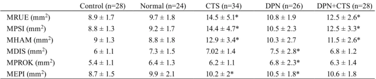
There was a significant increase in the median nerve CSA in the CTS and DPN+CTS groups when compared with the control, normal, and DPN groups at the radioulnar joint, pisiform, and hook of hamate hook levels (Table 1). The CSAs of the median nerve at the distal one-third of the forearm and proximal one-third of the forearm levels were higher in the DPN group compared with the control group (p=0.038 and p=0.035). There was a significant increase at the MEPI in the CTS, DPN, and DPN+CTS groups when compared with the control group (Table 1).
There was no statistically significant difference in the ulnar nerve at the radioulnar joint and distal one-third of the forearm levels between the groups. At the UPROX level, there was a significant increase in ulnar nerve CSA in the DPN group (6.5 ± 1.4) compared with the CTS group (5.5 ± 0.9). At the medial epicondyle level, the CSA of the ulnar nerve was significantly smaller in the control group (7.7 ± 1.1) compared with the CTS (9.2 ± 1.9), DPN (9.2 ± 1.6), and DPN+CTS (9.3 ± 1.4) groups (Table 2).
In the ROC analysis, cutoff values of 8.5 mm2 at the UEPI with 71.4% specificity and 70.4% sensitivity and 5.5 mm2 at
the UPROX, UDIS, and URUE levels with 25%, 32.1%, and 42.9% specificity and 75.9%, 74.1%, and 75.9% sensitivity, respectively, corresponding to the highest diagnostic accuracy for DPN in terms of ulnar nerve CSA (Table 3).
According to the above cutoff values, the most effective sonographic parameter for DPN diagnosis was the CSA at the UEPI. To compare the diagnostic utility of US with NCS, a new sonographic classification was applied to the groups (Table 4).
Table 1. Mean CSAs of the median nerve
Control (n=28) Normal (n=24) CTS (n=34) DPN (n=26) DPN+CTS (n=28) MRUE (mm2) 8.9 ± 1.7 9.7 ± 1.8 14.5 ± 5.1* 10.8 ± 1.9 12.5 ± 2.6* MPSI (mm2) 8.8 ± 1.3 9.2 ± 1.7 14.4 ± 4.7* 10.5 ± 2.3 12.5 ± 3.3* MHAM (mm2) 9 ± 1.3 8.8 ± 1.8 12.9 ± 3.4* 10.3 ± 2.7 11.5 ± 2.6* MDIS (mm2) 6 ± 1.1 7.3 ± 1.5 7.02 ± 1.4 7.5 ± 2.8* 6.8 ± 1.2 MPROK (mm2) 5.4 ± 1.1 6.4 ± 1.3 6.2 ± 1.1 6.8 ± 2.3* 6.3 ± 1.4 MEPI (mm2) 8.7 ± 1.5 9.9 ± 2.1 10.2 ± 2* 10.5 ± 1.8* 10.6 ± 1.8 CSAs: cross-sectional areas; MHAM: median nerve at the hamatum hook; MPSI: median nerve at the pisiform; MRUE: median nerve at the radioulnar joint; MDIS: median nerve at the distal one-third level of the forearm; MPROX: median nerve at the proximal one-third level of the forearm; MEPI: median nerve at the medial epicondyle.
*p<0.05
Table 2. Mean CSAs of the ulnar nerve
Control (n=28) Normal (n=24) CTS (n=34) DPN (n=26) DPN+CTS (n=28) URUE (mm2) 5.6 ± 0.8 5.5 ± 0.9 6.2 ± 1.6 6.4 ± 1.9 6.2 ± 1 UDIS (mm2) 5.9 ± 0.8 5.9 ± 1.1 5.6 ± 0.9 6.1 ± 1 6.1 ± 0.8 UPROX (mm2) 6.1 ± 1 5.9 ± 1.2 5.5 ± 0.9 6.5 ± 1.4* 6 ± 0.9 UEPI (mm2) 7.7 ± 1.1 8.1 ± 1.6 9.2 ± 1.9* 9.2 ± 1.6* 9.3 ± 1.4* Post hoc analysis: p=0.014 for DPN vs. CTS at the UPROX; p=0.01, p=0.003, and p=0.002 for DPN vs. control, CTS vs. control, and DPN+CTS vs. control at the UEPI, respectively.
CSAs: cross-sectional areas; URUE: ulnar nerve at the radioulnar joint; UDIS: ulnar nerve at the distal one-third level of the forearm; UPROX: ulnar nerve at the proximal one-third level of the forearm; UEPI: ulnar nerve at the medial epicondyle.
*p<0.05
Table 3. Cutoff values for the UEPI for the diagnosis of DPN Cutoff value,
mm2 Sensitivity Specificity Area under the ROC curve UEPI 8.5 70.4 71.4 0.787
UPROX 5.5 75.9 25 0.530
UDIS 5.5 74.1 32.1 0.554 URUE 5.5 75.9 42.9 0.649 UEPI: ulnar nerve at the medial epicondyle; UPROX: ulnar nerve at the proximal one-third level of the forearm; UDIS: ulnar nerve at the distal one-one-third level of the forearm; URUE: ulnar nerve at the radioulnar joint
DISCUSSION
In this study, the CSAs of the ulnar nerve were larger at epicondyle level in the patients with diabetic polyneuropathy. Additionally, median nerve CSAs were larger in diabetic patients with carpal tunnel syndrome at wrist levels compared with those of the healthy controls.
Diabetic distal symmetrical sensorimotor polyneuropathy is the most common form of neuropathy in diabetic patients and can cause many serious complications such as diabetic foot, Charcot joint, and flexion contractures, which reduce quality of life13). Upper limb symptoms are often associated with proximal spreading of the DPN; however, these symptoms can be
observed in other or adjunctive entrapment neuropathy components such as CTS and ulnar neuropathy at the elbow.
CTS is the most common entrapment neuropathy in diabetic patients14). A previous study showed that the LIMULD
difference can help to detect CTS in the background of DPN in NCSs10). Conversely, US has been used for the diagnosis of
entrapment neuropathies such as CTS and DPN11, 15–17). Wong et al.15) demonstrated that a median nerve CSA greater than
9.8 mm2 supports a diagnosis of CTS with 89% sensitivity and 83% specificity. Additionally, El-Miedany et al.11) suggested
that a median nerve CSA of 10 mm2 confirms a diagnosis of CTS with 97.9% sensitivity and 100% specificity. Watanabe et
al.3, 4) showed that the median nerve (at the wrist) and tibial nerve (at the ankle) CSAs were increased in diabetic patients
when compared with their control group and that CSAs were negatively correlated with motor nerve conduction velocity. In our study, we found a significant increase in the median nerve CSA at the epicondyle level in the CTS (10.1 ± 2), DPN (10.2 ± 2), and DPN+CTS (10.6 ± 1.7) groups when compared with the control group (8.7 ± 1.5). Additionally, the median nerve CSA was greater at wrist levels than in the control and DPN groups if there was CTS with or without DPN. These results support the finding that the median nerve enlarges at the wrist in carpal tunnel syndrome, as reported in previous stud-ies. In another study, Chen et al.17) compared median nerve CSAs among diabetic patients with CTS and DPN+CTS. They
found greater CSAs at the wrist and in the carpal tunnel in the latter group. However, median nerve CSAs are not helpful in the diagnosis of polyneuropathy, and they do not discriminate CTS in the background of DPN according to our results.
The US features of ulnar nerve CSAs have not been systematically studied to a sufficient extent in diabetic patients. In a study of 98 patients with DPN and 90 controls, the median and ulnar nerve CSAs were found to be increased in demyelinating neuropathies18). Arumugam et al.19) compared patients with DPN with normal controls and found that ulnar nerve CSAs
were significantly increased (p=0.016) at the level of mid-forearm. The ulnar nerve may become enlarged at the elbow due to structures that pressure the nerve (ganglia, anconeus epitrochlearis muscles, etc.), and this can lead to nerve entrapment. Furthermore, in addition to entrapment, peripheral nerve thickening can occur as a result of thickening of capillary walls in the endoneurium and increased intracellular hydration due to increased sorbitol in diabetic patients20, 21). To minimize the
ef-fect of local nerve entrapment, clinical and electrophysiological exclusion was performed. In the current study, we measured the ulnar nerve from four regions. We found increased CSAs at the epicondyle level in the DPN and DPN+CTS groups and as an interesting finding in CTS patients with diabetes mellitus when compared with the control group. These results showing that electrophysiological CTS can be a part of the polyneuropathy in diabetic patients should be interpreted carefully. Kim et al.22) also found increased CSAs of the median nerve in DPN-only patients compared with those of healthy controls at the
wrist. Therefore, ultrasonographic CTS diagnoses are inconsistent with electrophysiological results in patients with DPN. However, they did not examine the ulnar nerve. The current diagnostic criteria for CTS, DPN, and DPN+CTS should be reconsidered. It appears that, clinical, electrophysiological, and ultrasonographic diagnoses should be confirmed with a new gold standard technique by further studies with a larger population.
The cutoff values derived from ROC analysis and an ulnar CSA greater than 8.5 mm2 at the epicondyle level supported
the diagnosis of DPN with 70.4% sensitivity and 71.4% specificity in our study. Ulnar nerve entrapment is the second most common entrapment neuropathy in diabetic patients23). The usefulness of US in diagnosis of ulnar nerve entrapment was not
examined in this study; thus, these cutoff values patients who had clinical or electrodiagnostic ulnar nerve entrapment were excluded. In view of this cutoff and the literature, a sonographic classification can be done according to ulnar nerve CSAs at the medial epicondyle and median nerve CSAs at the wrist (Table 4).
According to our results, four normal subjects were diagnosed as CTS and five normal subjects were diagnosed as DPN by using US. Two possible reasons for these results are as follows: these patients might have had CTS or DPN but were electrophysiologically normal, or US might have exhibited some degree of false-positive results.
Table 4. Ultrasonographic classification
Normal: MPSI<10 mm2 and bilateral UEPI<8.5 mm2 CTS: MPSI>10 mm2 and bilateral UEPI<8.5 mm2 DPN: MPSI<10 mm2 and bilateral UEPI>8.5 mm2 DPN+CTS: MPSI>10 mm2 and bilateral UEPI>8.5 mm2
CTS: carpal tunnel syndrome; DPN: diabetic polyneuropathy; MPSI: median nerve at the pisiform; UEPI: ulnarnerve at the medial epi-condyle
The most important limitation of our study was that focal entrapment neuropathies might have been confused with DPN because they cannot be distinguished accurately. This bias was minimized by use of blinded researchers and standardized protocols.
In conclusion, neither US nor NCSs are a gold standard for the evaluation of peripheral nerves. NCS reflects nerve func-tion better than US and provides valuable informafunc-tion for differential diagnosis. US is a more comfortable technique for the patient and provides a dynamic evaluation. Increased CSAs of the ulnar nerve at the medial epicondyle level can be useful for the diagnosis of DPN, particularly for screening. In general, the relationship between generalized neuropathy and focal entrapment neuropathies is complex and difficult to clarify. We suggest that the combination of US and NCS may be used for the diagnosis of neuropathies in diabetic patients.
REFERENCES
1) Boulton AJ: Diabetic neuropathy: classification, measurement and treatment. Curr Opin Endocrinol Diabetes Obes, 2007, 14: 141–145. [Medline] [CrossRef] 2) Toyokura M: F-wave-duration in diabetic polyneuropathy. Muscle Nerve, 1998, 21: 246–249. [Medline] [CrossRef]
3) Watanabe T, Ito H, Morita A, et al.: Sonographic evaluation of the median nerve in diabetic patients: comparison with nerve conduction studies. J Ultrasound Med, 2009, 28: 727–734. [Medline]
4) Watanabe T, Ito H, Sekine A, et al.: Sonographic evaluation of the peripheral nerve in diabetic patients: the relationship between nerve conduction studies, echo intensity, and cross-sectional area. J Ultrasound Med, 2010, 29: 697–708. [Medline]
5) Hobson-Webb LD, Massey JM, Juel VC: Nerve ultrasound in diabetic polyneuropathy: correlation with clinical characteristics and electrodiagnostic testing. Muscle Nerve, 2013, 47: 379–384. [Medline] [CrossRef]
6) American Diabetes Association: Standards of medical care in diabetes–2010. Diabetes Care, 2010, 33: 11–61. [CrossRef]
7) Perkins BA, Olaleye D, Bril V: Carpal tunnel syndrome in patients with diabetic polyneuropathy. Diabetes Care, 2002, 25: 565–569. [Medline] [CrossRef] 8) Katz JN, Stirrat CR, Larson MG, et al.: A self-administered hand symptom diagram for the diagnosis and epidemiologic study of carpal tunnel syndrome. J
Rheumatol, 1990, 17: 1495–1498. [Medline]
9) Oken O, Catal SA, Ceceli E: An useful and simple method in diagnosis of carpal tunnel syndrome: hand diagram. Rheumatism, 2003, 18: 23–26.
10) Yagci I, Gunduz OH, Sancak S, et al.: Comparative electrophysiological techniques in the diagnosis of carpal tunnel syndrome in patients with diabetic poly-neuropathy. Diabetes Res Clin Pract, 2010, 88: 157–163. [Medline] [CrossRef]
11) El Miedany YM, Aty SA, Ashour S: Ultrasonography versus nerve conduction study in patients with carpal tunnel syndrome: substantive or complementary tests? Rheumatology (Oxford), 2004, 43: 887–895. [Medline] [CrossRef]
12) Bayındır O, Yagci I, Akyuz GD: Comparison of ultrasonography and electrophysiology in the diagnosis of carpal tunnel syndrome. Turk J Phys Med Rehab, 2011, 57: 334.
13) Terzi M, Cengiz N, Onar MK: Diabetic neuropathy. J Exp Clin Med, 2004, 21: 39–49.
14) Tracy JA, Dyck PJ, Harper CM: Hand symptomatology in diabetes usually due to mononeuropathy, not polyneuropathy. Ann Neurol, 2005, 58: 36. 15) Wong SM, Griffith JF, Hui AC, et al.: Discriminatory sonographic criteria for the diagnosis of carpal tunnel syndrome. Arthritis Rheum, 2002, 46: 1914–1921.
[Medline] [CrossRef]
16) Klauser AS, Halpern EJ, De Zordo T, et al.: Carpal tunnel syndrome assessment with US: value of additional cross-sectional area measurements of the median nerve in patients versus healthy volunteers. Radiology, 2009, 250: 171–177. [Medline] [CrossRef]
17) Chen SF, Lu CH, Huang CR, et al.: Ultrasonographic median nerve cross-section areas measured by 8-point “inching test” for idiopathic carpal tunnel syn-drome: a correlation of nerve conduction study severity and duration of clinical symptoms. BMC Med Imaging, 2011, 11: 22. [Medline] [CrossRef] 18) Zaidman CM, Al-Lozi M, Pestronk A: Peripheral nerve size in normals and patients with polyneuropathy: an ultrasound study. Muscle Nerve, 2009, 40:
960–966. [Medline] [CrossRef]
19) Arumugam T, Razali SN, Vethakkan SR, et al.: Relationship between ultrasonographic nerve morphology and severity of diabetic sensorimotor polyneuropa-thy. Eur J Neurol, 2016, 23: 354–360. [Medline] [CrossRef]
20) Zochodne DW: Diabetic polyneuropathy: an update. Curr Opin Neurol, 2008, 21: 527–533. [Medline] [CrossRef]
21) Kang S, Kim SH, Yang SN, et al.: Sonographic features of peripheral nerves at multiple sites in patients with diabetic polyneuropathy. J Diabetes Complica-tions, 2016, 30: 518–523. [Medline] [CrossRef]
22) Kim LN, Kwon HK, Moon HI, et al.: Sonography of the median nerve in carpal tunnel syndrome with diabetic neuropathy. Am J Phys Med Rehabil, 2014, 93: 897–907. [Medline] [CrossRef]