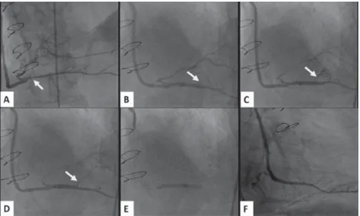
A sixty-four year old man with a previous history of CABG was referred for coronary angiography that revealed a critical stenosis of the right posterior descending artery at the anastomosis point of the saphenous vein graft (Figure A). Percutaneous intervention was initiated by engagement of the saphenous graft ostium with a 6F JR guiding catheter, followed by crossing the lesion with a hydrophilic guidewire. A drug-eluting stent, 2.25 x 23 mm in size, (Xience, Abbott Vascular, USA) was implanted at 14 atm pressure. Control angiography revealed a marked Ellis type IV coronary perforation with leakage of blood into the pericardial cavity (Figure B). Subsequently, the patient deteriorated hemodynamically with severe chest pain and hypotension. Echocardiography showed a marked pericardial effusion along with right ventricular collapse. Protamine sulfate was used to reverse anticoagulation followed by administration of IV fl uids and emergency pericardiocentesis. A 2.5 x 15 mm balloon (Sprinter Legend, Medtronic, USA) was advanced for 10 minutes of prolonged infl ation, which failed to heal the perforation (Figure C). A 2.80 x 19 mm covered stent (Graftmaster, Abbott Vascular, USA) was implanted at the level of the perforation. Control angiography showed persistence of the perforation with ongoing pulsatile fl ow (Figure D). A second covered stent, 2.80 x 16 mm in size, (Graftmaster, Abbott Vascular, USA) was implanted inside the fi rst stent and post-dilated for 5 minutes (Figure E). Fortunately, the perforation healed without leakage shown by control angiography, with good distal fl ow (Figure F). The patient was safely taken to the coronary care unit for hemodynamic stabilization and uneventfully discharged the next day after demonstration of no pericardial effusion on control echocardiography. This case appears to offer solutions to a life-threatening coronary perforation in the catheterization laboratory with its unique feature of implantation of two covered-stents.
İnatçı Koroner Perforasyon Olgusunda İç İçe Kaplı Stent Kullanımı
Oğuz Karaca1, Hacı Murat Güneş1, Beytullah Çakal1, İrfan Barutçu1, Muhsin Türkmen2 1 Medipol University Faculty of Medicine, Department of Cardiology, İstanbul2 Kartal Koşuyolu High Specialization Training and Research Hospital, Clinic of Cardiology, İstanbul, Turkey
Use of A Double-Covered Stent-in-Stent Technique
to Manage Persistent Coronary Perforation
Oğuz Karaca
E-mail: [email protected] Submitted: 09.04.2015 Accepted: 20.04.2015
@ Copyright 2015 by Koşuyolu Heart Journal. Available on-line at
www.kosuyoluheartjournal.com
155
Koşuyolu Heart Journal 2015;18(3):155 • DOI: 10.5578/khj.9946 ● ORIGINAL IMAGE
Correspondence
Figure 1. (A) Angiographic image demonstrating a critical stenosis of the right posterior descending artery at the anastomosis
point of the saphenous vein graft. (B) Ellis type IV coronary perforation with leakage of blood into the pericardial cavity. (C) Failure of prolonged balloon infl ation to heal coronary perforation. (D) Control angiographic image showing persistence of coronary perforation despite implantation of a covered stent. (E) Implantation of a second covered stent inside the fi rst one and post-dilatation for 5 minutes. (F) Control angiographic image demonstrating the disappearance of the perforation without leakage and with good distal fl ow.
View publication stats View publication stats