CdSe/CdMnS Nanoplatelets with Bilayer Core and Magnetically
Doped Shell Exhibit Switchable Excitonic Circular Polarization:
Implications for Lasers and Light-Emitting Diodes
Arman Najafi,
∥Steven Tarasek,
∥Savas Delikanli, Peiyao Zhang, Tenzin Norden, Sushant Shendre,
Manoj Sharma, Arinjoy Bhattacharya, Nima Taghipour, James Pientka, Hilmi Volkan Demir,
Athos Petrou, and Tim Thomay
*
Cite This:ACS Appl. Nano Mater. 2020, 3, 3151−3156 Read Online
ACCESS
Metrics & More Article Recommendations*
sı Supporting InformationABSTRACT: We utilized time-resolved photoluminescence (TRPL) spectroscopy to study the excitonic circular polarization (PX) from CdSe/CdMnS core/shell nanoplatelets (NPLs) with a bilayer core. This allows an extensive study of the emission dynamics as a function of magnetic field, temperature, doping concentration, and excitation wavelength. In the presence of an external magnetic field, pulsed excitation below the shell gap results in near-zero excitonic circular polarization PX at all time delays. In contrast, pulsed excitation with photon energy larger than the shell gap results in a rapid (100 ps) buildup of the excitonic circular polarization which subsequently remains constant at a level of up to 40%. We propose a model to describe the dynamics which takes into account the exchange
interaction between carrier and magnetic ion (Mn) spins. The studied system exhibits a fast switchable excitonic circular polarization, implying possible applications in lasers and light emitting diodes.
KEYWORDS: nanoplatelets, quasi-2D, time-resolved photoluminescence, spd exchange interaction, magneto-optical switch
S
emiconductor nanoplatelets (NPLs) have been continuing to garner attention due to their narrow emission spectra,1−5 potential for tunable emission,4,6,7 and atomically controlled thicknesses leading to quasi-2D structures8−11with very high quantum efficiency (QE).2,9,12,13NPLs have emerged as a promising platform for optoelectronic devices such as light emitting diodes (LEDs)4,14−17and lasers.3,7,9,14,18Recently, it has been demonstrated that the presence of charged excitons in NPLs19,20or doping with magnetic ions such as Mn either in the core or in the shell gives these structures novel magneto-optical properties.21−25A magneto-optical study of NPLs is a particularly powerful tool for the exploration and under-standing of their properties. This is realized by using the Mn ions as a“tracer”, which allows the study of the interaction of their spins with spins of the carriers. This provides the necessary information on the static and dynamic behaviors of the system, which is required for the design and performance evaluation of NPL-based devices.In this work, we study a magnetically doped CdSe/CdMnS core/shell NPL system with a significantly thinner core of only 2 monolayers (bilayer) compared to our previous works.21,22 In the presence of an external magnetic field, these NPLs exhibit circularly polarized emission that is dependent on the excitation wavelength. This gives the ability to switch between
low and high values of circularly polarized excitonic luminescence, which is a crucial feature for spin polarized lasers and LEDs.26Previous measurements on similar magnetic core/shell structures with thicker cores showed that an externally applied magnetic field causes a net circular polarization in the emission and that the degree of circular polarization saturates for magnetic fields above 3 T.21 The analysis of the photoluminescence (PL) from the thicker core NPLs showed the presence of two distinct spectral features, one corresponding to the excitonic emission of the system (observed in both PL and absorption) and a second feature associated with defect interface states that contributes to the asymmetry of the emission. The maximum in circular polarization occurred at the energy of the PL component associated with the interface states.22 However, we point out that in the bilayer core NPLs studied in this work, the circular polarization has a maximum at the exciton energy, and we Received: February 7, 2020
Accepted: March 30, 2020
Published: March 30, 2020
Letter
www.acsanm.org
Downloaded via BILKENT UNIV on February 12, 2021 at 06:24:40 (UTC).
show that the circular polarization of this recombination channel can be controlled by varying the excitation photon energy. We investigated the time evolution of the excitonic circular polarization PXin the presence of an external magnetic
field at T = 7 K. For excitation above the CdMnS shell gap, we observe strong excitonic circular polarization (PX = 40%); in
contrast, direct CdSe core excitation results in near-zero excitonic circular polarization (PX≈ 0%). For above shell gap
excitation, a fast (100 ps) switch from near-zero polarization to 40% is observed.
In Figure 1c, we plot the zero magnetic field transmission spectrum (magenta) from the high Mn concentration NPLs recorded at T = 7 K along with the PL at early times (green line, atΔt = 0 ns) and late times (cyan line, at Δt = 3.5 ns) excited using 400 nm pulses. The two features at 2.15 and 2.30 eV of the transmission spectrum are due to the e1hh1and e1lh1
heavy hole and light hole excitonic transitions, respectively.23 The e1hh1(e1lh1) exciton is formed by an electron occupying
thefirst confinement sub-band e1and a hole in thefirst heavy
hole confinement sub-band hh1 (light hole confinement
sub-band lh1). For the PL at early times, the emission peak
coincides with the e1hh1 exciton, whereas at later times, the
emission mostly contains components with energies below e1hh1; these are attributed to long-lived emission associated
with interface states. This separation in time is possible due to the different lifetimes of the excitonic and interface emissions.22
InFigures 1d and1e, we plot the left circularly polarizedσ+ (red line) and right circularly polarized σ− (black line) components of the time-integrated (0−5 ns) PL at B = 4 T which are excited using 400 and 515 nm pulses, respectively. The blue line in thesefigures represents the resulting circular polarization. Even though the PL spectra inFigure 1d and 1e look similar, the corresponding excitonic circular polarization, identified by the vertical green arrows, is quite distinct. Under 400 nm excitation (Figure 1d), the polarization has a maximum of 40% centered at the energy of e1hh1; however,
under 515 nm excitation (Figure 1e), the excitonic circular
polarization PX is vanishing and the maximum of circular
polarization of 30% coincides with the interface luminescence feature.
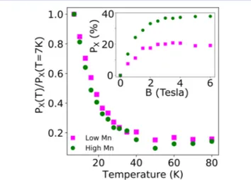
Figure 2 shows the magnetic field (inset, T = 7 K) and temperature (main figure, B = 4 T) dependence of the
excitonic circular polarization PXunder 400 nm excitation. The
PX in the inset increases monotonically with B and saturates around B = 3 T at 40 and 20% for the high Mn and low Mn doped NPLs, respectively. The ratio PX(T)/PX(7 K) at B = 4 T plotted in the main figure shows a significant decrease with increasing T. The B and T dependence of the excitonic circular polarization PX demonstrates that the shell exhibits Brillouin
Paramagnetic behavior. This leads to the observed PX, which is due to band splittings, where the splittings are proportional to the shell magnetization. The results of Figure 2 provide a strong indication on the existence of sp-d exchange interaction Figure 1.(a) Schematic diagram of the CdSe/CdMnS core/shell NPL samples used in this study. (b) TEM image of a NPL sample. (c) Magenta line depicts the optical transmission spectrum from the high Mn sample; green line: time slice of the TRPL spectrum at early times; cyan line: at later times. (d, e) Time-integrated PL analyzed asσ+(red) andσ−(black) and circular polarization (blue) as a function of photon energy at B = 4 T
with excitation under (d) 400 nm and (e) 515 nm linearly polarized pulsed laser.
Figure 2.Main: Excitonic circular polarization ratios PX(T)/PX(7 K)
plotted as a function of temperature at B = 4 T and with 400 nm excitation. Inset: Excitonic circular polarization PXas a function of
magneticfield with 400 nm excitation. Green circles and magenta squares refer to high and low Mn concentration samples, respectively.
between the spins of the electrons (s-symmetry) and holes (p-symmetry) and the spins of the Mn ions (in the d orbital) in the CdMnS shell.27
InFigure 3a (and3b) we plot the TRPL intensity sum IPL=
I+ + I−of the σ+ and σ− components for the e1hh1excitonic
feature, indicated by vertical arrows inFigure 1d and1e, under 400 nm (515 nm) pulsed excitation for the high Mn concentration sample. A single exponential least-square fit (not shown) yields comparable excitonic lifetimes of 0.6 ns for excitation at 400 nm and 0.5 ns for 515 nm excitation at B = 4 T. The excitonic circular polarization PXat a specific time delay
is indicated by the color below the IPLcurve (black line), where the area underneath for a particular time slice is proportional to the total number ofσ+andσ−emitted photons.Figure 3a and3b shows that the PXdepends critically on the excitation
photon energy. Under 515 nm excitation (Figure 3b), the polarization is low at all times. In contrast, using 400 nm excitation, PXincreases sharply during the rise time (100 ps) of IPL. During this time interval, a significant number of photons
with near-zero circular polarization is emitted. ForΔt > 100 ps, PXreaches a value of 40% and remains approximately constant.
Though we show this for only a single magneticfield (B = 4 T) and the high Mn concentration sample, we note that this behavior is exhibited for all magneticfield values greater than 3 T as well as for the low Mn concentration sample.
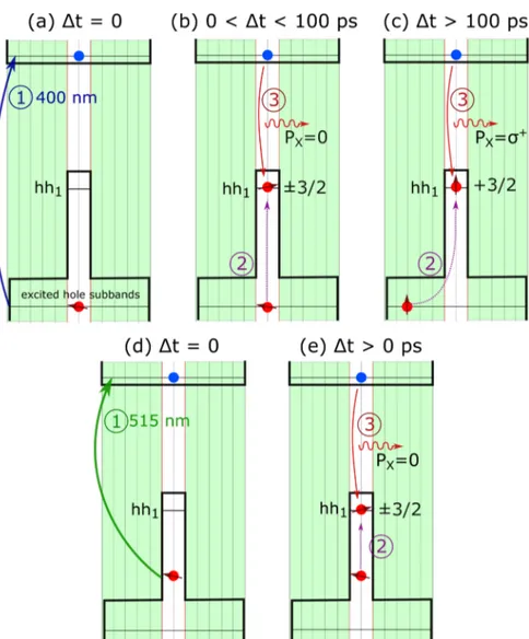
In this section, we discuss a possible mechanism that is responsible for the observed excitonic circular polarization dependence on excitation photon energy. The electron and hole wave functions in these structures play a crucial role in our model. We show calculated wave functions for the e1, hh1, and hh2states in the Supporting Information (Figure S2 and Figure S3). In II−VI diluted magnetic semiconductors, the exchange interaction between the hole and Mn spins is several times larger than the interaction between the electron and Mn spins,27allowing us to focus on the influence of the external magnetic field on the holes. The five panels in Figure 4 illustrate the dynamics of the excitonic circular polarization PX
according to the proposed model. Panels (a), (b), and (c) refer to an excitation above the core and shell band gaps (400 nm);
panels (d) and (e) refer to 515 nm excitation which has a photon energy below the shell but above the core band gap.
In panel (a), we indicate the photogeneration of electron− hole pairs at Δt = 0 by the incoming pulse. These holes populate excited hole sub-bands that have wave functions which extend throughout the magnetic CdMnS shell (Figure S2). In panel (b), we show the processes for 0 <Δt < 100 ps; during this time holes relax from their excited states to the hh1
ground state (step 2). During the first 100 ps, even though holes are delocalized, their spins do not have time to align antiferromagnetically with Mn spins that are aligned by the external magneticfield.28,29 Therefore, the photons from the e1hh1recombination (step 3) have near-zero PX. In panel (c), we show the processes forΔt > 100 ps. During this time, the delocalized holes which had enough time to align their spins antiferromagnetically with the Mn spins, are predominately in the +3/
2state. We assume that during nonradiative relaxation
of the holes to the hh1ground state, their spin state is at least
partially conserved. Now that the majority of holes are in the +3/2state, the emitted photons (step 3) are circularly polarized
asσ+due to the fact that we have more−1/
2to +3/2than +1/2
to −3/2 recombinations. We note that this picture is in
agreement with the data shown inFigure 3a.
In panel (d), we show the electron−hole pair generation under 515 nm excitation. In this case, the holes are strongly confined in the core and interact with the Mn only via the tail of the hole wave function that penetrates into the magnetic shell (Figure S3). In panel (e), we show the hole relaxation to the hh1 state (step 2). The difference in population between the +3/2 and −3/2 states is small; therefore, the emitted
photons (step 3) have a near-zero PX in agreement with the results ofFigure 3b.
We investigated the time evolution of the circular polar-ization PXfor the e1hh1exciton from CdSe/CdMnS core/shell
NPLs with bilayer cores doped with Mn in the shell. In the presence of a magneticfield, the excitonic circular polarization PXis strongly dependent on the excitation photon energy. For photons with an energy smaller than the CdMnS shell gap but bigger than the CdSe core gap, the excitonic circular polarization PXremains low for all times. However, excitonic
circular polarization PXusing an excitation with energy above the CdMnS shell gap results in 40% circular polarization of the e1hh1 excitonic ground state emission. In our time-resolved experiments, PXbuilds up from a low value following excitation
to a constant value within 100 ps. We developed a model that explains this behavior which takes into account the time required by the hole spins in the shell to align with the Mn spins. Such a system demonstrates the ability to switch the excitonic circular polarization depending on the excitation wavelength; this has potential applications in lasers and LEDs. The switching rate would be limited by the lifetime of the excitonic ground state which is in the order of a nanosecond. However, in utilizing the intrinsic switching time of 100 ps of the circular polarization under above shell gap excitation, a switching rate of tens of GHz would be feasible which could have applications in thefields of circularly polarized filters and polarizers.30,31
■
METHODSA schematic of the NPLs in this study is shown inFigure 1a. They are comprised of an undoped CdSe core (2 ML) surrounded by a Mn doped CdS shell (10 ML) with two different Mn concentrations, 3% (high) and 1% (low). The low temperature band gaps of CdSe core Figure 3. TRPL intensity sum IPL = I+ + I− for the high Mn
concentration sample; B = 4 T; T = 7 K (a) under 400 nm pulsed excitation and (b) under 515 nm pulsed excitation. The corresponding color scale under the plots represents the circular polarization of the e1hh1exciton (PX) for each delay time.
and CdMnS shell are 1.84 and 2.56 eV, respectively.23The samples were synthesized by slightly modifying a previously published procedure.23,32A TEM image of the Mn doped core/shell NPLs is presented inFigure 1b, showing lateral sizes of the NPLs on the order of 35 nm. Analysis of the small angle electron diffraction (SAED) pattern presented inFigure S1in the Supporting Information shows that our Mn doped core/shell NPLs have the zinc-blend structure.
Solution based NPLs were drop-cast either on silicon or sapphire substrates. For magneto-optical experiments, the samples were placed in a variable temperature optical cryostat equipped with a 7 T superconducting magnet. The measurements were performed in the Faraday geometry, with the direction of the appliedfield being parallel to the direction of the emitted light propagation. The transmission measurements were performed using a collimated white light beam from a tungsten-halogen lamp, with the transmitted light being collected and analyzed by a grating spectrometer equipped with a cooled CCD detector. The time-resolved photoluminescence (TRPL) was excited using a pulsed laser system with wavelengths of 400 and 515 nm, having a repetition rate of 250 kHz, a pulse width of <200 fs, and a pulse energy of approximately 4μJ. The linearly polarized 400 nm pulse was generated through second harmonic generation (SHG), while the linearly polarized 515 nm pulse was generated through an optical parametric amplification process. The NPLs emission was collected and spectrally/temporally analyzed using a spectrometer/ streak camera combination with a temporal resolution of 30 ps.
The time-resolvedσ+andσ−PL components were separated using
a combination of a quarter wave plate and a linear polarizer before the spectrometer/streak camera entrance slit. The degree of circular polarization at particular photon energy is defined as P = (I+− I−)/(I+
+ I−) where I+(I−) is the intensity of theσ+(σ−) component.
■
ASSOCIATED CONTENT*
sı Supporting InformationThe Supporting Information is available free of charge at https://pubs.acs.org/doi/10.1021/acsanm.0c00365.
Chemicals used, NPL sample synthesis/growth details, Figure S1: SAED of NPLs, Figure S2: wave function calculation for the electron ground state e1/heavy hole excited state hh2, Figure S3: wave function calculations
for the electron ground state e1/heavy hole ground state hh1(PDF)
■
AUTHOR INFORMATIONCorresponding Author
Tim Thomay− Department of Physics, State University of New York at Buffalo, Buffalo, New York 14260, United States; Figure 4. Schematic of the excitonic emission process. (a) Photoexcitation with the 400 nm pulse atΔt = 0, (b) relaxation/recombination processes for 0 <Δt < 100 ps, (c) relaxation/recombination processes for Δt > 100 ps, (d) photoexcitation with the 515 nm pulse at Δt = 0, and (e) relaxation/recombination processes for all timesΔt > 0 ps. The red arrows indicate the direction of the hole spin assuming that the magnetic field points up.
orcid.org/0000-0003-2271-6803; Email:thomay@ buffalo.edu
Authors
Arman Najafi − Department of Physics, State University of New York at Buffalo, Buffalo, New York 14260, United States Steven Tarasek− Department of Physics, State University of
New York at Buffalo, Buffalo, New York 14260, United States Savas Delikanli− Luminous! Centre of Excellence for
Semiconductor Lighting and Displays, School of Electrical and Electronic Engineering, School of Physical and Mathematical Sciences, School of Materials Science and Engineering, Nanyang Technological University, Singapore 639798 Singapore; Department of Electrical and Electronics Engineering, Department of Physics, UNAM-Institute of Materials Science and Nanotechnology, Bilkent University, Ankara 06800, Turkey Peiyao Zhang− Department of Physics, State University of New
York at Buffalo, Buffalo, New York 14260, United States Tenzin Norden− Department of Physics, State University of
New York at Buffalo, Buffalo, New York 14260, United States Sushant Shendre− Luminous! Centre of Excellence for
Semiconductor Lighting and Displays, School of Electrical and Electronic Engineering, School of Physical and Mathematical Sciences, School of Materials Science and Engineering, Nanyang Technological University, Singapore 639798 Singapore Manoj Sharma− Luminous! Centre of Excellence for
Semiconductor Lighting and Displays, School of Electrical and Electronic Engineering, School of Physical and Mathematical Sciences, School of Materials Science and Engineering, Nanyang Technological University, Singapore 639798 Singapore; Department of Electrical and Electronics Engineering, Department of Physics, UNAM-Institute of Materials Science and Nanotechnology, Bilkent University, Ankara 06800, Turkey; orcid.org/0000-0001-5215-9740
Arinjoy Bhattacharya− Department of Physics, State University of New York at Buffalo, Buffalo, New York 14260, United States
Nima Taghipour− Department of Electrical and Electronics Engineering, Department of Physics, UNAM-Institute of Materials Science and Nanotechnology, Bilkent University, Ankara 06800, Turkey
James Pientka− Department of Physics, St. Bonaventure University, St. Bonaventure, New York 14778, United States;
orcid.org/0000-0003-3167-4246
Hilmi Volkan Demir− Luminous! Centre of Excellence for Semiconductor Lighting and Displays, School of Electrical and Electronic Engineering, School of Physical and Mathematical Sciences, School of Materials Science and Engineering, Nanyang Technological University, Singapore 639798 Singapore; Department of Electrical and Electronics Engineering, Department of Physics, UNAM-Institute of Materials Science and Nanotechnology, Bilkent University, Ankara 06800, Turkey; orcid.org/0000-0003-1793-112X
Athos Petrou− Department of Physics, State University of New York at Buffalo, Buffalo, New York 14260, United States Complete contact information is available at:
https://pubs.acs.org/10.1021/acsanm.0c00365 Author Contributions
∥A.N. and S.T. contributed equally to this work. Notes
The authors declare no competingfinancial interest.
■
REFERENCES(1) Ithurria, S.; Tessier, M. D.; Mahler, B.; Lobo, R. P. S. M.; Dubertret, B.; Efros, A. L. Colloidal Nanoplatelets with Two-Dimensional Electronic Structure. Nat. Mater. 2011, 10, 936−941.
(2) Tessier, M. D.; Javaux, C.; Maksimovic, I.; Loriette, V.; Dubertret, B. Spectroscopy of Single CdSe Nanoplatelets. ACS Nano 2012, 6, 6751−6758.
(3) She, C.; Fedin, I.; Dolzhnikov, D. S.; Demortière, A.; Schaller, R. D.; Pelton, M.; Talapin, D. V. Low-Threshold Stimulated Emission Using Colloidal Quantum Wells. Nano Lett. 2014, 14, 2772−2777.
(4) Fan, F.; Kanjanaboos, P.; Saravanapavanantham, M.; Beauregard, E.; Ingram, G.; Yassitepe, E.; Adachi, M. M.; Voznyy, O.; Johnston, A. K.; Walters, G.; Kim, G.-H.; Lu, Z.-H.; Sargent, E. H. Colloidal CdSe
1−x S x Nanoplatelets with Narrow and Continuously-Tunable
Electroluminescence. Nano Lett. 2015, 15, 4611−4615.
(5) Shornikova, E. V.; et al. Addressing the Exciton Fine Structure in Colloidal Nanocrystals: The Case of CdSe Nanoplatelets. Nanoscale 2018, 10, 646−656.
(6) Delikanli, S.; Guzelturk, B.; Hernández-Martínez, P. L.; Erdem, T.; Kelestemur, Y.; Olutas, M.; Akgul, M. Z.; Demir, H. V. Continuously Tunable Emission in Inverted Type-I CdS/CdSe Core/Crown Semiconductor Nanoplatelets. Adv. Funct. Mater. 2015, 25, 4282−4289.
(7) She, C.; Fedin, I.; Dolzhnikov, D. S.; Dahlberg, P. D.; Engel, G. S.; Schaller, R. D.; Talapin, D. V. Red, Yellow, Green, and Blue Amplified Spontaneous Emission and Lasing Using Colloidal CdSe Nanoplatelets. ACS Nano 2015, 9, 9475−9485.
(8) Ithurria, S.; Dubertret, B. Quasi 2D Colloidal CdSe Platelets with Thicknesses Controlled at the Atomic Level. J. Am. Chem. Soc. 2008, 130, 16504−16505.
(9) Pedetti, S.; Ithurria, S.; Heuclin, H.; Patriarche, G.; Dubertret, B. Type-II CdSe/CdTe Core/Crown Semiconductor Nanoplatelets. J. Am. Chem. Soc. 2014, 136, 16430−16438.
(10) Nasilowski, M.; Mahler, B.; Lhuillier, E.; Ithurria, S.; Dubertret, B. Two-Dimensional Colloidal Nanocrystals. Chem. Rev. 2016, 116, 10934−10982.
(11) Yoon, D.-E.; Kim, W. D.; Kim, D.; Lee, D.; Koh, S.; Bae, W. K.; Lee, D. C. Origin of Shape-Dependent Fluorescence Polarization from CdSe Nanoplatelets. J. Phys. Chem. C 2017, 121, 24837−24844. (12) Tessier, M. D.; Mahler, B.; Nadal, B.; Heuclin, H.; Pedetti, S.; Dubertret, B. Spectroscopy of Colloidal Semiconductor Core/Shell Nanoplatelets with High Quantum Yield. Nano Lett. 2013, 13, 3321− 3328.
(13) Shendre, S.; Delikanli, S.; Li, M.; Dede, D.; Pan, Z.; Ha, S. T.; Fu, Y. H.; Hernández-Martínez, P. L.; Yu, J.; Erdem, O.; Kuznetsov, A. I.; Dang, C.; Sum, T. C.; Demir, H. V. Ultrahigh-Efficiency Aqueous Flat Nanocrystals of CdSe/CdS@Cd 1‑xZn xS Colloidal
Core/Crown@alloyed-Shell Quantum Wells. Nanoscale 2019, 11, 301−310.
(14) Bose, S.; Song, Z.; Fan, W. J.; Zhang, D. H. Effect of Lateral Size and Thickness on the Electronic Structure and Optical Properties of Quasi Two-Dimensional CdSe and CdS Nanoplatelets. J. Appl. Phys. 2016, 119, 143107.
(15) Cruguel, H.; Livache, C.; Martinez, B.; Pedetti, S.; Pierucci, D.; Izquierdo, E.; Dufour, M.; Ithurria, S.; Aubin, H.; Ouerghi, A.; Lacaze, E.; Silly, M. G.; Dubertret, B.; Lhuillier, E. Electronic Structure of CdSe-ZnS 2D Nanoplatelets. Appl. Phys. Lett. 2017, 110, 152103.
(16) Xiao, P.; Huang, J.; Yan, D.; Luo, D.; Yuan, J.; Liu, B.; Liang, D. Emergence of Nanoplatelet Light-Emitting Diodes. Materials 2018, 11, 1376−1398.
(17) Liu, B.; Delikanli, S.; Gao, Y.; Dede, D.; Gungor, K.; Demir, H. V. Nanocrystal Light-Emitting Diodes Based on Type II Nano-platelets. Nano Energy 2018, 47, 115−122.
(18) Li, Q.; Xu, Z.; McBride, J. R.; Lian, T. Low Threshold Multiexciton Optical Gain in Colloidal CdSe/CdTe Core/Crown Type-II Nanoplatelet Heterostructures. ACS Nano 2017, 11, 2545− 2553.
(19) Biadala, L.; Liu, F.; Tessier, M. D.; Yakovlev, D. R.; Dubertret, B.; Bayer, M. Recombination Dynamics of Band Edge Excitons in
Quasi-Two-Dimensional CdSe Nanoplatelets. Nano Lett. 2014, 14, 1134−1139.
(20) Shornikova, E. V.; et al. Electron and Hole g -Factors and Spin Dynamics of Negatively Charged Excitons in CdSe/CdS Colloidal Nanoplatelets with Thick Shells. Nano Lett. 2018, 18, 373−380.
(21) Delikanli, S.; Akgul, M. Z.; Murphy, J. R.; Barman, B.; Tsai, Y.; Scrace, T.; Zhang, P.; Bozok, B.; Hernández-Martínez, P. L.; Christodoulides, J.; Cartwright, A. N.; Petrou, A.; Demir, H. V. Mn2+-Doped CdSe/CdS Core/Multishell Colloidal Quantum Wells Enabling Tunable Carrier−Dopant Exchange Interactions. ACS Nano 2015, 9, 12473−12479.
(22) Murphy, J. R.; Delikanli, S.; Scrace, T.; Zhang, P.; Norden, T.; Thomay, T.; Cartwright, A. N.; Demir, H. V.; Petrou, A. Time-Resolved Photoluminescence Study of CdSe/CdMnS/CdS Core/ Multi-Shell Nanoplatelets. Appl. Phys. Lett. 2016, 108, 242406.
(23) Muckel, F.; Delikanli, S.; Hernández-Martínez, P. L.; Priesner, T.; Lorenz, S.; Ackermann, J.; Sharma, M.; Demir, H. V.; Bacher, G. Sp−d Exchange Interactions in Wave Function Engineered Colloidal CdSe/Mn:CdS Hetero-Nanoplatelets. Nano Lett. 2018, 18, 2047− 2053.
(24) Strassberg, R.; Delikanli, S.; Barak, Y.; Dehnel, J.; Kostadinov, A.; Maikov, G.; Hernandez-Martinez, P. L.; Sharma, M.; Demir, H. V.; Lifshitz, E. Persuasive Evidence for Electron−Nuclear Coupling in Diluted Magnetic Colloidal Nanoplatelets Using Optically Detected Magnetic Resonance Spectroscopy. J. Phys. Chem. Lett. 2019, 10, 4437−4447.
(25) Davis, A. H.; Hofman, E.; Chen, K.; Li, Z.-J.; Khammang, A.; Zamani, H.; Franck, J. M.; Maye, M. M.; Meulenberg, R. W.; Zheng, W. Exciton Energy Shifts and Tunable Dopant Emission in Manganese-Doped Two-Dimensional CdS/ZnS Core/Shell Nano-platelets. Chem. Mater. 2019, 31, 2516−2523.
(26) Holub, M.; Bhattacharya, P. Spin-Polarized Light-Emitting Diodes and Lasers. J. Phys. D: Appl. Phys. 2007, 40, R179−R203.
(27) Furdyna, J. K. Diluted Magnetic Semiconductors. J. Appl. Phys. 1988, 64, R29−R64.
(28) Schneiber, M.; Seufert, J.; Schoemig, H.; Bacher, G.; Forchel, A. W. B. Spin and Polarization Dynamics in Magnetic and Nonmagentic Semiconductor Quantum Dots. Ultrafast Phenomena in Semiconductors VII; 2003; pp 1−14.
(29) Kłopotowski, Ł.; Cywiński, Ł.; Wojnar, P.; Voliotis, V.; Fronc, K.; Kazimierczuk, T.; Golnik, A.; Ravaro, M.; Grousson, R.; Karczewski, G.; Wojtowicz, T. Magnetic Polaron Formation and Exciton Spin Relaxation in Single CdxMnxTe Quantum Dots. Phys. Rev. B: Condens. Matter Mater. Phys. 2011, 83, 081306.
(30) Kim, B. C.; Lim, Y. J.; Song, J. H.; Lee, J. H.; Jeong, K.-U.; Lee, J. H.; Lee, G.-D.; Lee, S. H. Wideband Antireflective Circular Polarizer Exhibiting a Perfect Dark State in Organic Light-Emitting-Diode Display. Opt. Express 2014, 22, A1725−A1730.
(31) Salerno, F.; Berrocal, J. A.; Haedler, A. T.; Zinna, F.; Meijer, E. W.; Bari, L. D. Highly Circularly Polarized Broad-Band Emission from Chiral Naphthalene Diimide-Based Supramolecular Aggregates. J. Mater. Chem. C 2017, 5, 3609−3615.
(32) Delikanli, S.; et al. Ultrathin Highly Luminescent Two-Monolayer Colloidal CdSe Nanoplatelets. Adv. Funct. Mater. 2019, 29, 1901028.