for the prevention of alar necrosis. However, no safe method of preventing instrument-related pressure sores during surgical treat-ment of mandibular condylar process fractures has been reported. Although some surgeons use gauze to prevent pressure sores of the skin near the wound, this narrows the operative field. In another craniofacial operation, the use of a rubber tube was described to help remove a retained screw after the head had broken off.5The rubber tube is therefore a convenient surgical tool, which can be used as described here to prevent the development of pressure sores and necrosis. This method is simple as well as cost-effective and can also be used during surgical procedures in other areas.
REFERENCES
1. Iwai T, Goto T, Maegawa J, et al. Use of a hydrocolloid dressing to prevent nasal pressure sores after nasotracheal intubation. Br J Oral Maxillofac Surg 2011;49:e65Ye66
2. Anand R, Turner M, Sharma S, et al. Use of a polyvinyl acetyl sponge (Merocel) nasal pack to prevent alar necrosis during prolonged nasal intubation. Br J Oral Maxillofac Surg 2007;45:601
3. Cherng CH, Chen YW. Using a modified nasotracheal tube to prevent nasal ala pressure sore during prolonged nasotracheal intubation. J Anesth 2010;24:959Y961
4. Huang TT, Tseng CE, Lee TM, et al. Preventing pressure sores of the nasal ala after nasotracheal tube intubation: from animal model to clinical application. J Oral Maxillofac Surg 2009;67:543Y551 5. Matsushita K, Kuribayashi K, Nagamine K, et al. Removal of broken
screws using a hollow rubber tube (Nelaton catheter). Br J Oral Maxillofac Surg 2011;49:672Y673
Evaluation of Preoperative
Model Surgery and the Use of a
Maxillary Sinus Surgical
Template in Sinus Floor
Augmentation Surgery
Ismail Doruk Kocyi?it, DDS, PhD,* Fatih Mehmet Coskunses, DDS, PhD,Þ
Ozkan Ozgul, DDS, PhD,þ Hakan Hifzi Tuz, DDS, PhD,* Yasemin Kartal, DDS, PhD, Muhlis Polat, DDS, PhD, Kaan Orhan, DDS, PhD§
Abstract: Maxillary sinus augmentation is an accepted technique for dental implant placement in presence of insufficient maxillary
bone. There are various techniques in the literature, either by crestal or lateral approach in maxillary sinus augmentation that have high percentage of success, while all have complications. Schneiderian membrane perforation is the most common complication encoun-tered during surgery. The aim of this study was to evaluate the benefits of preoperative model surgery and the ease of use of a maxillary sinus surgical template (MSST) during maxillary sinus augmentation surgery with a lateral approach. Ten patients included in the study needed rehabilitation of a partially or totally edentulous maxilla with an implant-supported fixed prosthesis and requiring sinus augmentation. A questionnaire was asked to performing sur-geons, and study results showed the use of an MSST was found to be effective in terms of adaptation (62.5%), window preparation (87.5%), ease of elevation (95.9%), ease of grafting (95.9%), re-duction of perforation risk (91.7%), and achieving immobility dur-ing the procedure (62.5%); however, the use of an MSST was also found to prolong the surgical procedure (100%) and restrict the view of the surgical area (79.2%). Maxillary sinus augmentation appears to be a useful tool for locating an appropriate entrance to the sinus cavity, allowing for safe elevation of the sinus membrane and ef-fectively grafting the sinus floor.
Key Words: Maxillary sinus, surgical template, augmentation
I
nsertion of a posterior dental implant in an edentulous maxilla requires sufficient distance between the alveolar crest and the maxillary sinus. In cases where alveolar crestal height is insufficient, the sinus floor membrane can be elevated and alveolar bone volume increased using various grafting material. First introduced by Tatum1 and Boyne and James,22 basic techniques for sinus elevation have been described to date, one through the lateral wall and the other through the alveolar crestal ridge. The lateral wall technique, which is performed as a single-stage procedure with simultaneous sinus augmentation and implant insertion, is preferred in situations where there is residual crestal height of at least 4 mm and sufficient support to provide implant stability. In cases where residual height is less than 4 mm or stability cannot be achieved, implant insertion should be delayed for 4 to 6 months after initial sinus augmentation surgery to allow for osteogenesis.3The lateral wall technique has a very high success rate when an attentive preoperative assessment is carried out and surgery is performed with care.The first step in the lateral approach involves the preparation of a window in the lateral wall of the maxillary sinus, taking care not to perforate the Schneiderian membrane. Once the window has been opened, the sinus floor membrane is carefully elevated medially, inferiorly, and anteriorly to create a space above the sinus floor. The space is then grafted, with the bony window used as either the floor for the elevated membrane or as a tap for the window created.
Intraoperative complications using the lateral approach to sinus augmentation include bleeding, laceration of the buccal flap, infra-orbital nerve damage, alveolar ridge fracture, damage to the adjacent tooth root, and membrane perforation.4Membrane damage, the most common complication, occurs in 10% to 40% of cases and can be attributed to anatomical variations in the maxillary sinus cavity, wall, and membrane thickness as well as iatrogenic-related conditions.5In addition to lengthening the time required to complete the surgical procedure, membrane perforation also increases the amount of re-quired biomaterials and the length of the overall treatment period.5,6
Computed tomography (CT) was first used for diagnostic purposes in the beginning of the 1980s.7Since then, technological and software improvements have led to an increase in the use of CT in presurgical From the *Department of Oral and Maxillofacial Surgery, Kirikkale
Uni-versity Faculty of Dentistry, Kirikkale; †Department of Oral and Max-illofacial Surgery, Kocaeli University Faculty of Dentistry, Kocaeli; ‡Department of Oral and Maxillofacial Surgery, Ufuk University, Ankara Turkey; and §Department of Oral and Maxillofacial Radiology, Near East University Faculty of Dentistry, Nicosia, Cyprus.
Received March 29, 2013.
Accepted for publication June 23, 2013.
Drs. Kartal and Polat are in private practice in Ankara, Turkey.
Address correspondence and reprint requests to Ismail Doruk Kocyi?it, DDS, PhD, Department of Oral and Maxillofacial Surgery, Kirikkale University Faculty of Dentistry, Kurtuluz Mahallesi, 692. Sokak, No. 31, 71100, Merkez/Kirikkale, Turkey; E-mail: [email protected] The authors report no conflicts of interest.
Copyright* 2013 by Mutaz B. Habal, MD ISSN: 1049-2275
planning using virtual or model surgery. Stereolithographic models constructed using numerical values obtained from CT images8can be used to create implant guides that allow for the identification of ap-propriate implant locations and axes before surgery, there by reducing intraoperative risks and patient discomfort by minimizing the invasive procedure and shortening the operation time.9,10Constructing a lateral wall approach template and testing it in model surgery make it possi-ble to properly localize the bony window, thereby reducing risks dur-ing augmentation of the sinus floor. This article evaluates the use of preoperative model surgery and a maxillary sinus surgical template (MSST) in maxillary sinus augmentation using a lateral approach.
MATERIALS AND METHODS
The study was conducted with 10 individuals who were referred to our clinic for rehabilitation of a partially or totally edentulous maxilla with an implant-supported fixed prosthesis and who re-quired sinus augmentation surgery (Table 1). All patients were informed about surgery and cone-beam CT examination, which is routine method in our clinic, and written consents were obtained before the operations. Cone-beam CT images were used to construct a three-dimensional model of each patient; the number, dimension, location, and vector of implants to be inserted were digitally planned
TABLE 1. Patients and Operations
Patient Age, y Sex
Operated Sinus Simultaneous/ 2-Stage Surgery Implant Astra Tech Osseospeed Diameter/Length Intraoperative Complication Postoperative Complication Maxillary sinus surgical template group
1 45 F Bilateral Simultaneous 15 4.0/13 None None 16 4.5/11
25 4.0/13 27 5.0/11
2 54 M Bilateral Left sinus (2-stage) 15 4.0/13 None None 16 5.0/11
25 4.0/11 26 4.5/13
3 65 F Right Simultaneous 14 4.0/11 None None 15 4.0/13
4 48 M Left Simultaneous 24 4.0/11 None None 25 4.0/13
26 4.0/11
5 51 M Bilateral Simultaneous 16 4.5/13 None None 25 4.0/13
26 4.5/11 Surgical implant
guide group
6 48 M Bilateral Simultaneous 17 5.0/11 None None 16 5.0/11
7 49 M Left Simultaneous 26 4.0/11 None None 27 4.5/11
8 55 F Bilateral Simultaneous 16 5.0/11 None None 17 5.0/11
27 4.0/13
9 59 F Left Simultaneous 25 4.0/13 None None 26 4.5/11
10 63 M Right Simultaneous 16 4.5/11 None None 17 4.0/11
F indicates female; M, male.
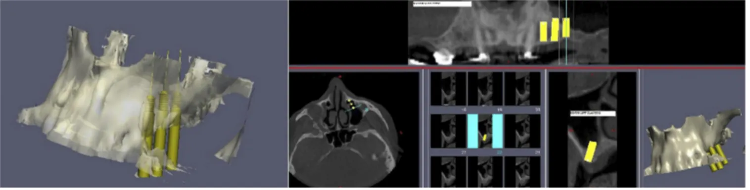
(Implant 3D Rel.3.0.8; Media Lab Software 2007, La Spezia, Italy), and MSSTs were constructed accordingly (Fig. 1).
Stereolithographic models were constructed using a rapid proto-typing machine. Sinus osteotomy borders were outlined on the models of 5 randomly selected patients, and implant localization of all pa-tients was simulated in model surgery. Maxillary sinus surgical tem-plates and stereolithographic surgical implant guides were constructed in the laboratory based on the findings of model surgery (Fig. 2).
Following routine surgical preparations, a full-thickness muco-periosteal flap was raised to expose the lateral wall of the maxillary sinus, and an MSST was located at the lateral wall. Under guidance of the MSST, an osteotomy line was outlined on the cortical bone using a fine bur, and a bony window was removed (Fig. 3). Using the same template, implants were inserted in accordance with the model surgery. In the control group, implants were inserted using stereolithographic surgical implant guide, whereas localization of lateral sinus wall and osteotomy was performed in traditional surgical procedure. Primary closure of the surgical wound was achieved, and postsurgery antibiotics, analgesics, antibacterial mouthwash, and an-tihistaminic decongestants were prescribed. All patients were recalled
for follow-up visits on 3, 6, and 10 days after surgery. Fixed prosthetic restorations were completed between 6 and 10 months after implant surgery (average, 8 months) (Fig. 4). Astra Tech Osseospeed TX dental implants (Mo¨lndal, Sweden) were inserted in diameters of 4.0, 4.5, and 5.0 mm and in lengths of 11 and 13 mm in all patients.
Immediately following the procedure, an oral questionnaire was administered to the 3 participating surgeons to evaluate the fol-lowing: adaptation of the guide to the area, immobility of the tem-plate during the procedure, ease of creation of the lateral window, sufficiency of the window for elevation and grafting, and the effects of template use on the duration of the procedure.
RESULTS
Sinus elevations were performed in 10 patients (6 men, 4 women; age range, 45Y65 years). In total, 15 sinus elevations were per-formed (5 bilateral elevations and 5 unilateral elevations). In 14 of 15 surgeries, sinus elevation and implant insertion were performed simultaneously, whereas in 1 case, implant insertion was performed
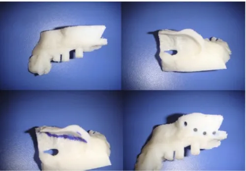
FIGURE 2. Complete or partially maxillary model was produced by using a fast prototyping machine.
FIGURE 3. Full-thickness mucoperiosteal flap was elevated, and the MSST was located on the area. Sinus window was prepared for both implant locations and lateral sinus wall over the same template.
6 months after sinus augmentation. No operative or postoperative complications were observed in any patient (Table 1).
The use of an MSST was found to be effective with regard to adaptation (62.5%), window preparation (87.5%), ease of elevation (95.9%), ease of grafting (95.9%), reduction of perforation risk (91.7%), and immobility during the procedure (62.5%). However, the use of an MSST was also observed to prolong surgery (100%) and restrict the view of the surgical area (79.2%). Overall, model surgery was found to be an effective means of preparing for actual sinus elevation surgery (87.5%) (Table 2).
DISCUSSION
Implant-supported fixed prosthetic restorations in the posterior maxilla require an adequate degree of bone. In cases where residual bone height in the posterior maxilla is insufficient for maintaining the support required by fixed restorations, maxillary sinus elevation must be performed before or in conjunction with implant surgery. A lateral wall approach is generally preferred in sites with inadequate residual bone height.11However, intraoperative complications may
sometimes occur including bleeding, membrane perforation, alve-olar crest fracture, damage to adjacent teeth, and infraorbital nerve damage.9 Membrane perforation, the most frequently reported complication in the literature may occur as a result of variations in anatomic structures, difficulties in accessing the surgical area, in-direct and/or blind manipulation, failure to clearly identify a lateral window, instrument-related complications during elevation, and the thin nature of the membrane itself.11,12
Membrane perforation may lead to delay or termination of the surgery as well as postoperative complications such as bacterial infection in the osteomeatal region and unfavorable bone healing.13 The risk of membrane perforation can be avoided through the op-timal positioning of a lateral window of an appropriate width. In some patients, the lateral wall of the maxillary sinus is very thin, and even experienced surgeons may find it difficult to identify the sinus mucosa border.
The literature contains several reports of maxillary sinus anterior osteotomy surgical guides that were designed and constructed using software and prototyping machines.12In the present study, an MSST was created in the laboratory on a three-dimensional model and used in surgery. Model surgery was found to be an effective means for the surgeon to become familiar with the surgical field before actual surgery, and the templates that were developed allowed for optimum osteotomies with no perforations or other operative or postoperative complications.
Following tooth loss, there is a decrease in the amount of bone available in the posterior maxilla, which causes the maxillary sinus to expand inferiorly. Given the influence of the anatomic interaction between the sinus floor and antrum on the pneumatization and health of the sinus, it is important to maintain a smooth floor, with no ‘‘blind holes’’ and a good structural relationship with the antrum. The success of sinus elevation and augmentation depends on ade-quate elevation of the membrane on the medial, inferior, and anterior aspects of the sinus cavity. Failure to achieve proper elevation will inhibit grafting and increase the risk of infection. An MSST helps to delineate the osteotomy line before augmentation, thereby facilitat-ing the preparation of the window to secure adequate elevation, which indirectly helps to eliminate blind holes and ease the place-ment of graft material in blind corners of the maxillary sinus. The use of an MSST also helps to minimize bone loss by preventing excessive window preparation.
FIGURE 4. Panoramic x-ray of the patient 6 to 10 months after implant surgery.
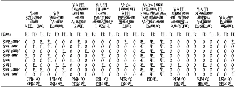
TABLE 2. MSST Evaluated Side by Side From the Surgeons With the Questionnaire, Who Were Engaged in the Operations
Patient Is the Adaptation of the MSST Easy? Is It Immobile During Surgery? Is MSST Restricting the View of the Operation Area? Is MSST a Useful Tool for Preparation of the Lateral Window? Do You Think That MSST Reduces the Perforation Risk? Do You Think That the MSST Alters (Increases/ Decreases) the Duration of the Surgery? Is MSST Facilitating the Elevation of the Sinus Membrane? Is MSST Facilitating the Sinus Grafting Procedure? Is Model Surgery Effective? S1 S2 S3 S1 S2 S3 S1 S2 S3 S1 S2 S3 S1 S2 S3 S1 S2 S3 S1 S2 S3 S1 S2 S3 S1 S2 S3 Case 1 (right) Y Y N N Y Y Y N Y Y Y Y Y N Y INC INC INC Y Y Y Y Y Y Y Y N Case 1 (left) Y N N N Y Y Y N Y Y Y Y Y Y Y INC INC INC Y Y Y Y Y Y Y Y Y Case 2 (right) Y Y Y Y Y N Y Y N Y Y Y Y Y Y INC INC INC Y Y Y Y Y Y Y Y Y Case 2 (left) N Y Y N Y Y Y Y Y Y N Y Y Y Y INC INC INC Y Y Y Y Y Y Y Y Y Case 3 Y Y Y N Y Y N Y Y Y Y Y Y Y Y INC INC INC Y Y Y Y Y Y Y Y Y Case 4 Y N Y Y N N Y Y Y Y N Y Y N Y INC INC INC Y N Y Y N Y Y Y Y Case 5 (right) Y N N Y Y N Y Y Y Y Y Y Y Y Y INC INC INC Y Y Y Y Y Y Y Y N Case 5 (left) N Y N Y Y N Y Y N Y N Y Y Y Y INC INC INC Y Y Y Y Y Y Y Y N 62.5%, Y 62.5%, Y 79.2%, Y 87.5%, Y 91.7%, Y 100 INC 95.9%, Y 95.9%, Y 87.5%, Y 37.5%, N 37.5%, N 20.8%, N 12.5%, N 8.3%, N 4.1%, N 4.1%, N 12.5%, N Y indicates yes; N, no; INC, increase.
Maxillary sinus augmentation requires a graft volume of approxi-mately 2 to 3 mL.14The amount of graft required can be calculated during model surgery. Moreover, not only can model surgery using an MSST prevent excessive graft use and potentially improve cost-effectiveness,12 it can also help inexperienced clinicians and junior surgeons to familiarize themselves with sinus anatomy and the surgical procedure.
The high cost of model and template production involved in the use of an MSST may be discounted, given the important benefits an MSST can provide, such as achieving a more ideal lateral window and reducing the risk of infection.
Recent studies in computer-guided implant dentistry have fo-cused on planning, drill guides, and immediate restoration.10The need for sinus guides has increased with the increase in popularity of sinus lift procedures; however, the literature still includes only limited reports on the use of surgical templates for maxillary sinus augmentation osteotomies. From the authors’ experiences, an MSST appears to be an effective tool for locating an appropriate entrance to the sinus cavity, achieving safer membrane elevation and success-fully grafting the sinus floor.
REFERENCES
1. Tatum H Jr. Maxillary and sinus implant reconstructions. Dent Clin North Am 1986;30:207Y229
2. Boyne PJ, James RA. Grafting of the maxillary sinus floor with autogenous marrow and bone. J Oral Surg 1980;38:613
3. Woo I, Le BT. Maxillary sinus floor elevation: review of anatomy and two techniques. Implant Dent 2004;13:28Y32
4. Schwartz-Arad D, Herzberg R, Dolev E. The prevalence of surgical complications of the sinus graft procedures and their impact on implant survival. J Periodontol 2004;75:511Y517
5. Zijderveld SA, van den Bergh JP, Schulten EA, et al. Anatomical and surgical findings and complications in 100 consecutive maxillary sinus floor elevation procedures. J Oral Maxillofac Surg 2008;66:1426Y1438
6. Wallace SS, Mazor Z, Froum SJ, et al. Schneiderian membrane perforation rate during sinus elevation using piezosurgery: clinical results of 100 consecutive cases. Int J Periodontics Restorative Dent 2007;27:413Y419
7. Banta HD. The diffusion of the computed tomography (CT) scanner in the United States. Int J Health Serv 1980;10:251Y269
8. Bouyssie´ JF, Bouyssie´ S, Sharrock P, et al. Stereolithographic models derived from x-ray computed tomography reproduction accuracy. Surg Radiol Anat 1997;19:193Y199
9. Sarment D, Sukovic P, Clinthorne N. Accuracy of implant placement with a stereolithographic surgical guide. Int J Oral Maxillofac Implants 2003;18:571Y577
10. van Steenberghe D, Ericsson I, Van Cleynenbreugel J, et al. High precision planning for oral implants based on 3-D CT scanning. A new surgical technique for immediate and delayed loading. Appl Osseo Int Res 2004;4:27Y31
11. Pjetursson BE, Tan WC, Zwahlen M, et al. A systematic review of the success of sinus floor elevation and survival of implants inserted in combination with sinus floor elevation. J Clin Periodontol 2008;35:216Y240
12. Mandelaris GA, Rosenfeld AL. Alternative applications of guided surgery: precise outlining of the lateral window in antral sinus bone grafting. J Oral Maxillofac Surg 2009;67:23Y30
13. Johansson B, Wannfors K, Ekenba¨ck J, et al. Implants and sinus-inlay bone grafts in a 1-stage procedure on severely atrophied maxillae: surgical aspects of a 3-year follow-up study. Int J Oral Maxillofac Implants 1999;14:811Y819
14. Uchida Y, Goto M, Katsuki T, et al. A cadaveric study of maxillary sinus size as an aid in bone grafting of the maxillary sinus floor. J Oral Maxillofac Surg 1998;56:1158Y1163
Clinical and Pathological
Features of the Giant,
Invasive Basal Cell Carcinoma
of the Scalp
Vesna S. Rakic, MD,* Dimitrije C. Brasanac, MD, PhD,Þ Zlata M. Janji(, MD, PhD,þ
Slobodan M. Bogdanovic, MD, PhD,* Mazen A. Arafeh, MD* Background: The giant, invasive basal cell carcinoma of the scalp is a rare clinical form of this tumor that appears on the skin, but may spread to some of the following structures: soft tissues of the scalp, bones, meninges, and the brain. In literature, so far, it is known as the GBCC. It is caused by aggressive BCC subtypes.
Methods: We will present here a research of clinical and patho-logical features of 47 pathopatho-logical specimens in 31 patients where the following features were examined: the dimension of the tumor, the dimension of the tissue segment, tumor area, segmentation area, resection margin width, microscopic resection margin status, tumor invasion level, and the outcome.
Results and Conclusions: We have concluded that microscopic resection margin dimensions from 1 to 10 mm are safe and that relapse occurrences in giant, invasive BCCs of the scalp depend on microscopic resection margin dimensions, resection margin sta-tus, tumor invasion levels, risky occupation, and risky behavior of the patient.
Key Words: Giant, invasive basal cell carcinoma, scalp, relapse, resection margins
Abbreviations: AJCC, American Joint Committee of Cancer, NMSC, Non-melanoma skin cancer, MIN, Microscopic dimensions of resection margins, INH KCS, Institute for neurosurgery of the Clinical Center of Serbia, KOPRH KCS, Clinic for Burns, Plastic and Reconstructive Surgery of the Clinical Center of Serbia, GBCC, Giant basal cell carcinoma, BCC, Basal cell carcinoma, BSC, Carcinoma basosquamosum, BCCs, Superficial form of BCC, BCNS, Basal cell nevus syndrome, 5-FU, 5-Fluorouracil (Efudix), EHO, Echosonography, NMR, Nuclear magnetic resonance imaging, CT, computed tomography
B
asal cell carcinoma (BCC) is the most frequent malignant skin tumor.1Y4It is characterized by high incidence and low mortal-ity rate (G0.1%),2with rare occurrence of metastasis (G0.1%).5,6
From the *Clinic for Burns, Plastic and Reconstructive Surgery, Clinical Center of Serbia, Belgrade; †Institute of Pathology, School of Medicine, University of Belgrade, Belgrade; and ‡Clinic for Plastic and Recon-structive Surgery, Clinical Center of Vojvodina, Novi Sad, Serbia. Received March 31, 2013.
Accepted for publication June 23, 2013.
Address correspondence and reprint requests to Dr Vesna S. Rakic, MD, Zvecanska 9, Belgrade, Serbia; E-mail: [email protected] The authors report no conflicts of interest.
Copyright* 2013 by Mutaz B. Habal, MD ISSN: 1049-2275