position. Besides, this situation may depend also on ex-perience level of the radiology technician. As a conse-quence, operation room staff may be exposed to high level of ionizing radiation during surgery. In this study, we have aimed to evaluate the effect of use of the dou-ble-fluoroscopy on the level of radiology emission and duration of surgery in the patients who underwent os-teosynthesis by cannulated screw fixation for femoral neck fracture. Our hypothesis to conduct this study is that use of double fluoroscopy reduces both duration of surgery and level of the exposure to ionizing radiation. MATERIAL AND METHODS
Our retrospective study included 35 patients who were performed osteosynthesis by cannulated screw fixation for femoral neck fracture between December 2013 and INTRODUCTION
Proximal femoral fracture demonstrates high morbidity and mortality rates and one of the most commonly per-formed trauma surgeries (14). It is estimated that ap-proximately annually 4.5 million femoral fractures will occur worldwide in 2050 (21). Femoral neck fractures make an important part of the hip fractures. Cannulated screw fixation by closed repositioning is one of the leading surgical techniques performed in femoral neck fractures (2, 4, 19, 24). In general, repositioning is achieved using the traction table before sterile staining during the surgery. In repositioning and osteosynthesis by single-fluoroscopy; images should be obtained in two different planes thus taking multiple fluoroscopic images is required. During this procedure, numerous unnecessary images may be taken due to inaccurate fo-cusing of the fluoroscopy on the targeted localization or inability to reposition the fluoroscopy on the targeted
An Effective Technique in Treatment of Femoral
Neck Fractures – Ostheosynthesis under Double
Fluoroscopic Guidance
Název česky ...
B. GÜLENÇ1, B. GÜNAYDIN2, S. ÇAMUR3, M. A. TALMAÇ4, I. GÜLER1, A. KARA1 1Istanbul Medipol University, Orthopaedics and Traumatology Department, Istanbul, Turkey
2Namik Kemal University, Orthopaedics and Traumatology Department, Tekirdag, Turkey
3Umraniye Training and Research Hospital, Orthopaedics and Traumatology Department, Istanbul, Turkey 4Sisli Etfal Training and Research Hospital, Orthopaedics and Traumatology Department, Istanbul, Turkey
ABSTRACT
PURPOSE OF THE STUDY
The most commonly used technique for fixation of the femoral neck fractures is percutaneous osteosynthesis with cannulated screw under fluoroscopy guidance. Osteosynthesis performed under single-fluoroscopic guidance requires continuous repositioning the fluoroscope to obtain biplane imaging. This study aimed to evaluate whether there is a statistically significant difference between the osteosynthesis procedures performed under guidance of single-fluoroscopy and double fluoroscopy with respect to radiation emission safety and treatment parameters for fixation of femoral neck fractures. MATERIAL AND METHODS
This retrospective study included 35 patients operated for femoral neck fracture between December 2013 and October 2017. Osteosynthesis was performed in 21 of these patients using cannulated screw under guidance of single-fluoroscopy whereas other 14 patients underwent same type of osteosynthesis assisted by double-fluoroscopy. The parameters of these two groups such as duration of surgery preparation, duration of the surgery, duration of radiation exposure, collo-diaphyseal angle and Harris hip score values were recorded.
RESULTS
No significant difference was found between the groups regarding time for surgery preparation (p > 0.05). On the other hand, duration of surgery and overall duration of radiation exposure were found 14 (p < 0.05) and 15 (p < 0.05) minutes shorter in the double-fluoroscopy group, respectively. No significant difference was found between the groups in terms of collo-diaphyseal angle and Harris hip score values (p > 0.05).
CONCLUSIONS
Use of double-fluoroscopy significantly reduces time of surgical duration and also level of radiation exposure in surgery of femoral neck fractures.
October 2017. Mean age of the pa-tients included in the study was 33.39 (20–46) years. Preoperative radio graphies of the patients were taken and all fractures were classified according to Garden’s Classification (10). The patients whose preoperative radiographies could not be obtained and patients with an inserted fixation material except cannulated screw, a medical history of previous open surgery, pathological fractures and polytraumas were excluded from the study. Data of the patients such as age, time for surgery preparation (time required from the end of anaes-thesia-related procedures to reduction is achieved), duration of surgery and overall radiation duration were recorded. It was determined that all
the patients were operated within the first 24 hours
after application to the hospital. Collo-diaphyseal angle values of all the patients in both groups were recorded based on their postoperative control radiographies. Mean follow-up duration was 11.8 (6–20) months. The study group included 25 male and 11 female patients. All the pa-tients were undergone functional evaluation based on Harris hip scores in their last control examinations.
The study has been reported in line with the STROCSS criteria (1). Surgical technique
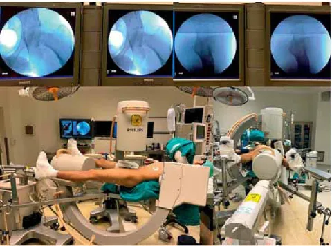
All the patients were operated on the fracture table. Following anaes-thesia induction, the broken leg was placed in the neutral position on the coronal plane after traction apparatus was connected to both feet. Both in the single-fluoroscopy and double-fluoroscopy groups, the opposite leg was taken into abduction to allow the best entrance of the device (Fig. 1). Philips (BV Endura, An-dover, MA) fluoroscopy device was used in the patients.
In the single-fluoroscopy group, the device was placed into the operation site through both legs after connecting traction apparatus and positioning the patient. Prior to skin preparation for surgery, fracture repositioning was performed after obtaining images on the antero-posterior (AP) and lateral planes. Following, the device was with-drawn and sterile draping was performed. Then, the device was placed into the same position and the cannu-lated screws were forwarded by controlling on the AP and lateral planes.
In the double-fluoroscopy group, one of the fluoroscopes was inserted by maximally approximating to the hip by making a tilt of 15 degrees through both legs in the sagittal plane. While the other fluoroscope was positioned perpendicularly to the patient by making an angle of 90 degrees in the operation site. In the same manner, prior to skin preparation for surgery, fracture repositioning was performed by controlling on the AP and lateral planes. After marking the position of the devices on the basement using medical plaster following reduction,
Fig. 1. Preoperative preparation of patient and flouroscopies.
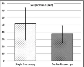
types; Garden Type 2, Type 3 and Type 4 fractures were determined and no significant difference was found in terms of fracture type be-tween the groups (p > 0.05) (Table 1). Accor ding to the comparison with respect to time for surgery prepara-tion, this time interval was 20.5 (15– 26) minutes in the double-fluoro -scopy group whereas that was 18.2 (12–27) minutes in the singlefluoro -scopy group. There was a difference of 2 minutes in favor of the single-fluoroscopy group between the groups but this difference was not statistically significant (p > 0.05). In terms of surgical duration; mean surgery duration was 37.7 (28–50) minutes in the double-fluoroscopy group whereas this interval was 51.9 (30–75) minutes in the single-fluoroscopy group. There was a difference of 14 minutes between the groups but this difference was statistically significant (p > 0.05) (Table 2). With respect to overall duration of radiation exposure, there was a difference of 25 seconds between the groups and this difference was statistically significant the devices were withdrawn to perform skin preparation
for surgery and sterile draping. Need for technician was minimized by obtaining AP and lateral images simul -taneously during the surgery (Fig. 2). In both groups, 3 pieces of 6.5 mm spongios cannulated screws were used in accordance with the principle of three point fixation (Fig. 3). All of the operations were performed by two senior surgeons (AK, SÇ). It was recorded that 3 distinct fluoroscopy technicians with an experience of minimum 3 years functioned during the operations. Statistical analysis
Kolmogorov-Smirnov test was used for distribution of the variables and descriptive statistical analysis. Stu-dent’s t test was used in analysis of the differences between the groups with normally distributed data while comparison between the non-normally distributed data were analyzed by Mann-Whitney U test. Alpha signifi-cance level was accepted 0.05 and all the tests performed were two-tailed tests.
RESULTS
Ages of the patients in the groups were compared and no significant difference was found between the groups (Table 1) (p > 0.05). In analysis of the fracture
Fig. 3. A – preoperative X-ray of an 23-year-old patient who had collum femoris fracture; B – preoperative reduction views which are obtained by double flouroscopy; C – final flouroscopy view of patient after surgery.
Table 1. Outcomes of a study comparing double fluoroscopy with single fluoroscopy
Single flouroscopy Double flouroscopy p
Mean. s.s./n-% Median Mean. s.s./n-% Median
Age 32.6 ± 7.3 34.0 34.2 ± 7.0 35.0 0.434 m
Operation time (min) 52.0 ± 12.9 50.0 37.8 ± 9.1 33.5 0.001 t
Preparation time (min) 18.3 ± 4.6 18.0 20.6 ± 3.4 20.0 0.122 t
Radiation time (min) 67.4 ± 14.5 66.0 42.9 ± 8.6 45.0 0.000 t
Collo-diaphyseal angle 126.1 ± 3.9 125.0 127.9 ± 3.9 127.5 0.200 t
Fracture type G2 5 23.8% 3 25.0% 0.960 X
(Garden classification) G3 8 38.1% 6 50.0%
G4 8 38.1% 5 41.7%
t t test / m Mann-Whitney U test / X Chi-Square test
Table 2. Comparison of surgery time between two flouroscopy groups
(p > 0.05) (Table 3). No significant difference was found between the groups in terms of collo-diaphyseal angle values determined based on the postoperative control radiographies (p < 0.05). Of the parameters tested at the last control examination; mean Harris hip score of the single-fluoroscopy group was 90.2 (84–96) whereas that value of the double-fluoroscopy group was 90.1 (83.8–96). This difference between the groups was not statistically significant.
DISCUSSION
We have suggested in this study that use of double-fluoroscopy shortens level of the radiation exposure during the osteosynthesis by cannulated screw fixation performed for femoral neck fractures and duration of surgery.
Anatomic reposition and rigid fixation in the femoral neck fractures reduces incidence of the complications that may occur after surgical procedures (8, 22, 23, 27). Sliding hip screw and cannulated screw are the most commonly preferred materials (7, 9, 16). Its availability for closed application and thus low amounts of haemor-rhage and minimal levels of tissue loss are the most im-portant advantages of the fixation by cannulated screws.
One of the complications during closed osteosynthesis is exposure of the surgical team to a high level of ionizing radiation. It has been shown that exposure to radiation increases seriously in the cases that duration of fluoroscopy application lasted longer than 1.7 minutes (20). The subjects who performed operations based on radiation emission for a long time interval are under risk for tumor development. It has been reported in a comparative analysis study that incidence of breast cancer was found higher in the female orthopaedic sur-geons than normal population (6). It has been reported in another study which compared orthopaedic surgeons
with normal population that orthopaedic surgeons which perform radiation-emitting therapies were found under higher risk for tumor development (17).
Not only healthcare professionals, but also patients who undergo operations requiring use of fluoroscopy are under risk for radiological hazards. It has been reported that risk for tumor development was found higher in the patients who underwent surgery involving radiation exposure (3). Because of all these reasons; level of radiation exposure during orthopaedic inter-ventions should be reduced. We conclude that use of double fluo roscopy may prevent high level of radiation exposure during surgery.
There are some studies conducted on use of double fluoroscopy in the field of vertebral surgery. Li et al. have performed vertebroplasty on 43 osteoporotic vertebral fractures under guidance of single- and dou-ble-fluoroscopy and stated that duration of surgery was shortened and that they have encountered a lower fre-quency of bone cement leakage (15).
Kotil et al. have applied biplanar fluoroscopy during cervical transpedicular fixation due to reasons such as trauma or degenerative and reported that surgical success rate by this technique was 100%. On the other hand, no comparison on the level of radiation exposure in these two studies (13).
Another study has evaluated fixation surgery using ilio sacral screw for unstable pelvic fractures under gui -dance of single-and double-fluoroscopy and reported that duration of surgery was shortened approximately by 65%. At the same time, duration of radiation exposure was also decreased significantly (18).
Xu et al. have analyzed surgical duration and level of the radiation exposure in their study on vertebroplasty for 143 osteoporotic vertebral fractures under guidance of single-and double-fluoroscopy. They have reported that both parameters were found significantly lower in the double-fluoroscopy group (26).
There is a limited number of studies on use of double-fluoroscopy in the operations performed for lower-ex-tremities. Wesberry et al. have performed in-situ pinning technique using double-fluoroscopy in 77 cases with slipped capital femoral epiphysis (SCFE) and reported no significant difference with respect to duration of surgery preparation between the groups. They have re-ported no difference between the groups regarding also duration of the surgery (25). This study has performed a similar fluoroscopic positioning with our study. On the other hand, this study has emphasized especially insertion of the screws and demonstrated that placement of the screws was more optimal under guidance of double fluo roscopy. It has been reported in a review on 6 patients which analyzed duration of fluoroscopy that duration of fluoroscopy was reduced by 34% during in-situ pinning accompanied by double flouroscopy (12). There is a limited number of study which analyzed double-scopy performed in the fractures of hip (5, 11). The durations of radiation exposure and surgery were compared in a study on cephalomedullary nailing in 44 cases with intertrochanteric femoral fractures. This study
Table 3. Comparison of radiation time between two flouroscopy groups
demonstrated a significant reduction in both durations of radiation exposure and surgery in the group of double fluoroscopy. No significant difference was found regarding duration of surgery preparation in both groups (5). Both studies have applied similar evaluation criteria and had similar outcomes with our study.
Major limitation of our study is small sampling size. Another limitation was absence of a precise data on the level of cumulative radiation due to inability to measure radiation using dosimetry.
On the other hand, being first study on use of double-fluoroscopy in femoral neck fractures and emphasizing the importance of level of radiation during surgery are the essential prominent features of our study.
CONCLUSIONS
In femoral neck fractures, cannulated screw fixation under guidance of double-fluoroscopy significantly reduces duration of surgery and level of radiation emission to environment compared with single-fluo-roscopy. We have concluded that exposure of both surgical team and the patients to high doses of radiation emission can be prevented thanking to use of double-fluoroscopy.
Level of evidence: Level IV, Retrospective case series.
Conflicts of interest: All named authors hereby declare that they
have no conflicts of interest to disclose.
Funding: This research received no specific grant from any funding
agency in the public commercial, or not-for-profit sectors.
Ethical approval: The medical ethics committee of the Istanbul
Medipol Univercity.(Istanbul-Turkey) approved this study with 10840098-604.01.01-E.21946 number. Consent to participate was obtained from the participants.
References
1. Agha RA, Borrelli MR, Vella-Baldacchino M, Thavayogan R, Orgill DP, STROCSS Group. The STROCSS statement: strengthening the reporting of cohort studies in surgery. Int J Surg. 2017;46:198– 202.
2. Asnis SE, Wanek-Sgaglione L. Intracapsular fractures of the femoral neck. Results of cannulated screw fixation. J Bone Joint Surg Am. 1994;76:1793–1803.
3. Beebe MJ, Jenkins P, Rothberg DL, Kubiak EN, Higgins TF. Pro-spective assessment of the oncogenic risk to patients from fluoroscopy during trauma surgery. J Orthop Trauma. 2016;30:e223–229. 4. Bout CA, Cannegieter DM, Juttmann JW. Percutaneous cannulated
screw fixation of femoral neck fractures: the three point principle. Injury. 1997;28:135-139.
5. Brin YS, Palmanovich E, Aliev E, Laver L, Yaacobi E, Nyska M, Kish BJ. Closed reduction and internal fixation for intertrochanteric femoral fractures is safer and more efficient using two fluoroscopes simultaneously. Injury. 2014;45:1071–1075.
6. Chou LB, Chandran S, Harris AH, Tung J, Butler LM. Increased breast cancer prevalence among female orthopedic surgeons. J Womens Health (Larchmt). 2012;21:683–689.
7. Dargan DP, Callachand F, Diamond OJ, Connolly CK. Three-year outcomes of intracapsular femoral neck fractures fixed with sliding hip screws in adults aged under sixty-five years. Injury. 2016;47:2495– 2500.
8. Florschutz AV, Langford JR, Haidukewych GJ, Koval KJ. Femoral neck fractures: current management. J Orthop Trauma. 2015;29:121– 129.
9. Gadegone WM, Chandak RM, Lokhande VR. DHS osteosynthesis with internal bone grafting in unstable delayed presented intracapsular neck femur fractures. Injury. 2017;48(Suppl 2):44–49.
10. Garden RS. Low-angle fixation in fractures of the femoral neck. J Bone Joint Surg Br. 1961;43:647–663.
11. Kara A, Celik H, Seker A, Uzun M, Sonmez MM, Erdil M. Proce-dural outcomes of double vs. single fluoroscopy for fixing inter-trochanteric femur fractures. Arch Orthop Trauma Surg. 2016;136:929–934.
12. Klug R, McCarthy JJ, Eilert RE. The use of a two C-arm technique in the treatment of slipped capital femoral epiphysis. Orthopedics. 2004;27:1041–1042.
13. Kotil K, Sengoz A, Savas Y. Cervical transpedicular fixation aided by biplanar fluoroscopy. J Orthop Surg (Hong Kong). 2011;19:326– 330.
14. La Velle DG. Fractures of hip. In: Canale ST (ed.). Campbell’s operative orthopaedics, 11th edition, :Mosby, Phialdelphia, 2008, pp 3237–3308.
15. Li YY, Huang TJ, Cheng CC, Hsu RW. A comparison between one- and two-fluoroscopic techniques in percutaneous vertebroplasty. BMC Musculoskelet Disord. 2008;9:67.
16. Ma JX, Kuang MJ, Xing F, Zhao YL, Chen HT, Zhang LK, Fan ZR, Han C, Ma XL. Sliding hip screw versus cannulated cancellous screws for fixation of femoral neck fracture in adults: a systematic review. Int J Surg. 2018;52:89–97.
17. Mastrangelo G, Fedeli U, Fadda E, Giovanazzi A, Scoizzato L, Saia B. Increased cancer risk among surgeons in an orthopaedic hospital.Occup Med (Lond). 2005;55:498–500.
18. Peng KT, Huang KC, Chen MC, Li YY, Hsu RW. Percutaneous placement of iliosacral screws for unstable pelvic ring injuries: comparison between one and two C-arm fluoroscopic techniques. J Trauma. 2006;60:602–608.
19. Rehnberg L, Olerud C. Subchondral screw fixation for femoral neck fractures. J Bone Joint Surg Br. 1989;71:178–180. 20. Sanders R, Koval KJ, DiPasquale T, Schmelling G, Stenzler S,
Ross E. Exposure of the orthopaedic surgeon to radiation. J Bone Joint Surg Am. 1993;75:326–330.
21. Veronese N, Maggi S. Epidemiology and social costs of hip fracture. Injury. 2018;49:1458–1460.
22. Wang C, Xu GJ, Han Z, Jiang X, Zhang CB, Dong Q, Ma JX, Ma XL. Correlation between residual displacement and osteonecrosis of the femoral head following cannulated screw fixation of femoral neck fractures. Medicine (Baltimore). 2015;94:e2139.
23. Wang T, Sun JY, Zha GC, Jiang T, You ZJ, Yuan DJ. Analysis of risk factors for femoral head necrosis after internal fixation in femoral neck fractures. Orthopedics. 2014;37:e1117–1123. 24. Weil YA, Qawasmi F, Liebergall M, Mosheiff R, Khoury A. Use
of fully threaded cannulated screws decreases femoral neck shortening after fixation of femoral neck fractures. Arch Orthop Trauma Surg. 2018;138:661–667.
25. Westberry DE1, Davids JR, Cross A, Tanner SL, Blackhurst DW. Simultaneous biplanar fluoroscopy for the surgical treatment of slipped capital femoral epiphysis. J Pediatr Orthop. 2008;28:43– 48.
26. Xu RJ, Yan YQ, Chen GX, Zou TM, Cai XQ, Wang DL. A method of percutaneous vertebroplasty under the guidance of two C-arm fluoroscopes. Pak J Med Sci. 2014;30:335–338.
27. Zhang YL, Chen S, Ai ZS, Gao YS, Mei J, Zhang CQ. Osteonecrosis of the femoral head, nonunion and potential risk factors in Pauwels grade-3 femoral neck fractures: A retrospective cohort study. Medicine (Baltimore). 2016;95:e3706.
Corresponding author: Barış Gülenç M.D.
Istanbul Medipol Univercity
Orthopaedics and Traumatology Department Göztepe mah. Tem Yanı
Bagclar/Istanbul, Turkey E-mail: [email protected]