25
Jinekoloji - Obstetrik ve Neonatoloji Tıp Dergisi 2016 Olgu Sunumları Sayısı; Sayfa: 25 - 26
ACET M. ve ark.
Yazışma Adresi/ Correspondence Address: Mustafa Acet, Ebru Öztürk
Medipol MEGA Hastanesi TEM Avrupa Otoyolu Göztepe Çıkışı No:1 Bağcılar / ISTANBUL 34214 Tel/Phone:+90533 344 17 02
E-mail : [email protected]
Geliş Tarihi/ Received:26/03/2015 Kabul Tarihi/ Accepted: 30/05/2015
Successful Pregnancy In A Patient With Unusual Ovarian Localization And Mullerian
Duct Anomalies: A Case Report
Bir Olgu Sunumu : Olağandışı Over Lokalizasyonu Ve Müller Kanalı Anomalili Hastada
Başarılı Gebelik
Mustafa ACET
1, Leyla ALTUNTAŞ
1, Sema OVALI
1, Tuba YELKE
2, Betül YORGUNLAR
1,
Hale Aktün TAMER
1, Ebru ÖZTÜRK
11
Medipol University, Medical School, Department of Obstetrics and Gynecology, İstanbul, Turkey
2
Medipol University, Medical School, Department of Embryology, İstanbul, Turkey
ABSTRACT
In this article we aimed to describe the clinical findings in a patient with subhepatic right ovary and müllerian duct anomalies who was successfully treated with in vitro fertilization (IVF) due to male factor infertility. Ovarian maldescent is a rare condition, and ovary localization can be seen up to the subhepatic region. This unusual ovarian localization suggests ovarian maldescent can be anywhere between the liver and pelvic brim When required, transabdominal ultrasound-guided oocyte retrieval and IVF can be performed successfully.
Keywords: Maldescent ovary, mullerian duct anomalies, in vitro fertilization.
ÖZ
Sağ over lokalizasyonu subhepatik alanda izlenen ve Müller kanal anomalileri bulunan bu olguda male faktör sebebiyle ıvf uygulanmıştır. İnmemiş over lokalizas-yonu nadir görülmekle birlikte , görüldüğünde sıklıkla pelvis giriminde olmaktadır. Bu olguda literaütrde ilk defa bu kadar yüksek seviyede yani subhepatik bölgede yerleşmiş bir over lokalizasyonu belirtilmiştir. Male faktör sebebiyle IVF uygulanan hastaya abdominal oosit toplama işlemi yapılmış, inmemiş over olgularında overin pelvik giriminden subhepatik alana kadar herhangi bir alanda olabileceği fikri savunulmuştur.
Anahtar Kelimeler: İnmemiş over, mülleriyan kanal anomalisi, in vitro fertilizasyon.
Introduction
A wide range of malformations of mullerian duct system are some of the more challenging disorders of obstetricians and gynecologists (1).
In the literature ovarian maldescent has been defined as the attachment of the superior pole of the ovary above the level of pelvic brim (2). Although the embryologic cause of ovarian maldescent is uncertain, the incidence of ova-rian maldescent is increased in patients with mulleova-rian duct anomalies (MDA) (3).
In this case, we present an infertile woman with uterus didelphys and comple-te vaginal longitudinal septum together with maldescended right ovary locacomple-ted high in the subhepatic region. Maldescent of ovary with MDA was reported rarely in the literature, this is the first case of subhepatic localization of ovary with MDA in an infertile patient who underwent in vitro fertilization.
Case Report
A 36 year-old woman applied to our clinic with 6 years of primary infertility. On her pelvic examination, a complete longitudinal vaginal septum and two cervices were detected. Transvaginal ultrasound revealed two uterine horns and left ovary with naturel appearance. The right ovary could not be visualized.
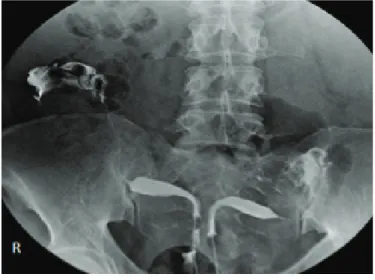
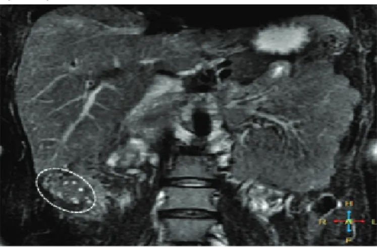
A hysterosalphingogram was ordered which demonstrated a very long right tuba uterina just about to liver (Figure 1). We performed magnetic resonance imaging (MRI) in order to clarify the anomaly and surprisingly it revealed right ovary located high in the subhepatic region (Figure 2).
Figure 1. Hysterosalpingography (HSG) revealing uterus didelphys and very long right tuba uterina lying under the liver
26
Jinekoloji - Obstetrik ve Neonatoloji Tıp Dergisi 2016 Olgu Sunumları Sayısı; Sayfa: 25 - 26
ACET M. ve ark.
Figure 2. Magnetic resonance imaging (MRI) revealing unusually localized right ovary
The ovarian tissue with multiple follicles was noticed inferior to the liver, pos-terior to the assending colon in coronal and transverse MRI images. A hemor-rhagic cyst which had a high signal on the T1-weighted MRI image, and no suppression of the signal with fat-saturation was detected. Hormonal analysis of the patient was normal. Sperm analysis showed severe oligoasthenoterato-zoospermia. Cytogenetic analysis of the couple and Y-chromosome microde-letion analysis were all normal. Female one had a family history of pulmonary embolism and heterozygote Factor V Leiden gene mutation.
Three IVF cycles were carried out for male factor infertility following longitudi-nal vagilongitudi-nal septum resection. The first and second IVF cycles were performed with antagonist protocol and recombinant follicle-stimulating hormone at a 150 IU/ day dose was used. While transvaginal oocyte retrieval was applied for the left ovary, percutaneous ultrasound guided aspirations was performed from the right ovary. In the first IVF cycle 5 oocytes from the left ovary and 11 oocytes from the right ovary were aspirated and one grade I embryo was transferred to the left uterine cavity on day three. Pregnancy could not be achieved.
In the second IVF cycle, 7 oocytes from the left ovary and 14 oocytes from the right ovary were aspirated and two blastocyst stage embryos were transferred to the left uterine cavity and three more blastocyst stage embryos were frozen. Again, no pregnancy could be achieved in the second cycle. In both cycles, we observed that left ovary was less respondant to the gonadotropins compared to the right one and follicles were smaller. Therefore, follicular aspiration from the right ovary was also performed. Two months later fozen embryo transfer cycle was started. In a natural cycle, a 19 mm dominant follicle was observed in the right ovary on cycle day 12. Endometrial thickness was 12 mm in the right cavity and 10 mm in the left. Serum estradiole level was 320 pg/ml, LH level was 50 mIU/ml. HCG at a dose of 10000 IU was administered on day 12. Luteal phase support was not performed. Low moleculer weight heparin 0,4ml/day and acetylsalicylic asid 100 mg/day were initiated two days after hCG.
In the third treatment cycle, frozen embryo transfer cycle, two embryos were transferred, one to each uterine cavity. Beta hCG level was 220 mIU/mL ten days after embryo transfer and, one gestational sac with fetal heart beat in the right uterine cavity was observed on ultrasound three weeks after embryo transfer. Antenatal follow-up of the patient was uneventful and a 47 cm, 2910 g boy baby was delivered by caesarean section at 36,5 gestational week. No
complications occurred in the postpartum period and the patient was dischar-ged on postoperative day two.
Discussion
In the third month of fetal life, the developing ovaries descend from a position near the kidneys to their final position in the pelvis. Failure of the ovaries to enter the true pelvis has been reported rarely, in 5 of 2,025 operative ob-servations of patients evaluated and treated for infertility (2).In the current case, we present for the first time a patient with maldescended ovary whi-ch is located high in the subhepatic region. This unusual ovarian localization suggests ovarian maldescent can be anywhere between the liver and pelvic brim. Although, there is no clear association between ovarian maldescent and infertility, implications of ovarian maldescent for infertility treatment have not been discussed.
In vitro fertilization methods used in those patients often involve ovarian sti-mulation and oocyte retrieval. Typical oocyte retrieval via transvaginal route might be technically difficult for highly located, malpositioned ovaries, and alternative procedures such as laparoscopic or abdominal ultrasound guided aspirations might be needed (3). Some authors suggest laparoscopic ova-rian transposition in order to facilitate transvaginal oocyte retrieval (4). In this case, we performed oocyte retrival from the right maldescended ovary under transabdominal ultrasound guidance and usual transvaginal route was used for the left follicular aspiration. Interestingly, better ovarian response was observed in the maldescended ovary compared to the normally localized left ovary in both ovarian stimulation cycles. Therefore, a higher number of oo-cytes were retrieved from the maldescended right ovary. In previous reports, investigators have suggested that the incidence of polycystic ovarian appea-rance is higher in patients with malpositioned ovaries (5).
In summary, we presented for the first time a woman with a maldescended right ovary located high in the subhepatic region and congenital uterine ano-maly successfully treated with IVF for male factor infertility. Maldescended ovary is a rare condition which might be localized anywhere between the liver and pelvic brim and, when required transabdominal ultrasound-guided oocyte retrieval and IVF can be performed successfully.
References
1. Jones HW Jr. Mullerian anomalies. Hum Reprod. 1998;13:789-91. 2. Verkauf BS and Bernhisel MA. Ovarian maldescent. Fertil Steril
1996;65:189-92.
3. Allen JW, Cardall S, Kittijarukhajorn M, Siegel CL. Incidence of ovarian maldescent in women with mullerian duct anomalies: evaluation by MRI. AJR Am J Roentgenol. 2012;198:381-5.
4. Goodman DM, Yalcinkaya TM, Ragland C. Ovarian transposition: a novel surgical technique to maximize oocyte retrieval in women with mullerian anomalies. Fertil Steril 2013;100:395.
5. Dabirashrafi H, Mohammad K, Moghadami Tabrizi N. Ovarian malposition in women with uterine anomalies. Obstet Gynecol 1994;83:293–4.