J Cell Biochem. 2020;121:508-523. wileyonlinelibrary.com/journal/jcb
508
|
© 2019 Wiley Periodicals, Inc.R E S E A R C H A R T I C L E
Palbociclib, a selective CDK4/6 inhibitor, restricts cell
survival and epithelial
‐mesenchymal transition in Panc‐1
and MiaPaCa
‐2 pancreatic cancer cells
Ozge Rencuzogulları
1,2|
Pınar Obakan Yerlikaya
1|
Ajda Çoker Gürkan
1|
Elif Damla Arısan
1|
Dilek Telci
2 1Department of Molecular Biology andGenetics, Science and Literature Faculty, Istanbul Kultur University, Istanbul, Turkey
2Department of Genetics and
Bioengineering, Faculty of Engineering, Yeditepe University, Istanbul, Turkey
Correspondence
Elif Damla Arısan, Department of Molecular Biology and Genetics, Science and Literature Faculty, Istanbul Kultur University, Atakoy Campus, 34156 Istanbul, Turkey.
Email: [email protected]
Funding information
Istanbul Kultur University Scientific Support Center
Abstract
The mortality rate of pancreatic cancer has close parallels to its incidence rate because of limited therapeutics and lack of effective prognosis. Despite various novel chemotherapeutics combinations, the 5‐year survival rate is still under 5%. In the current study, we aimed to modulate the aberrantly activated PI3K/ AKT pathway and epithelial‐mesenchymal transition (EMT) signaling with the
treatment of CDK4/6 inhibitor PD‐0332991 (palbociclib) in Panc‐1 and
MiaPaCa‐2 pancreatic cancer cells.
It was found that PD‐0332991 effectively reduced cell viability and proliferation dose‐dependently within 24 hours. In addition, PD‐0332991 induced cell cycle arrest at the G1 phase by downregulation of aberrant expression of CDK4/6 through the dephosphorylation of Rb in each cell lines. Although PD‐0332991 treatment increased epithelial markers and decreased mesenchymal markers,
the nuclear translocation of β‐catenin was not prevented by PD‐0332991
treatment, especially in MiaPaCa‐2 cells. Effects of PD‐0332991 on the regulation of PI3K/AKT signaling and its downstream targets such as GSK‐3 were cell type‐dependent. Although the activity of AKT was inhibited in both cell lines, the phosphorylation of GSK‐3β at Ser9 increased only in Panc‐1.
In conclusion, PD‐0332991 induced cell cycle arrest and reduced the cell
viability of Panc‐1 and MiaPaCa‐2 cells. However, PD‐0332991 differentially affects the regulation of the PI3K/AKT pathway and EMT process in cells due to its distinct influence on Rb and GSK‐3/β‐catenin signaling. Understanding the effect of PD‐0332991 on the aberrantly activated signaling axis may put forward a new therapeutic strategy to reduce the cell viability and metastatic process of pancreatic cancer.
K E Y W O R D S
1
|
I N T R O D U C T I O N
The high mortality rate affecting both male and female populations with equal incidence in worldwide due to
increased incidence of aggressive pancreas cancer.1
Despite the positive achievements in combined therapy options, 5‐year survival is still low.2 Pancreatic cancer cells exert extensive dysregulation in the cell cycle machinery. Especially, aberrant induction of K‐Ras due to mutational alterations in the majority of pancreatic cancer cells is associated with active PI3K/AKT/mTOR signaling axis leading to an impaired cyclin D‐CDK4/6
expression profile.3 Cell growth and metabolism are
orchestrated by PI3K/AKT/mTOR pathway, which acts as a cellular sensor for nutrients and growth factors, and activated in the downstream of receptors such as IGFR, EGFR, and other RTKs.4Given that aberrant activation of PI3K/AKT/mTOR signaling due to loss of PTEN is associated with the poor progression and
chemoresis-tance phenotype of pancreatic cancer,5,6 molecular
targeted approach against this pathway can be encoura-ging. Previous studies indicate that treatment of pan-creatic cancer cells with mTOR inhibitors such as everolimus and temsirolimus in long time periods triggers the activation of PI3K pathway via insulin receptor substrate, resulting in the secondary AKT
activation and cyclin D1 expression.7,8 AKT is also
critical in the transition of epithelial to the mesenchymal profile (EMT), which is associated with an increased cell motility and invasion through transcriptional activation of EMT‐associated genes such as SNAI2, vimentin, and Zeb1.9AKT can directly phosphorylate (serine 552) and stimulate the nuclear translocation of β‐catenin into the nucleus to induce transcriptional activation of EMT markers.10 In addition, active AKT can simultaneously phosphorylate GSK‐3β (Ser9), which prevents the GSK‐3β mediated degradation of β‐catenin and Snail. Increased cellular availability ofβ‐catenin leads to the transcription of cyclins and promotes cell cycle machinery. Cyclin D1 is the target gene of β‐catenin/LEF signaling and over-expression of D‐type mammalian cyclins stabilizes β‐ catenin to stimulate cell cycle. Concomitantly, cyclin D1 upregulation and the loss of negative control over Cdk4/6 results in the excessive activation of cyclin D‐Cdk4/6 complex formation and enhance cell proliferation. It is well known that EMT‐related Notch, Sonic Hedgehog, and Wnt signaling pathways also effectively regulate cyclin D1 as a downstream target. Because cyclin D1 overexpression leads to poor prognosis of pancreatic cancer, targeted inhibition of cyclin D‐Cdk4/6 may possess a significant therapeutic potential for pancreatic cancer treatment.
For this purpose, targeting of cell cycle regulatory proteins in the treatment of pancreatic cancer gains more importance to remarkably reduce the cell proliferation rate. Palbociclib (PD‐0332991), a potent CDK4/6 inhibi-tor, has received US Food and Drug Administration approval for the treatment of advanced postmenopausal
breast cancer in combination with letrozole.11 PD‐
0332991 is also a highly selective inhibitor for CDK2 and it can downregulate target genes of E2F transcription factor due to the dephosphorylation of Rb.12 Thereby, PD‐0332991 behaves as a potent inhibitor of cell proliferation by inducing cell cycle arrest at G1. The potential of PD‐0332991 in clinical utilization with paclitaxel for the treatment of metastatic pancreatic cancer is under investigation at phase I clinical trials.13 As recent studies highlight that the administration of CDK4/6 inhibitor as a single agent is not sufficient to
reduce poor progression pancreatic cancer cells
(Shiau),14-16the clarification of molecular targets of PD‐ 0332991 in different pancreatic cancer cells possess importance to design combinational therapeutic strate-gies, In this study, we aim to investigate the effect of PD‐ 0332991 on the cell proliferation and invasiveness of pancreatic cancer cells via the regulation of the PI3K/ AKT pathway and EMT signaling.
2
|
M A T E R I A L S A N D M E T H O D S
2.1
|
Cell lines and reagents
Both pancreatic cancer cells were obtained from American Type Culture Collection (Rockville, MD). Cells were incubated at 37°C and 5% CO2 incubator (Hera Cell 150i,
Thermo Lab Systems, Beverly, MA) and maintained in Dulbecco's modified Eagle medium (DMEM; GIBCO‐Life Technologies, Carlsbad, CA) medium supplemented with 1% penicillin/streptomycin (GIBCO, Invitrogen Co, Carlsbad, CA) and 10% fetal bovine serum (FBS; Pan Biotech GmbH,
Aidenbach, Germany). PD‐0332991 was purchased from
Selleck Chemicals, were dissolved in dimethyl sulfoxide. The following antibodies; total Rb, p‐Rb (S780), CDK6, CDK4, p21, c‐Myc, p‐CHK‐1 (S317), cyclin D3, β‐catenin, E‐ cadherin, N‐cadherin, vimentin, PI3K, AKT, P‐AKT (S473), p‐GSK‐3β (Ser9) polyclonal anti‐rabbit/mouse antibodies and β‐actin, Histon 3 were purchased from Cell Signaling Technology (CST; Danvers, MA). Each antibody (dilution ratio: 1:500‐1:1000) was prepared in superblock T20 (Thermo Fisher Scientific) reagent (Beverly, MA). According to manufacturer instructions, HRP‐conjugated secondary anti‐ rabbit or anti‐mouse antibodies from CST (dilution rate: 1:3000) were used for detection.
2.2
|
Cell viability, colony formation,
and cell survival assay
Cell viability response of pancreatic cancer cells to PD‐ 0332991 (0‐12.5 µM) was determined for 24 hours using 3‐(4,5‐dimethylthiazol‐2‐yl)‐2,5‐diphenyltetrazolium bro-mide cell viability assay as described previously.17 Cells
were treated with PD‐0332991 for 14 days, and colony
formation was analyzed with 0.1% of crystal violet staining as described before.17 Cell survival assay was performed to investigate the effect of PD‐0332991 (2 and 3 µM) on cell growth. A total of 25 × 103 cells per well were seeded into 12‐well plates in the presence of PD‐ 0332991 in the cells. Following incubation for 0 to 96 hours, cells were counted by using automatic cell counter from NanoEnTek (Cambridge, UK) after staining with trypan blue dye (0.4% w/v).
2.3
|
Cell cycle and Western blot
analysis
Cells (seeding density 5 × 10⁵in each well/six‐well plate) were treated with PD‐0332991 (2 and 3 µM) for 24 hours. Cells fixed in 70% ethanol were treated with RNase A and propidium iodide staining solution was analyzed by flow cytometer using C6 software (BD Bioscience).
Cells were harvested with 1X phosphate buffered saline (PBS) and were lysed on ice with a protein lysis solution.17 For, nuclear‐cytoplasmic isolation was
per-formed following the NE‐PER Nuclear and Cytoplasmic
Extraction protocol (Thermo Fisher Scientific). Total
protein lysate (30‐50 μg) was separated on a 10%‐12%
sodium dodecyl sulfate‐polyacrylamide gel electrophor-esis and then transferred onto polyvinylidene fluoride membranes. Five percent nonfat milk was used to block the membranes. After that membrane was incubated with proper primary antibodies and secondary antibodies for overnight at 4°C. Following the addition of lab‐made
enhanced chemiluminescence reagent,17 membranes
were examined with ChemiDoc MP (Bio‐Rad).
2.4
|
Wound healing assay
Cells were seeded at a density of 6 × 10⁵cells/well in six‐ well plates until cells reached monolayer confluence. After the treatment of cells with 2 and 3 µM concentra-tion of PD‐0332991, a straight scratch on the cell monolayers was created.18 Cells scratched to obtain a
wound layer. Detached‐cells were washed with 1X PBS
and cells were then supplemented with renewed medium and observed for 0 hours. Cells were treated with indicated drug concentrations and incubated at 37°C until monitoring for 24 hours by using a light microscope
(Olympus, Japan) (×10). Images were taken and analyzed using Olympus Micro DP Manager Image Analysis program at different time points and presented by bar graphics using the Graph Pad software (4.04 version).
2.5
|
Immunofluorescence analysis
For immunofluorescence examination, cells were seeded to the coverslip. After drug treatment for 20 minutes, −20°C cold methanol/acetone (1:1) was used as a fixative ., and 1X PBS containing 3% bovine serum albumin (BSA) was used to prevent unspecific binding. Cells were incubated for 1 hour at 37°C with anti‐β‐catenin which is diluted in PBS + 0.3% BSA (1/1000). After that cell was incubated with a secondary Alexa 488‐conjugated antibody (1/3000). Cells were visua-lized by a fluorescence microscope (Olympus, Japan) (×40). The same protocol was performed to visualize the E‐cadherin and focal adhesion‐related proteins (vinculin, F‐actin).
2.6
|
Soft agar assay
The base agar was prepared to create a mixture of 2X DMEM medium (20% FBS and 2% penicillin/streptomy-cin‐[GIBCO, Invitrogen Co]) and 0.5% agarose in 1X PBS at 1:1 ratio. After the dispersal of the 1 mL mixture into each well of six‐well plates, 2.5 × 10⁵
cells/mL in a 1:1 mix of 0.3% agarose and 2X DMEM medium was placed on top of the base gel. After, 500 µL medium in the presence or absence of drugs was placed into the wells for 14 days. At Day 14, colonies (>50μm) were counted and photographed under an inverted microscope at ×40 objective. Results were also analyzed by fluorescence microscopy following DAPI staining (Olympus IX70, Tokyo, Japan).
2.7
|
Data analysis
Graph Pad software (4.04 version) was used to analyze the statistical data were obtained from the averages of at least three independent experimentations. Image J gel Analysis program was used for the analysis of Western blot results and values obtained in the graph were
normalized to β‐actin. Two‐way analysis of variance
Bonferroni's multiple comparisons test was used for evaluation findings.
3
|
R E S U L T S
3.1
|
PD
‐0332991 reduced cell viability
and cell proliferation
PD‐0332991 treatment was effective to reduce cell viability and proliferation in a dose‐dependent manner
in both pancreatic cancer cells. Increasing concentrations of PD‐0332991 at 2 and 3 µM for 24 hours decreased cell viability by approximately 25%‐40% in Panc‐1 and MiaPaCa‐2 cells, respectively (Figure 1A). As 2 and 3 µM of PD‐0332991 showed moderate cytotoxicity and cytostatic effects, these concentrations were selected for the subsequent experiments for 24 hours treatments. The effect of long‐term treatment was also determined for
evaluation if the response of the PD‐0332991 were
maintained over time by the colony formation assay. Although the cell viability assay suggested that MiaPaCa‐ 2 cells were more sensitive to the PD‐0332991 treatment for 24 hours than Panc‐1 cells, PD‐0332991 treatment induced a significant decrease in the colony numbers as compared with control cells. In addition, Panc‐1 cells
were more sensitive to PD‐0332991 compared with
MiaPaCa‐2 cells (Figure 1B).
Cell survival assay using trypan blue was also performed to understand the effect of PD‐0332991 on
cell growth and proliferation. Cell growth was inhibited during 24 hours PD‐0332991 treatment for each concen-tration. However, 3 µM PD‐0332991 showed a cytotoxic effect in Panc‐1 cells, but not in MiaPaCa‐2 cells for 48 hours treatment. Knudsen et al15previously indicated that MiaPaCa‐2 cells were resistant to PD‐0332991 treatment due to its expression profile of Rb and cyclin E. The effect of PD‐0332991 on cell growth inhibition was reduced in 72 and 96 hours especially in MiaPaCa‐2 cells. These results were correlated with the long‐term effect of 3 µM PD‐0332991 in MiaPaCa‐2 cells (Figure 1C,D).
3.2
|
PD
‐0332991 induced cell cycle
arrest
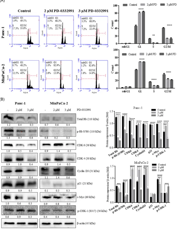
The cells were sensitive to PD‐0332991 with a significant blockade in the G1 phase of the cell cycle (Figure 2A). However, the different response of PD‐0332991 on cell
F I G U R E 1 The effect of cell viability of PD‐0332991 on Panc‐1 and MiaPaCa‐2. Cells (1 × 10⁴) were exposed to PD‐0332991 (0.5‐
12.5 µM) for 24 hours. After drug treatment, MTT cell viability assay proceeded. Columns presented the average ± SD of with three separate experiments with at least four replicates. B, Long‐term responses to PD‐0332991 in both cell lines were assessed using crystal violet staining after 10 days of treatment with media replenishment every 3 days. C, D, Cell survival assay for PD‐0332991‐treated Panc‐1 (C) and MiaPaCa‐ 2 (D) cells. Cells were seeded at 5 × 10⁴cells/well and treated with PD‐0332991 (2 and 3 µM) for 24‐96 hours. The data shown exemplify the mean ± SD from two experiments with three replicates. Data were assessed by two‐way ANOVA test. Asterisks show statistically significant variances between control and 3 µM PD‐0332991 for 24 hours treatment (*P < .05), control and 3 µM PD‐0332991 for 48 hours treatment in Panc‐1 (**P = .002), control vs 2 and 3 µM PD‐0332991 for 48 hours treatment in MiaPaCa‐2 cells (***P < .001). ANOVA, analysis of variance; MTT, 3‐(4,5‐dimethylthiazol‐2‐yl)‐2,5‐diphenyltetrazolium bromide
proliferation was determined via examination of the various expression profile of cell cycle regulatory proteins particularly Rb and CDK4/6 (Figure 2B). Panc‐1 and MiaPaCa‐2 cells displayed a different response to PD‐ 0332991 treatment as Panc‐1 cells showed higher expression of Rb than MiaPaCa‐1 cells. Phosphorylation of Rb has various effects on the cell cycle regulation through the ability of interaction with its different partner proteins. Phosphorylation of Rb at S780 causes to lose its ability to bind to E2F.17PD‐0332991 inhibited phosphorylation of RB at S780 in Panc‐1 cells; however, MiaPaCa‐2 cells did not display similar results. As previously demonstrated in breast cancer cells that Rb status is important to factor for the response of cells to PD‐0332991.19
CDK6 were downregulated by 2 and 3 µM PD‐0332991 in Panc‐1 cells. Although, 2 µM PD‐0332991 treatment upregulated CDK6 expression, 3 µM PD‐ 0332991 treatment downregulated CDK6 expression in MiaPaCa‐2 cells. Two‐micrometer PD‐0332991 treatment upregulated CDK4 and cyclin D3 in each cell line. Interestingly, 3 µM PD‐0332991 treatment significantly reduced the CDK4 and cyclin D3 protein levels in each cell line. p21 plays an important role in the regulation of the 1CDK4/6‐cyclin D complex during cell division, such as stabilizing the CDK4/6‐cyclin D complex or inhibiting CDK4/6 activity. Three‐micrometer PD‐0332991 treat-ment decreased the basal levels of p21 in Panc‐1 cells. This decrease in p21 might also result in the accumula-tion of cyclin D3 which is known as an early marker of senescence.20Another regulator of the cell cycle is c‐Myc that contains E2F‐binding sites, was downregulated by 3 µM PD‐0332991 in each cell lines. However, 2 µM PD‐ 0332991 treatment did not affect the expression of c‐Myc in MiaPaCa‐2 cells, which consisted of the expression levels of CDK4/6 and cyclin D.21 Chk1 is activated by ataxia telangiectasia mutated and Rad 3‐related kinase at S317 phosphorylation site to inhibit the CDKs and leads
to checkpoint arrest.22 Three‐micrometer PD‐0332991
treatment increased the Chk1 levels in MiaPaCa‐2 cells,
but there was no significant effect in Panc‐1 cells.
Activation of Chk1 might lead to the control of DNA damage response and prevention of apoptotic process which triggered by PD‐0332991 treatment.
3.3
|
PD‐0332991 increased the epithelial
markers but could not prevent nuclear
translocation of
β‐catenin
To investigate the role of PD‐0332991 on the regulation EMT process, immunofluorescence and immunoblotting assays were performed. EMT is a critical process and plays important roles during tumor progression. To determine the expression profile and localization of E‐cadherin, an epithelial marker, immunofluorescence analysis was per-formed. DAPI staining was performed to visualize the nuclei. MiaPaCa‐2 cells had higher expression of E‐cadherin than Panc‐1 cells after PD‐0332991 treatment (Figure 3A). In agreement with the immunofluorescence data, PD‐0332991 treatment increased the expression of E‐cadherin in both cell lines (Figure 3B). While downregulation of total β‐catenin expression level was evident following PD‐0332991 treatment in the Panc‐1 cell, an increase of β‐catenin expression levels in MiaPaCa‐2 cells was detected in the Western blot experiments. Interestingly, the protein levels of mesenchymal marker N‐cadherin showed an increase in response to 2 µM PD‐0332991, while a slight reduction was observed in response to 3 µM PD‐0332991 treatment in Panc‐1 cells. However, each concentration of PD‐0332991 exerted a significant decrease of N‐cadherin in MiaPaCa‐2 cells. Finally, EMT marker vimentin expression was not changed upon PD‐0332991 treatment in Panc‐1 cells, but a dose‐ dependent downregulation was detected in its levels for MiaPaCa‐2 cells (Figure 3B).
A high level of intracytoplasmic and/or nuclear
expres-sion of β‐catenin in cancer cells was associated with
metastasis.23 To investigate the localization of
β‐catenin, immunofluorescence analysis was performed
(Figure 3C). As shown in Figure 3C, β‐catenin protein
staining got weaker after PD‐0332991 treatment in each cell line. However, β‐catenin staining was observed in nuclei after 3 µM PD‐0332991 treatment in MiaPaCa‐2 cells. Cytoplasmic and nuclear localization of c‐Myc and β‐catenin levels are associated with poor prognosis and metastasis in various cancer types.24To analyze the effect of PD‐0332991 on pancreatic cancer cell metastasis, the expression levels of β‐catenin and c‐Myc was determined in the nuclear and cytoplasmic fractions of cells (Figure 3D). A significant
F I G U R E 2 The effect of PD‐0332991 (PD) on the cell cycle in a dose‐dependent manner. A, A total of 5 × 10⁵cells were seeded to six‐
well plates, following indicated drug treatment for 24 hours cells were harvested with trypsin and washed three times with 1X PBS. Cells were fixed with 70% ethanol for a week in +4°C. PI staining was performed and then cells were analyzed with flow cytometry. Control vs 2 and 3 µM PD‐033299 in Panc‐1 cells: G1: ****P < .0001, S: **P = .002; G2/M: ****P < .0001; control vs 2 and 3 µM PD‐033299 in MiaPaCa‐2 cells: G1, S, G2/M: ****P < .0001. B, The role of PD‐0332991 on the regulation of cell cycle‐related proteins. Total 30 µg of whole cell lysate was loaded to 12% SDS‐PAGE gels and probed with listed antibodies. Expression profile of Rb, p‐Rb (S780), CDK6, CDK4, cyclin D3, p21, c‐Myc, and p‐CHK‐1 (S317) were analyzed by immunoblotting following indicated PD‐0332991 treatments in cells. Expression levels of proteins were analyzed. **P < .01; ***P = .002; ****P < .0001.β‐Actin was used as a loading control. PBS, phosphate buffered saline; PI, propidium iodide; SDS‐PAGE, sodium dodecyl sulfate‐polyacrylamide gel electrophoresis
F I G U R E 3 The role of PD‐0332991 on the levels and localization of E‐cadherin and β‐catenin. Cells were grown on coverslips and proceed for immunofluorescence procedure. A, E‐cadherin (green) antibodies were used at concentrations for 1/1000 dilution. Nuclei were stained with DAPI (blue). B, Following PD‐0332991 treatment for 24 hours, total protein was isolated. Expression levels ofβ‐catenin, E‐cadherin, N‐cadherin, and vimentin were analyzed. **P < .01; ***P < .005; ****P < .0002 β‐actin was used as a loading control. C,β‐Catenin (green) antibodies were used at concentrations for 1/1000 dilution. Nuclei were stained with DAPI (blue). Scale bar is 10 µM. D, Following PD‐0332991 treatment for 24 hours nuclear‐cytoplasmic protein isolation procedure was proceeded in Panc‐1 and MiaPaCa‐2 cells. Expression levels of c‐Myc and β‐catenin were analyzed. β‐Actin was used as a cytoplasmic extraction loading control. Histon 3 was used as a nuclear extraction loading control. *P < .05, **P < .01, ***P = .002
decrease in the cytoplasmic/nuclear accumulation of c‐Myc levels was detected. The cytoplasmic β‐catenin levels were downregulated after 3 µM PD‐0332991 treatment in both cell lines. Although the nuclear β‐catenin levels were decreased in MiaPaCa‐2 cells, there was no change in that of the Panc‐1 cells.
3.4
|
The role of PD
‐0332991 on actin
cytoskeleton/focal adhesion
As the actin cytoskeleton organization is the driving force in the acquisition of mesenchymal migratory phenotype in the EMT process, the dynamicity of actin cytoskeleton/
focal adhesion was investigated in PD‐033299‐treated
pancreatic cancer cells (Figure 4A,B). F‐actin and vinculin have been shown to play critical roles in the adhesion of cells to the extracellular matrix (ECM) and the formation of adhesions. Thus, we examined the localization of F‐actin and vinculin after PD‐0332991 treatment (Figure 4A,B). Cell adhesion was triggered by
PD‐0332991 treatment through recruitment of vinculin
from the cytoplasm to the focal adhesion complex in Panc‐1 cells. Especially 3 µM PD‐0332991‐treated Panc‐1 cells displayed strong F‐actin and vinculin staining at the cell periphery at the cell adhesion sites.25However, each concentration of PD‐0332991 did not significantly induce the recruitment of vinculin/F‐actin in MiaPaCa‐2 cells. In addition, the concentration of vinculin‐F‐actin complex was more abundant in control cells than treated‐ MiaPaCa‐2 cells.
The potential role of PD‐0332991 on wound healing was also determined. In Figure 4C, wound healing assay was performed following PD‐0332991 treatment for 24 hours. Cells were stained with DiOC6 to better
visualize the wound closure. Although 2 µM PD‐
0332991 halted cell migration of pancreatic cancer cells
into the wound area, the presence of 3 µM PD‐0332991
completely abrogated the wound bed closure capability of Panc‐1 and MiaPaCa‐2 cells (Figure 4D).
3.5
|
Effects of PD‐0332991 on the cell
survival
‐signaling axis
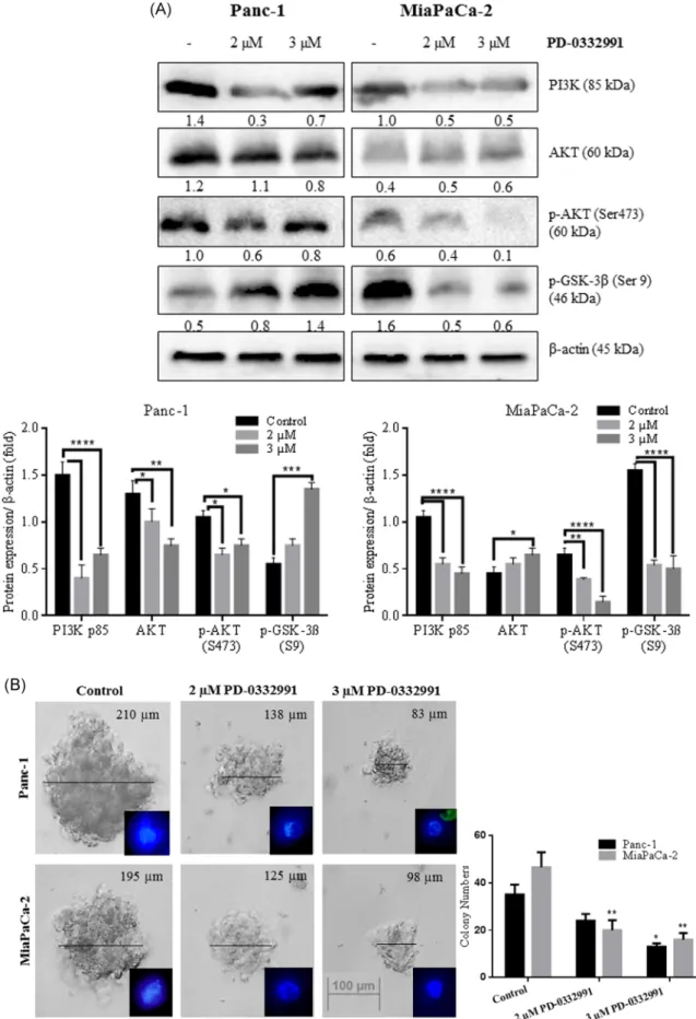
We analyzed the role of PD‐0332991 on the cell survival‐ signaling axis by investigating the downstream targets of
the PI3K/AKT pathway (Figure 5A). PD‐0332991
treat-ment significantly decreased the PI3K levels while little change in the total AKT levels was detected in 3 µM PD‐ 0332991‐treated cells. The level of active p‐AKT at Ser473 was only decreased in a dose‐dependent manner in MiaPaCa‐2 cells after PD‐0332991 treatment. PD‐0332991 was potent to suppress the AKT phosphorylation in Panc‐ 1 cells. Once activated at Ser473 residue, AKT phosphor-ylates and inhibits GSK‐3β (Ser9), thereby stimulating
glycogen synthesis under rich nutrient conditions.26
p‐GSK‐3β (Ser9) protein levels in response to PD‐
0332991 was increased in Panc‐1 cells. Conversely, PD‐ 0332991 significantly decreased the phosphorylation of GSK‐3β at Ser9 in MiaPaCa‐2 cells. As previous reports
indicated that AKT dependent phosphorylation of GSK‐
3β led to the translocation of β‐catenin to nucleus triggering EMT process, soft agar assay was performed
(Figure 5B) to investigate whether PD‐0332991 had an
effect on the anchorage‐independent cell proliferation. PD‐0332991 treatment at both concentrations induced a significant decrease in the colony formation ability of
cells. Evaluation of colony numbers by inverted
microscopy demonstrated an important reduction in the colony cell number for both pancreatic cancer lines.
4
|
D I S C U S S I O N
In the current study, we demonstrated that targeting CDK4/6 is remarkable therapeutic strategy due to its ability on the decreasing cell viability through regulation of cell survival and metastasis‐related signaling axis in Panc‐1 and MiaPaCa‐ 2 cells. Although each selected concentration of PD‐0332991 induced cell cycle arrest at the G1 phase and decreased cell viability effectively, the Rb expression profile showed an important regulatory role to evaluate the sensitivity of cells against PD‐0332991 treatment. This finding was similar to previous understanding of pancreatic cancer therapy models. Recent studies indicated that pancreatic cancer cells, which exerted higher Rb expression levels, were more sensitive to PD‐0332991, but the cells with low expression of Rb were resistant to PD‐0332991. In our study, we confirmed this data by cell viability assay. The cell viability ratio was more reduced in MiaPaCa‐2 compared with Panc‐1 cells following PD‐0332991 treatment for 24 hours. However, the long‐term effect of PD‐0332991 showed that Panc‐1 cells were more sensitive than MiaPaCa‐2 cells. In addition, biological evidence from immunoblotting results confirmed that PD‐ 0332991 decreased the Rb expression levels, which was higher in Panc‐1 cells than MiaPaCa‐2 cells. In addition, phosphorylation of Rb, which is significantly higher in Panc‐
1 cells than MiaPaCa‐2 was decreased by PD‐0332991
treatment. In contrary to various tumor models, pancreatic cancer models have a disparate response to PD‐0332991 due to increased expression of cyclin D1.27 PD‐0332991 was reported to induce cell cycle arrest at the G1 phase, which led to cyclin D1 accumulation.15Another study in breast cancer cells with various expression profile for Rb showed that PD‐ 0332991‐treated breast cancer cells with high Rb expression showed an increased ratio in cell cycle arrest and resulted with a delay of cell metastasis.19PD‐0332991 suppressed of E2F target genes in breast cancer cells, but pancreatic cancer showed different response through cell cycle regulatory process. c‐Myc is one of the targets of E2F, which is crucial on the G1/S transition, and accumulation of c‐Myc is observed in arrested‐cell at G1 phase.28 In our study, we observed that c‐Myc was highly expressed in Panc‐1 and MiaPaCa‐2 cells. According to previous studies, downregula-tion of c‐Myc is an important therapeutic strategy due to its ability on the regulation of cell survival‐signaling axis. The
data here showed that PD‐0332991 downregulated the
expression level of c‐Myc. However, PD‐0332991‐treated MiaPaCa‐2 cells remained c‐Myc expression, which showed correspondence with a high number of colonies even after treatment. It was well clarified that transcriptional activation
F I G U R E 4 The role of PD‐0332991 on the EMT process. A, Panc‐1 and B, MiaPaCa‐2 cells were grown on coverslips and proceed for immunofluorescence procedure for F‐actin (red) and vinculin (green) antibodies were used at concentrations for 1/500 dilution. Nuclei were stained with DAPI (blue). Scale bar is 10 µM. C, Wound healing assay was performed to evaluate the migration of Panc‐1 and MiaPaCa‐2 cells after 2 and 3 µM PD‐0332991 treatment. A total of 6 × 10⁵
cells/well were seeded. At this density, cells reached at monolayer confluency after 24 hours. A straight scratch was created with a sterile pipette tip. Then, cells exposed to PD‐0332991. Wound closure was imaged by fluorescence microscopy after DiOC6 staining (×20). D, Wound closure was determined by the graphic. EMT, epithelial‐mesenchymal transition. ****P < .0001
of c‐Myc promoted cell proliferation and anchorage‐inde-pendent colony growth in pancreatic cancer cells.29 There-fore, suppression of c‐Myc might also indirectly down-regulate the E2F‐targeted genes and Rb in PD‐0332991‐ treated Panc‐1 and MiaPaCa‐2 cells.
Cell cycle regulatory genes are related to the expression of various genes, which are included in metastasis.30 Therefore, targeting the cell cycle reg-ulatory genes might increase the efficacy of pancreatic cancer therapy. Cancer cells exhibit to mesenchymal characteristics before invasion.31 The earlier clinical study in pancreatic cancer patients with CDKN2A loss and mutation concluded that the monotherapy of PD‐ 0332991 did not have clinical activity.32In our study, the Panc‐1 and MiaPaCa‐2 cells which have homo-zygote deletion in CDKN2A were used to understand the underline mechanism of the PD‐0332991 on the regulation of cell proliferation and metastasis. The effect of PD‐0332991 on EMT signaling should be
clearly investigated because of the aberrant activity of cell cycle regulatory genes such as cyclin D and cyclin E, regulated by the EMT‐related transcription factors. In light of the investigation, new therapeutic strate-gies might be developed through the cell survival related signaling axis in PD‐0332991‐treated Panc‐1 and MiaPaCa‐2 cells. A phase I clinical trial for small molecule antagonist which has an inhibitory effect on
the interaction between β‐catenin and its
transcrip-tional coactivator CREB‐binding protein suggested
that targeting Wnt signaling by modulatingβ‐catenin
have a promising therapeutic strategy in pancreatic cancer patients.33 Therefore, investigation of epithe-lial to mesenchymal transition‐related genes is neces-sary to understand the effect of PD‐0332991 on pancreatic cancer cells. Our results revealed that 3 µM PD‐0332991 increased E‐cadherin expression
and downregulated N‐cadherin, EMT‐associated
pro-tein. Vimentin expression levels were downregulated
by PD‐0332991 treatment in MiaPaCa‐2 cells, but PD‐ 0332991 exerted no significant effect in Panc‐1 cells, which has lower expression profile. The increasing rate of E‐cadherin expression is significant to prevent cell proliferation and metastasis due to an increase in apoptosis. This process is provided upregulation of cell adhesion by binding of E‐cadherin to β‐catenin in MiaPaCa‐2 pancreatic cancer cells.34
A contrary study
showed that PD‐0332991 enhanced cell invasion
through activating transforming growth factor‐beta (TGF‐β) signaling and Smad transcriptional activity in COLO‐357 and Panc‐1 cells, but not in
TGF‐β–resis-tant AsPC‐1 cells.35 Therefore, TGF‐β and Smad
activity is should be considered during anti‐CDK4/6
therapy in pancreatic cancer cells. β‐Catenin is
aberrantly activated in various cancer types such as breast, pancreatic cancers.36,37 In a clinical study, patients with pancreatic tumors showed that in-creased‐β‐catenin protein levels, cytoplasmic and
nuclear localization.37 In our study,
immunofluores-cence analysis determined that PD‐0332991 decreased
β‐catenin levels in Panc‐1 cells in the cytoplasm as well as nuclear localization in Panc‐1 cells. However,
nuclear protein levels of β‐catenin did not effect by
PD‐0332991 treatment in Panc‐1 cells. In addition, PD‐0332991 treatment did not prevent the nuclear
localization of β‐catenin in MiaPaCa‐2 cell confirmed
by immunofluorescence assay. It has been reported that E‐cadherin‐β‐catenin association, which provide cell contact, could be disrupted during tumor progres-sion. Therefore, dissociation of both E‐cadherin and β‐ catenin resulted in an increase of their protein levels
in tumors.38 Inhibition of β‐catenin signaling
sup-pressed the pancreatic tumor growth through
dis-sociation of nuclear β‐catenin/TCF‐1 complex.
Dis-ruption of the nuclear β‐catenin complex led to a
decrease inβ‐catenin‐targeted proteins such as c‐Myc
and cyclin D1.39Our results revealed that PD‐0332991 treatment decreased cytoplasmic c‐Myc levels, was associated with the inhibition of cell proliferation in
pancreatic cell lines. The actin cytoskeletons, α‐
actinin, vinculin directly related with the cytoplasmic region of E‐cadherin regulates cell‐cell adhesion via
stabilizing the cadherin‐catenin complex.40
In
addi-tion, E‐cadherin composes a complex with the death
receptors DR4 and DR5 that induces the apoptotic signals. Another study showed that binding of actin to vinculin regulates vinculin activation and is included
in stiffness of ECM and cell migration.41 In the
current study, we observed that PD‐0332991 treatment
increased the F‐actin and vinculin levels. However,
the colocalization of vinculin and F‐actin was more
prominent in the edge of Panc‐1 cells than MiaPaCa‐2 cells after PD‐0332991 treatment. Because, focal adhesions are the pivotal points for the connection of the cytoskeleton with the ECM, the regulation of its components is important for maintaining the meta-static profile of cancer cells.42 Wound healing assay
also confirmed the effect of PD‐0332991 on EMT in
our experimental cell models. EMT is regulated by various signaling pathways such as Wnt, PI3K/AKT signaling axis. PI3K/AKT signaling is a downstream target of K‐Ras, which is the most frequently mutated
gene in pancreatic cancer cells. Targeting the K‐Ras
unfortunately is not sufficient for the therapy of pancreatic cancer. Therefore, PI3K/AKT signaling has been focused as a key therapeutic target to stop increasing cell proliferation and then, triggered apoptosis. Our results, which were consisted of the previous studies, claimed that Panc‐1 cells expressed
the PI3K/AKT signaling‐related proteins higher than
MiaPaCa‐2 cells.43,44 Panc‐1 cells were resistant and
MiaPaCa‐2 cells were sensitive to PI3K‐targeted
therapy. PD‐0332991 treatment decreased the PI3K levels in each cell lines. Although total AKT levels were not affected by PD‐0332991 treatment, p‐AKT at Ser473 was decreased by PD‐0332991 treatment in both cell lines. PD‐0332991 more efficiently inhibited AKT signaling in MiaPaCa‐2 cells than Panc‐1 cells. Studies in pancreatic tumors revealed that novel therapies might be overcome the challenges of pancreatic cancer at the molecular level through combining therapy by targeting both PI3K and mitogen‐activated protein kinase pathways. However, the various mutations in pancreatic cancer cells and late diagnosis of disease are the major obstacles in the
F I G U R E 5 PD‐0332991 mediated anti‐survival mechanism and limit EMT progression in a cell type‐dependent manner. A, Total 30 µg of whole cell lysate was loaded to 12% SDS‐PAGE gels and probed with listed antibodies. A, Expression profile of PI3K, AKT, p‐AKT (Ser473), and p‐GSK‐3β (Ser9) were determined by immunoblotting following indicated PD‐0332991 treatments. β‐Actin was used as a loading control. *P < .05; **P < .01; ***P < .005; ****P < .0001. B, A representative colony in soft agar was formed by cells only, or by coculturing with 2 and 3 µM PD‐0332991 14 days, DAPI stained‐cells were visualized by fluorescence microscopy. The size bar shows equivalent magnification in both images (×10). At Day 14, colonies larger than 50μm were counted under an inverted microscope and photographed (×40). The graphic was represented the colony numbers larger than 50μm in each cell line. SDS‐PAGE, sodium dodecyl sulfate‐polyacrylamide gel electrophoresis
efficacy of inhibitors. GSK‐3 is also a key component
in AKT and β‐catenin pathways, which is observed in
different expression levels in pancreatic cancer cells.45 In a study in Panc‐1 and BxPC3 pancreatic cancer cells supported that GSK‐3β regulated the radiation‐ triggered inhibition of cell viability and proliferation
through β‐catenin dependent mechanism. Xenografts
of each Panc‐1 and BxPC3 showed silencing of GSK‐3β
displayed resistance. However, silencing of β‐catenin
resulted in sensitization to cell death.46In the current study, we observed that MiaPaCa‐2 cells expressed higher GSK‐3 than Panc‐1 cells. Therefore, PD‐ 0332991 exerted the opposite effect on GSK‐3
phos-phorylation. Phosphorylation of GSK‐3β at Ser9 leads
to the inactivation of GSK‐3, which is resulted with the stabilization and induction ofβ‐catenin activity.47 In our results, an increase in p‐GSK‐3β appeared to be compensatory in the mechanism because of the
expression levels of β‐catenin in Panc‐1 cells. PD‐
0332991 had a synergistic effect with an inhibitor of hypoxia to induce cell death through increasing of
GSK‐3 in the colorectal cancer cell.48
We observed
that PD‐0332991 effectively halted the anchorage‐
independent growth of cells. Thus, PD‐0332991 can also be successful therapy option in the treatment of lung, breast and ovarian cancer stem cell population
via prevention the spheroid formation.49 Anchorage‐
independence associates intensely with invasiveness and tumorigenicity in several cells types.50
In conclusion, PD‐0332991 decreased cell viability and induced cell cycle arrest at G1 phase in Panc‐1 and MiaPaCa‐ 2 cells. Panc‐1 cells, which have higher Rb expression, were sensitive to PD‐0332991, but the MiaPaCa‐2 cells with low expression of Rb were resistant to PD‐0332991. We observed that each pancreatic cancer cell lines had increased survival and metastasis capacity due to various expressions of PI3K/
AKT and β‐catenin. Especially attenuated the nuclear
localization of β‐catenin could be at least one of the mechanism that prevents the EMT‐related gene expression in pancreatic cancer cells. Although PD‐0332991 treatment efficiently reduced spheroid formations of cells, the ther-apeutic efficacy of PD‐0332991 might be increased via inhibition ofβ‐catenin.
A C K N O W L E D G M E N T
The authors are thankful to Istanbul Kultur University Scientific Projects Center.
C O N F L I C T O F I N T E R E S T S
The authors declare that there are no conflict of interests.
A U T H O R C O N T R I B U T I O N S
DT and EDA supervised the study. The experimental work was undertaken by OR. The manuscript and figures were produced by OR, DT, and EDA with input from POY and ACG.
O R C I D
Ozge Rencuzogulları
http://orcid.org/0000-0002-2157-1289
Pınar Obakan Yerlikaya
http://orcid.org/0000-0001-7058-955X
Ajda Çoker Gürkan
http://orcid.org/0000-0003-1475-2417
Elif Damla Arısan
http://orcid.org/0000-0002-4844-6381
Dilek Telci http://orcid.org/0000-0002-2793-6533
R E F E R E N C E S
1. Siegel RL, Miller KD, Jemal A. Cancer statistics, 2017. CA Cancer J Clin. 2017;67(1):7‐30.
2. Hariharan D, Saied A, Kocher HM. Analysis of mortality rates for pancreatic cancer across the world. HPB (Oxford). 2008;10(1):58‐62.
3. Ji Z, Mei FC, Xie J, Cheng X. Oncogenic KRAS activates hedgehog signaling pathway in pancreatic cancer cells. J Biol Chem. 2007;282(19):14048‐14055.
4. Osaki M, Oshimura Ma, Ito H. PI3K‐Akt pathway: its functions and alterations in human cancer. Apoptosis. 2004;9(6):667‐676. 5. Lee S, Choi E‐J, Jin C, Kim D‐H. Activation of PI3K/Akt pathway by PTEN reduction and PIK3CA mRNA amplification contributes to cisplatin resistance in an ovarian cancer cell line. Gynecol Oncol. 2005;97(1):26‐34.
6. Xu J, Zhou J‐Y, Wei W‐Z, Wu GS. Activation of the Akt survival pathway contributes to TRAIL resistance in cancer cells. PLoS One. 2010;5(4):e10226.
7. Yao JC, Lombard‐Bohas C, Baudin E, et al. Daily oral everolimus activity in patients with metastatic pancreatic neuroendocrine tumors after failure of cytotoxic chemotherapy: a phase II trial. J Clin Oncol. 2010;28(1):69‐76.
8. Yao JC, Shah MH, Ito T, et al. Everolimus for advanced pancreatic neuroendocrine tumors. N Engl J Med. 2011;364(6):514‐523. 9. Singh A, Settleman J. EMT, cancer stem cells and drug
resistance: an emerging axis of evil in the war on cancer. Oncogene. 2010;29(34):4741‐4751.
10. Xu W, Yang Z, Lu N. A new role for the PI3K/Akt signaling pathway in the epithelial‐mesenchymal transition. Cell Adh Migr. 2015;9(4):317‐324.
11. Finn RS, Aleshin A, Slamon DJ. Targeting the cyclin‐dependent kinases (CDK) 4/6 in estrogen receptor‐positive breast cancers. Breast Cancer Res. 2016;18(1):17.
12. VanArsdale T, Boshoff C, Arndt KT, Abraham RT. Molecular pathways: targeting the cyclin D‐CDK4/6 axis for cancer treatment. Clin Cancer Res. 2015;21:2905‐2910.
13. Dickson M, Schwartz G. Development of cell‐cycle inhibitors for cancer therapy. Curr Oncol. 2009;16(2):36‐43.
14. Asby D, Bienemann A, Wright L, et al. Dipg‐42. The combination of the CDK4/6 inhibitor palbociclib with the rapalogue temsirolimus inhibits Dipg cell proliferation via synergistic attenuation of cell cycle regulators. Neuro‐Oncology. 2017;19(Suppl 4):iv14‐iv14.
15. Franco J, Witkiewicz AK, Knudsen ES. CDK4/6 inhibitors have potent activity in combination with pathway selective ther-apeutic agents in models of pancreatic cancer. Oncotarget. 2014;5(15):6512.
16. Loibl S, Turner NC, Ro J, et al. Palbociclib combined with fulvestrant in premenopausal women with advanced breast cancer and prior progression on endocrine therapy: PALOMA‐3 results. Oncologist. 2017;22:1028‐1038.
17. Berrak Ö, Akkoç Y, Arısan ED, Çoker‐Gürkan A, Obakan‐ Yerlikaya P, Palavan‐Ünsal N. The inhibition of PI3K and NFκB promoted curcumin‐induced cell cycle arrest at G2/M via altering polyamine metabolism in Bcl‐2 overexpressing MCF‐7 breast cancer cells. Biomed Pharmacother. 2016;77:150‐160. 18. Rencüzoğullari Ö, Arısan ED, Obakan Yerlikaya P, Çoker
Gürkan A, Keskin B, Palavan Ünsal N. Inhibition of extracellular signal‐regulated kinase potentiates the apoptotic and antimetastatic effects of cyclin‐dependent kinase inhibitors on metastatic DU145 and PC3 prostate cancer cells. J Cell Biochem. 2018;120:5558‐5569.
19. Finn RS, Dering J, Conklin D, et al. PD 0332991, a selective cyclin D kinase 4/6 inhibitor, preferentially inhibits prolifera-tion of luminal estrogen receptor‐positive human breast cancer cell lines in vitro. Breast Cancer Res. 2009;11(5):R77.
20. Paternot S, Colleoni B, Bisteau X, Roger PP. The CDK4/CDK6 inhibitor PD0332991 paradoxically stabilizes activated cyclin D3‐CDK4/6 complexes. Cell Cycle. 2014;13(18):2879‐2888. 21. Cretella D, Ravelli A, Fumarola C, et al. The anti‐tumor efficacy of
CDK4/6 inhibition is enhanced by the combination with PI3K/ AKT/mTOR inhibitors through impairment of glucose metabolism in TNBC cells. J Exp Clin Cancer Res. 2018;37(1):72.
22. Benada J, Macurek L. Targeting the checkpoint to kill cancer cells. Biomolecules. 2015;5(3):1912‐1937.
23. Iwaya K, Ogawa H, Kuroda M, Izumi M, Ishida T, Mukai K. Cytoplasmic and/or nuclear staining of beta‐catenin is associated with lung metastasis. Clin Exp Metastasis. 2003;20(6):525‐529. 24. Zhang Y, Liu B, Zhao Q, Hou T, Huang X. Nuclear localization
of β‐catenin is associated with poor survival and chemo‐/ radioresistance in human cervical squamous cell cancer. Int J Clin Exp Pathol. 2014;7(7):3908‐3917.
25. Yu H, Gao M, Ma Y, Wang L, Shen Y, Liu X. Inhibition of cell migration by focal adhesion kinase: Time‐dependent difference in integrin‐induced signaling between endothelial and hepato-blastoma cells. Int J Mol Med. 2018;41(5):2573‐2588.
26. Cross DA, Alessi DR, Cohen P, Andjelkovich M, Hemmings BA. Inhibition of glycogen synthase kinase‐3 by insulin mediated by protein kinase B. Nature. 1995;378(6559):785‐789. 27. Chou A, Froio D, Nagrial AM, et al. Tailored first‐line and second‐line CDK4‐targeting treatment combinations in mouse models of pancreatic cancer. Gut. 2018;67(12):2142‐2155. 28. Bracken AP, Ciro M, Cocito A, Helin K. E2F target genes:
unraveling the biology. Trends Biochem Sci. 2004;29(8):409‐417.
29. Buchholz M, Schatz A, Wagner M, et al. Overexpression of c‐myc in pancreatic cancer caused by ectopic activation of NFATc1 and the Ca2+/calcineurin signaling pathway. EMBO J. 2006;25(15):3714‐3724.
30. Schaal C, Pillai S, Chellappan SP. The Rb–E2F transcriptional regulatory pathway in tumor angiogenesis and metastasis. Adv Cancer Res. 2014;121:147‐182.
31. Lee JM, Dedhar S, Kalluri R, Thompson EW. The epithelial–mesenchymal transition: new insights in signal-ing, development, and disease. J Cell Biol. 2006;172(7): 973‐981.
32. Mangat PK, Halabi S, Bruinooge SS, et al. Rationale and design of the targeted agent and profiling utilization registry (TAPUR) study. JCO Precis Oncol. 2018;2018:1‐14.
33. Ko AH, Chiorean EG, Kwak EL, et al. Final results of a phase Ib dose‐escalation study of PRI‐724, a CBP/beta‐catenin modulator, plus gemcitabine (GEM) in patients with advanced pancreatic adenocarcinoma (APC) as second‐line therapy after FOLFIRINOX or FOLFOX. J Clin Oncol. 2016;34(15_sup-15_suppl):e15721‐e15721.
34. Lowy AM, Knight J, Groden J. Restoration of E‐cadherin/β‐ catenin expression in pancreatic cancer cells inhibits growth by induction of apoptosis. Surgery. 2002;132(2):141‐148.
35. Liu F, Korc M. Cdk4/6 inhibition induces epithelial ‐mesench-ymal transition and enhances invasiveness in pancreatic cancer cells. Mol Cancer Ther. 2012;0562:2012.
36. Geyer FC, Lacroix‐Triki M, Savage K, et al. β‐Catenin pathway activation in breast cancer is associated with triple‐negative phenotype but not with CTNNB1 mutation. Mod Pathol. 2011;24(2):209‐231.
37. Zeng G, Germinaro M, Micsenyi A, et al. Aberrant Wnt/β‐ catenin signaling in pancreatic adenocarcinoma. Neoplasia. 2006;8(4):279‐289.
38. Kamei T, Matozaki T, Sakisaka T, et al. Coendocytosis of cadherin and c‐Met coupled to disruption of cell‐cell adhesion in MDCK cells–regulation by Rho, Rac and Rab small G proteins. Oncogene. 1999;18(48):6776‐6784.
39. Pramanik KC, Fofaria NM, Gupta P, Ranjan A, Kim S‐H, Srivastava SK. Inhibition ofβ‐catenin signaling suppresses pancreatic tumor growth by disrupting nuclearβ‐catenin/TCF‐1 complex: critical role of STAT‐3. Oncotarget. 2015;6(13):11561.
40. Lu M, Marsters S, Ye X, Luis E, Gonzalez L, Ashkenazi A. E‐ cadherin couples death receptors to the cytoskeleton to regulate apoptosis. Mol Cell. 2014;54(6):987‐998.
41. Omachi T, Ichikawa T, Kimura Y, Ueda K, Kioka N. Vinculin association with actin cytoskeleton is necessary for stiffness‐ dependent regulation of vinculin behavior. PLoS One. 2017;12(4):e0175324.
42. Gattazzo F, Urciuolo A, Bonaldo P. Extracellular matrix: a dynamic microenvironment for stem cell niche. Biochim Biophys Acta. 2014;1840(8):2506‐2519.
43. Wong MH, Xue A, Julovi SM, et al. Cotargeting of epidermal growth factor receptor and PI3K Overcomes PI3K–Akt oncogenic dependence in pancreatic ductal adenocarcinoma. Clin Cancer Res. 2014;20:4047‐4058.
44. Zhong H, Sanchez C, Spitrzer D, et al. Synergistic effects of concurrent blockade of PI3K and MEK pathways in pancreatic cancer preclinical models. PLoS One. 2013;8(10):e77243.
45. Noda S, Kishi K, Yuasa T, et al. Overexpression of wild‐type Akt1 promoted insulin‐stimulated p70S6 kinase (p70S6K) activity and affected GSK3beta regulation, but did not promote insulin‐stimulated GLUT4 translocation or glucose transport in L6 myotubes. J Med Invest. 2000;47(1/2):47‐55.
46. Watson RL, Spalding AC, Zielske SP, et al. GSK3beta and beta‐ catenin modulate radiation cytotoxicity in pancreatic cancer. Neoplasia. 2010;12(5):357‐365.
47. Huang J, Guo X, Li W, Zhang H. Activation of Wnt/β‐catenin signaling via GSK3 inhibitors direct differentiation of human adipose stem cells into functional hepatocytes. Sci Rep. 2017;7:40716.
48. Zhang J, Zhou L, Zhao S, Dicker DT, El‐Deiry WS. The CDK4/6 inhibitor palbociclib synergizes with irinotecan to promote colorectal cancer cell death under hypoxia. Cell Cycle. 2017;16(12):1193‐1200.
49. Bonuccelli G, Peiris‐Pages M, Ozsvari B, Martinez‐Out-schoorn UE, Sotgia F, Lisanti MP. Targeting cancer stem
cell propagation with palbociclib, a CDK4/6 inhibitor: Telomerase drives tumor cell heterogeneity. Oncotarget. 2017;8(6):9868.
50. Kim WY, Jang J‐y, Jeon YK, Chung DH, Kim YG, Kim C‐W. Syntenin increases the invasiveness of small cell lung cancer cells by activating p38, AKT, focal adhesion kinase and SP1. Exp Mol Med. 2014;46(4):e90.
How to cite this article: Rencuzogulları O, Yerlikaya PO, Gürkan AÇ, Arısan ED, Telci D. Palbociclib, a selective CDK4/6 inhibitor, restricts cell survival and epithelial‐mesenchymal transition in Panc‐1 and MiaPaCa‐2 pancreatic cancer cells. J Cell Biochem. 2020;121:508‐523.