http://journals.tubitak.gov.tr/veterinary/ © TÜBİTAK
doi:10.3906/vet-1307-11
A new approach to Neospora caninum infection epidemiology: neosporosis
in integrated and rural dairy farms in Turkey
Naci ÖCAL1, Hasan Tarık ATMACA2, Metin Koray ALBAY3, Ahmet DENİZ4, Hakan KALENDER5, Kader YILDIZ6, Oğuz KUL2,*
1Department of Internal Medicine, Faculty of Veterinary Medicine, Kırıkkale University, Kırıkkale, Turkey 2Department of Pathology, Faculty of Veterinary Medicine, Kırıkkale University, Kırıkkale, Turkey 3Department of Internal Medicine, Faculty of Veterinary Medicine, Mehmet Akif Ersoy University, Burdur, Turkey
4Department of Parasitology, Etlik Central Veterinary Control and Research Institute, Ankara, Turkey 5Department of Obstetrics and Gynecology, Faculty of Veterinary Medicine, Kırıkkale University, Kırıkkale, Turkey
6Department of Parasitology, Faculty of Veterinary Medicine, Kırıkkale University, Kırıkkale, Turkey
1. Introduction
Neospora caninum is an apicomplexan protozoon first described in dogs in 1988, the morphological and biological features of which display great similarity to those of Toxoplasma gondii (1–3). Neosporosis is a major infection of dogs and cattle in almost every country across the globe. Neospora caninum has been reported from many countries on the Australian, American, European, and Asian continents, and epidemiological investigations have shown that in 12%–42% of cows infected with N. caninum, major economic losses occur due to abortion (4–8). The only clinical finding observed in adult cattle is abortion and it is reported that, in some holdings, the rate of abortion may reach a level as high as 87% (4,9,10). Abortion may be observed in pregnant cattle of all ages after the first trimester of gestation, and mainly during the fifth and sixth months (3,11). Endemic and epidemic neosporosis is characterized by environmental contamination as a result of the shedding of oocysts in the
feces of dogs and the occurrence of abortions above a level of 10% within a 6- to 8-week period in pregnant cattle. It has been indicated that, in infected regions, such a disease pattern may result in the occurrence of abortions in 33% of the pregnant cattle population (5,11). Calves below the age of 2 months that are born infected with N. caninum display symptoms of emaciation, difficulty in standing up, and flexion or hyperextension of the fore and rear limbs (5,10). Nevertheless, some animals that are congenitally infected but do not show any clinical symptoms may cause the birth of further congenitally infected calves, if selected as breeders (10). Due to the transmission of the disease to the next generations by calves congenitally infected with N. caninum that do not show any clinical symptoms until the age of breeding, the incidence of neosporosis is in fact much higher than estimated (11,12).
To date, the known definitive hosts of N. caninum are the dog, coyote, and red fox, and the oocysts of the parasite have been identified in the feces of naturally infected dogs
Abstract: Both cattle and dogs were examined in modern and rural dairy farms that had a history of abortion over 5%. The blood samples
were collected from 427 aborted cattle and the sera were tested using a commercial ELISA test kit. Additionally, a necropsy procedure was carried out on the fetuses and calves dead within 2 months after birth; the tissue samples were evaluated by histopathologic, immunoperoxidase, and PCR techniques. Eighteen dogs in close contact with the cattle in the same field were included in the study and blood and feces samples were collected. The feces samples were analyzed by copro-PCR and the sera were tested by indirect fluorescent antibody test. As a result, 161 out of 427 sera samples (37.7%) were found positive for N. caninum. In cattle, the lowest seropositivity was 6.7% and the highest seropositivity was 74.24%. Neosporosis seroprevalence in integrated holdings was lower than those of rural dairy cattle facilities (66.7%). The seropositivity for N. caninum in dogs was determined as 72.7% in rural holdings and 28.6% in integrated holdings. According to the risk analysis, N. caninum-seropositive cows had greater exposure to N. caninum-seropositive dogs in rural family holdings and integrated holdings (P = 0.054, odds ratio = 0.929; and P= 0.008, odds ratio = 0.986, respectively).
Key words: Abortion, cattle, dog, neosporosis, Neospora caninum, epidemiology, Turkey
Received: 09.07.2013 Accepted: 27.09.2013 Published Online: 28.02.2014 Printed: 28.03.2014
(13–16). Outside the host, the oocysts sporulate within 24 h, and a sporulated oocyst, which measures 10–11 µm in size, contains 2 sporocysts, each including 4 sporozoites (14). Ingesting feed with N. caninum oocyst-contaminated canine feces generally infects intermediate hosts susceptible to the infection. Previous studies point to high levels of seropositivity in cattle in contact with infected dogs capable of shedding oocysts (17,18). Similarly, a higher incidence has been reported in dogs from dairy farms compared to dogs living in urban areas (18). Neospora caninum may also be transmitted from the dam to the calf by transplacental route. It has been demonstrated that congenitally infected calves with subclinical neosporosis, when raised in herds, display abortion in late gestation (3,5).
Neosporosis is characterized by necrosis, perivascular mononuclear cell infiltration, and demyelination and multifocal gliosis in the cerebrum, cerebellum, and white and gray matter of the medulla spinalis (5,19). Epizootic infections are frequently associated with hepatitis. The placenta may also present with lesions, yet there are very few reports available on the presence of the parasite in placental tissue (5).
The present study is aimed at the investigation of the epidemiological relationship between dogs and cattle, based on neosporosis prevalence in integrated and rural dairy farms.
2. Materials and methods
2.1. Dairy holdings with abortion problems and material collection
Within the scope of the present study, dairy farms located in Kırıkkale, Aksaray, and Burdur provinces of Turkey that had abortion levels above 5% within the last 2 years were visited, and both the dairy cattle and dogs on these farms were examined for N. caninum infection. On the farms, detailed anamnesis was first taken concerning the level
of abortion, vaccination status, and disease background, and serum samples were collected from pregnant cattle and animals with a history of abortion within the last 2 years. Data pertaining to the 6 dairy farms visited during the study period and the animals included in the present study are shown in Table 1. Sera, aborted fetuses and placenta samples of 427 cows, and blood, feces, and tissue samples taken from 18 dogs either raised on these dairy farms or in close contact with these farms constituted the material of the study. Fetal and placental samples were taken from cows that had aborted and tissue samples were taken at necropsy from calves that had died within the first 2 months after birth. These samples were examined by pathological and immunohistochemical methods and by polymerase chain reaction (PCR). For use in PCR, fresh tissue samples were stored, without being fixed, at –20 °C until analysis.
2.2. Parasitological examinations
Serum was extracted from blood samples collected from cattle and dogs and was stored at –20 °C until analysis. For the detection of the presence of N. caninum antibodies in cattle sera, a commercial ELISA test kit (VMRD Inc., USA) was used. For the detection of N. caninum antibodies in canine sera, commercial indirect fluorescent antibody test kits (Fuller Lab., USA) were used. The detection of the presence of oocysts and their identification according to their characteristic morphological features were performed by microscopy. Due to the similarity between Hammondia heydorni oocysts and N. caninum oocysts, both found in canine feces, suspect oocysts were sporulated and assessment was based on their micrometric measurements (5,17).
2.3. Pathological examinations
Calves and aborted fetuses collected during the study period were necropsied. At necropsy, tissue samples were taken from all organs and these tissue samples and the
Table 1. Data pertaining to dairy farms with a history of abortion and confirmed Neospora caninum infection and the seroprevalence of
neosporosis in dogs and cattle from these farms.
Farm no. Province Type of business Number of animals N. caninum seroprevalence Cows Dogs Cows (%) Dogs (%) Farm 1 Kırıkkale Rural family holding 66 7 74.2 (49/66) 57.1 (4/7) Farm 2 Kırıkkale Rural family holding 9 4 11.1 (1/9) 100 (4/4) Farm 3 Burdur Integrated holding 15 2 6.7 (1/15) 50 (1/2) Farm 4 Burdur Integrated holding 9 1 0 (0/9) 0 (0/1) Farm 5 Burdur Integrated holding 67 4 28.4 (19/67) 25 (1/4) Farm 6 Aksaray Integrated holding 261 0 34.9 (91/261) 0 (0/0)
placenta were fixed in 10% buffered formalin. After being passed through graded series of alcohol and xylene, the tissue samples were embedded in paraffin. Three cross-sections of 4–5 µm thick were cut from each paraffin block with a microtome and were examined under a light microscope after being stained with hematoxylin and eosin.
2.4. Immunoperoxidase staining
Tissue sections mounted on electrostatic adhesive slides were subjected to routine processing in xylene for the elimination of paraffin and in graded alcohol series for rehydration. Furthermore, the sections were boiled in citrate buffer for 30 min for the retrieval of N. caninum antibodies. Subsequently, endogenous peroxidase activity was inhibited by the use of 3% hydrogen peroxide solution prepared in methanol, and nonspecific staining was prevented by the use of normal goat serum. Incubation was performed with N. caninum monoclonal antibodies (210/70 NC, VMRD Inc.) at a dilution of 1/10,000 for 60 min. Next, staining with secondary antibodies marked with horse-radish peroxidase and staining with AEC/DAB chromogen was done. In the immunoperoxidase test, a brain section (10) pertaining to a calf known to be infected with neosporosis was used as a positive control, and for the control of nonspecific background staining, a control slide with phosphate buffered saline instead of primary antibodies was used.
2.5. PCR analysis
In PCR analyses, for DNA isolation from fresh tissues and tissues with lesions, which were impregnated with formaldehyde and embedded in paraffin, commercial kits (QIAGEN FFPE DNA Isolation Kit) were used. The presence of isolated DNA was confirmed by running it on 2% agarose gel, and the DNA concentration was measured spectrophotometrically. In PCR analyses, seminested PCR procedures were applied using the NC-5 gene and the Np21-Np6 and Np9-Np10 primers (20,21). Accordingly, for the first stage of the seminested PCR procedure, a PCR reaction mix of a volume of 50 µL, comprising 150 ng of target DNA, 2 mM MgCl2, 10X reaction solution [50 mM KCl, 10 mM Tris-HCl (pH 8.3),0.1% Triton X-100], 10 pmol of each primer, 200 µM of each dNTP, and 2 U of Taq DNA polymerase, was prepared. After a first denaturation at 95 °C for 5 min, 35 cycles of denaturation were performed, each at 95 °C for 30 s, followed by annealing at 57 °Cfor 30 s and 2 extension stages, the first at 72 °C for 60 s and the second again at 72 °C but for 7 min. For the second stage of the seminested PCR procedure, a PCR reaction mix was prepared using 2 µL of the first PCR product and the aforementioned constituents at the proportions indicated above. The second stage of the procedure involved an initial denaturation at 95 °C for 5 min, 35 denaturation cycles at 95 °C for 30 s, annealing at 56 °Cfor 30 s, an initial
extension phase at 72 °C for 60 s, and a second extension phase at 72 °C for 7 min. The PCR products were analyzed by running them on a 1.8% agarose gel. The detection of the 224-bp region by the use of the Np21-Np6 primers in the first PCR and the Np9-Np10 primers in the next PCR ruled out the suspicion of Toxoplasma, Sarcocystis, and Hammondia sp. and confirmed the samples to be Neospora-positive.
2.6. Data analysis
Data were analyzed statistically with SPSS 17.0 (SPSS Inc., USA). Statistical significance was determined by Pearson chi-square tests. For the purpose of chi-square analysis, P < 0.05 was accepted as statistically significant (15). The odds ratios for seropositivity in cows and dogs in animal holdings were determined.
3. Results
3.1. Clinical findings
3.1.1. Clinical neosporosis in dairy cattle
The most significant clinical finding observed in cattle was abortion during the last trimester of gestation; another finding of major significance was the observation of an infertility problem in the herd. In one of the dairy farms included in the study, apart from stillbirth, a calf born infected with N. caninum displayed incoordination, hyperextension of the fore and rear limbs, shaking of the head, opisthotonos, exophthalmos, and, as a major neurological sign, weakening of the patellar, pedal, anal, and pupillary reflexes.
3.1.2. Clinical neosporosis in dogs
Although the rate of seropositivity for N. caninum in the 18 dogs found on the dairy farms included in the study was determined as 55.5% (10/18), all dogs appeared to be clinically healthy. However, on the farm in which a case of clinical neosporosis was diagnosed in a calf (Farm 1), anamnesis revealed that a pregnant dog had aborted. However, no sample could be taken from this case. Furthermore, on Farm 1, a 6-week-old puppy that had died with signs of gastroenteritis was found to be positive for N. caninum by PCR analysis and pathological examination.
3.2. Neospora caninum infection in cattle and dogs from dairy farms
3.2.1. Neosporosis seroprevalence in cattle
Throughout the study period, in total 6 dairy farms were visited, and results obtained for both cattle and dogs from these farms are shown in Table 1. Of the 427 bovine sera tested, 161 (37.7%) were found to be positive for N. caninum. In terms of seropositivity, the lowest rate in cattle was detected on Farm 3 (6.7%), while the highest rate in cattle was measured on Farm 1 (74.2%). It was observed that, in rural family holdings, neosporosis seroprevalence (66.7%) was above the average. On the other hand, the rate
of 31.5% determined for integrated holdings was below the average (Table 1).
3.2.2. Neosporosis seroprevalence in dogs
Of the 18 dogs raised in the presence of cattle on these farms, 10 (55.5%) were found to be seropositive for N. caninum. In dogs, the lowest seropositivity rate was detected on Farm 5 (25%), while the highest seropositivity rate was detected on Farm 2 (100%). Farm 6 was a professional integrated holding that raised breeder cattle, and it was not allowed to keep companion dogs within the borders of this holding. However, as the area of the farm was rather large and the surroundings were not under control, it is assumed that breeder cattle on this farm may have come into contact with stray dogs from surrounding villages. As it was not possible to sample stray dogs for blood or feces, no data for canine animals were obtained from this farm. On the basis of mean values, it was observed that the seroprevalence of neosporosis was higher in dogs compared to cattle. The seropositivity for N. caninum in dogs was determined as 72.7% in rural holdings and 28.6% in integrated holdings.
In this study we report that N. caninum-seropositive cows had a greater exposure to N. caninum-seropositive dogs in rural family holding and integrated holding (P = 0.054, odds ratio = 0.929; and P = 0.008, odds ratio = 0.986, respectively). Cows on both of the farms at a high risk of infection due to dogs were detected with anti-N. caninum antibodies. Statistical analysis and risk estimates are given in Tables 2 and 3.
3.3. Pathological findings
Throughout the study period, fetuses (n = 6) and precolostral serum samples pertaining to N. caninum-seropositive cows that had aborted were also found to be positive. Tissue samples taken from a calf that had died after displaying clinical nervous symptoms were examined pathologically. In the cases examined, multifocal pale areas were observed in the myocardium, and skeletal muscle also displayed paleness and resembled poultry meat in appearance. The main histopathological findings observed were nonpurulent interstitial myocarditis and Zenker’s necrosis in the heart, and multifocal gliosis, neuronal degeneration, necrosis, and focal liquefaction necrosis in the brain. In some of the cases, multifocal hyaline degeneration and necrosis and interstitial myositis were observed in particular in the fore and rear limbs. The mediastinal lymph nodes presented with a central reaction, and in the sinuses macrophage infiltration was detected. Immunoreactions positive for the N. caninum antigen were observed in the form of necrotic areas in the cytoplasm of neurons and neuroglia of the central nervous system, and they were also detected in the cytoplasm of macrophages in the myocardium and in granular form in skeletal muscle (Table 4; Figures 1–4).
At the necropsy of a 1.5-month-old Anatolian shepherd dog of the Kangal breed, which had died due to parvoviral enteritis on Farm 1, greyish white consolidated areas in the lungs; pale areas in the myocardium and skeletal muscle; and segmental hemorrhage, fibrinous enteritis, and necrosis of the Peyer’s patches in the small
Table 2. Statistical analysis of seroprevalence of neosporosis in dogs and cattle from
rural family and integrated holdings.
Rural family holding Integrated holding
Value Value
Pearson chi-square 0.054 (b) 0.008 (c) Continuity correction (a) 0.005 0.000
Likelihood ratio 0.054 0.008
Fisher’s exact test
linear-by-linear association 0.054 0.008
No. of valid cases 172 718
a: Computed only for a 2 × 2 table.
b: Zero cells (0.0%) had expected count of less than 5. The minimum expected count was 28.73.
c: Zero cells (0.0%) had expected count of less than 5. The minimum expected count was 113.54.
Table 3. Risk estimate of seropositivity in rural family and integrated holdings.
Rural family holdings Integrated holdings
Value 95% Confidence interval Value 95% Confidence interval Lower Upper Lower Lower Upper Lower Odds ratio for dogs (dogs/cows) 0.929 0.496 1.737 0.986 0.720 1.350 For cohort cows = dogs 0.974 0.781 1.216 0.991 0.800 1.227 For cohort cows = cows 1.049 0.700 1.573 1.004 0.909 1.110
No. of valid cases 172 718
Table 4. Histopathological and immunohistochemical findings in calves.
Case
No. Heart(HP/IHC) Skeletal muscle (HP/IHC) Brain(HP/IHC) (HP/IHC)Lymph node Lun (HP/IHC) Spleen(HP/IHC) Liver(HP/IHC) Tongue(HP/IHC)
1 +/+++ ++/++ +++/+++ ++/++ -/- ++/++ ++/+++ -/-2 +/++ ++/++ ++/+++ +/+++ -/- +/++ ++/+++ -/-3 -/- +/+ ++/++ +/+ +/+ +/+ +/+ -4 +/+ -/- -/+ +/++ ++/+ -/+ -/+ -5 +/+ -/- +/++ +/++ -/- +/++ -/- -/-6 -/+ -/- +/++ ++/++ +++/+++ +/+ +/+ -7 +/+ +++/++ +/++ +/++ -/++ -/- +/- ++/+++ HP/IHC: Histopathology/immunohistochemistry.
intestines were observed. Histopathological examination revealed findings suggestive of parvoviral enteritis as well as necrotic and purulent bronchopneumonia, myocarditis, and nonpurulent meningoencephalitis. Immunohistochemically, positive immunoreactions for N. caninum antigen were detected in the lesions in the brain, lungs, and heart (Figure 4). It was ascertained that the mother of the puppy was also N. caninum-seropositive. This case constitutes the first clinically fatal neosporosis case described in dogs from Turkey.
3.4. PCR results
The 224-bp bands obtained by running the products of the PCR reaction on agarose gel were accepted as specifically positive for N. caninum (Figure 5). DNA samples isolated from the aborted fetus, dead calf, and dog tissues were determined as PCR-positive for N. caninum.
4. Discussion
Neospora caninum has been reported to cause a high level of abortion in pregnant cattle (4,6,22–24) and has been listed as the primary cause of cattle abortions in some countries (25). The number of seropositive animals on
dairy farms varies greatly, but it can reach a level as high as 87%. In a study conducted in a herd characterized by epidemic abortions, economic loss due to a single aborted cow was calculated as 2053 euro. This cost was a result of the mortality of the offspring, the elongation of the period between parturitions, the increased number of artificial inseminations required for the conception of animals, reduced milk yield, and diagnosis and treatment (7). In a similar study conducted in Canada, in a herd of 50 heads with 20% herd prevalence, the annual economic loss was calculated as US$ 2304.98, which towered up to US$ 1,909,794.00 in 3 provinces (26).
Dogs are infected by ingesting calf tissues containing the tissue cysts of the parasite, and they shed the oocysts in their feces. In view of the life cycle of the parasite, to prevent disease transmission on dairy farms, it is of the utmost importance to control dogs and keep aborted bovine fetuses from being eaten by these animals. In the present study, holdings were divided into 2 groups, namely rural family holdings and modern integrated holdings. In rural holdings, free-roaming dogs were found, which frequented the feed stores and stalls of cattle. On the other
hand, in integrated modern dairy holdings, 1 or 2 dogs were kept for security, and these animals were generally kept tethered at the entrance of the holding. In rural holdings, N. caninum seroprevalence in both cattle (66.7%) and dogs (72.7%) was higher than that in integrated holdings. However, the statistical analysis emphasized that the risk of N. caninum infection is high when the dogs are
seropositive in both types of holdings. The most striking finding determined in rural holdings was the infection rate of dogs being significantly higher compared to that in integrated holdings. This result suggests that these dogs may have eaten aborted fetuses and the placental tissue expelled by dairy cows infected with neosporosis. Furthermore, pregnant dogs infected with neosporosis may
1 2
3 4
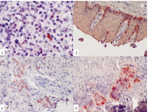
Figure 1-4. Positive reactions for Neospora caninum antigen, the ABC immunoperoxidase
technique, Mayer’s hematoxylin counterstaining:1-lymph node, granular immunoreactive stainings for N. caninum in the cytoplasm of a macrophage, calf; 2-cutaneous mucosa of the tongue, dense immunopositive areas in germinative epithelial cells, calf; 3-liver, immunopositivity for N. caninum in hepatocytes, calf; 4-intense positive reaction in the periphery of necrotic areas in the lungs, dog.
Figure 5. The appearance of the agarose gel after Neospora caninum-specific PCR. In the pathways marked as 80/10
also contribute to a prevalence increasing from generation to generation by transmitting the disease to their offspring. For the prevention of infection, dogs should be examined for N. caninum infection before bringing them onto farms.
The seropositivity rate determined in dogs (55.5%) in the present study is a rather high percentage and is above the rates previously reported for different provinces in Turkey [Bursa, 10% (27); Kırıkkale, 26.4% (10); Kars, 25.2% (28)]. This may be interpreted as dogs found on dairy farms being subjected to a higher rate of infection sources. In dogs, neosporosis mainly causes progressive neurological disorders and results in peracute mortality within 1 week in young animals (29,30). On 1 of the farms included in the study (Farm 1), anamnesis revealed that 1 of the pregnant dogs had aborted. PCR results demonstrated that the 6-week-old puppy that had died with signs of gastroenteritis was Neospora caninum-positive. Previous studies conducted on the seroprevalence of the parasite in Turkey have revealed data suggesting that dogs are infected with this particular agent. However, there is not sufficient information available on the epidemiology and routes of transmission of the disease in Turkey. The main routes of infection for cattle are transplacental transmission from the dam to the calf and the ingestion of oocysts that are shed in the feces of infected dogs through the consumption of contaminated feed. In the present study, the seropositivity rate of the dogs found on the farms where Neospora caninum-seropositive cattle (6.7%–37.7%) and abortions were detected (Table 1) was measured as 55.5%. This finding draws attention to the importance of dogs in the epidemiology of the disease. In a study conducted in dogs in Bursa Province, out of the 150 dogs examined, 15 had antibodies against Neospora caninum (27). In a serological survey conducted in aborted cows in Kars Province and its vicinity, 1 of the local cows yielded suspect results, while out of 73 imported Simmental cows, 6 (8.2%) were determined to be N. caninum-seropositive
(30). The immunopathological examination of a 20-day-old Simmental calf revealed the presence of the parasite in the brain, heart, thymus, skeletal muscle, and lymph nodes (10).
In the fecal examination of seropositive dogs, nonsporulated oocysts measuring 7.5-10 µm, which did not resemble the morphology of any known protozoon, were observed. These oocysts were not able to be isolated, given their small number and further feces samples taken being devoid of them. The period during which dogs shed oocysts in their feces is limited to 3–15 days, and, in general, a dog infected with neosporosis sheds oocysts only once in its lifetime. This reduces the chance of the parasite being isolated from the definitive host. Nevertheless, in the present study, the first clinical N. caninum infection reported in dogs from Turkey was described both clinically and pathologically.
In conclusion, in the present study, the relationship between neosporosis in cattle and dogs was clearly demonstrated. The findings obtained in the present study suggest that, for the prevention of N. caninum infection, which causes major economic losses in cattle due to abortions and the birth of congenitally infected calves, it is of great significance that infected cattle be culled. Furthermore, in order to prevent dogs, which play an important role in the transmission and dissemination of the disease, from feeding on aborted fetuses and expelled placenta, it is important that dogs be kept tethered on farms.
Acknowledgments
The authors would like to thank Assistant Professor Serkan Erat from Kırıkkale University, Faculty of Veterinary Medicine, for his help with statistical analysis. This project was supported by the Kırıkkale University Scientific Research Council (Project number: 2008/44).
References
1. Conrad PA, Barr BC, Sverlow KW, Anderson M, Daft B,
Kinde H, Dubey JP, Munson L, Ardans A. In vitro isolation and characterisation of a Neospora sp. from aborted bovine foetuses. Parasitology 1993; 106: 239–249.
2. Björkman C, Holmdahl J, Uggla A. An indirect enzyme-linked
immunoassay (ELISA) for demonstration of antibodies to
Neospora caninum in serum and milk of cattle. Vet Parasitol
1997; 68: 251–260.
3. Dubey JP. Neosporosis - the first decade of research. Int J Parasitol 1999; 29: 1485–1488.
4. Reichel MP. Neospora caninum infections in Australia and New
Zealand. Aust Vet J 2000; 78: 258–261.
5. Dubey JP. Review of Neospora caninum and neosporosis in animals. Korean J Parasitol 2003; 41: 1–16.
6. Armengol R, Pabon M, Adelantado C, Lopez-Gatius F, Almeria
S. First report of Neospora caninum abortion in a beef cow-calf herd from Andorra, Europe. J Parasitol 2006; 92: 1361–1362.
7. Bartels CJ, Hogeveen H, van Schaik G, Wouda W, Dijkstra T. Estimated economic losses due to Neospora caninum infection in dairy herds with and without a history of Neospora caninum associated abortion epidemics. In: Proceedings of the 11th International Symposium on Veterinary Epidemiology and Economics; 2006. Available at http://www.sciquest.org.nz/ node/63884.
8. Hornok S, Edelhofer R, Hajtos I. Seroprevalence of neosporosis
in beef and dairy cattle breeds in Northeast Hungary. Acta Vet Hung 2006; 54: 485–491.
9. Jardine JE, Wells BH. Bovine neosporosis in Zimbabwe. Vet Rec 1995; 137: 223.
10. Kul O, Kabakci N, Yildiz K, Ocal N, Kalender H, Ilkme NA.
Neospora caninum associated with epidemic abortions in
dairy cattle: the first clinical neosporosis report in Turkey. Vet Parasitol 2009; 159: 69–72.
11. Reitt K, Hilbe M, Voegtlin A, Corboz L, Haessig M, Pospischil A. Aetiology of bovine abortion in Switzerland from 1986 to 1995--a retrospective study with emphasis on detection of
Neospora caninum and Toxoplasma gondii by PCR. J Vet Med
A Physiol Pathol Clin Med 2007; 54: 15–22.
12. Dubey JP, Schares G. Diagnosis of bovine neosporosis. Vet Parasitol 2006; 31: 1–34.
13. McAllister MM, Dubey JP, Lindsay DS, Jolley WR, Wills RA, McGuire AM. Dogs are definitive hosts of Neospora caninum. Int J Parasitol 1998; 28: 1473–1478.
14. Lindsay DS, Upton SJ, Dubey JP. A structural study of the
Neospora caninum oocyst. Int J Parasitol 1999; 29: 1521–1523.
15. Ferroglio E, Pasino M, Ronco F, Bena A, Trisciuoglio A. Seroprevalence of antibodies to Neospora caninum in urban and rural dogs in north-west Italy. Zoonoses Public Health 2007; 54: 135–139.
16. Paradies P, Capelli G, Testini G, Cantacessi C, Trees AJ, Otranto D. Risk factors for canine neosporosis in farm and kennel dogs in southern Italy. Vet Parasitol 2007; 145: 240–244.
17. Dubey JP. Recent advances in Neospora and neosporosis. Vet Parasitol 1999; 84: 349–367.
18. Aguiar DM, Cavalcante GT, Rodrigues AA, Labruna MB, Camargo LM, Camargo EP, Gennari SM. Prevalence of
anti-Neospora caninum antibodies in cattle and dogs from Western
Amazon, Brazil, in association with some possible risk factors. Vet Parasitol 2006; 30: 71–77.
19. Lindsay DS, Dubey JP. Immunohistochemical diagnosis of
Neospora caninum in tissue sections. Am J Vet Res 1989; 50:
1981–1983.
20. Müller N, Zimmermann V, Hentrich B, Gottstein B. Diagnosis of Neospora caninum and Toxoplasma gondii infection by PCR and DNA hybridization immunoassay. J Clin Microbiol 1996; 34: 2850–2852.
21. Liddell S, Jenkins MC, Dubey JP. Vertical transmission of Neospora caninum in BALB/c mice determined by polymerase chain reaction detection. J Parasitol 1999; 85: 550– 555.
22. Osawa T, Wastling J, Maley S, Buxton D, Innes EA. A multiple antigen ELISA to detect Neospora-specific antibodies in bovine sera, bovine foetal fluids, ovine and caprine sera. Vet Parasitol 1998; 79: 19–34.
23. Atkinson RA, Cook RW, Reddacliff LA, Rothwell J, Broady KW, Harper PAW, Ellis JT. Seroprevalence of Neospora caninum infection following an abortion outbreak in a dairy cattle herd. Aust Vet J 2000; 78: 262–266.
24. Bartels CJ, Arnaiz-Seco JI, Ruiz-Santa-Quitera A, Bjorkman C, Frossling J, von Blumroder D, Conraths FJ, Schares G, van Manen C, Wouda W et al. Supranational comparison of
Neospora caninum seroprevalences in cattle in Germany, the
Netherlands, Spain and Sweden. Vet Parasitol 2006; 137: 17– 27.
25. Haddad JPA, Dohoo IR, John A, Van Leewen JA. A review of Neospora caninum in dairy and beef cattle — a Canadian perspective. Can Vet J 2005; 46: 230–224.
26. Chi J, VanLeeuwen JA, Weersink A, Keefe GP, Chi J. Direct production losses and treatment costs from bovine viral diarrhoea virus, bovine leukosis virus, Mycobacterium avium subspecies paratuberculosis, and Neospora caninum. Prev Vet Med 2002; 55: 137–153.
27. Coskun SZ, Aydin L, Bauer C. Seroprevalence of Neospora
caninum infection in domestic dogs in Turkey. Vet Rec 2000;
146: 649–654.
28. Mor N, Akca A. Epidemological studies upon Neospora
caninum in cattle and dogs in the province of Kars, Turkey:
a cross-sectional study. Kafkas Univ Vet Fak Derg 2012; 18: 193–199 (article in Turkish with abstract in English).
29. Björkman C, Lunden A, Holmdahl J, Barber J, Trees AJ, Uggla A. Neospora caninum in dogs: detection of antibodies by ELISA using an iscom antigen. Parasite Immunol 1994; 16: 643–648. 30. Akca A, Gokce HI, Guy CS, McGarry JW, Williams DJ.
Prevalence of antibodies to Neospora caninum in local and imported cattle breeds in the Kars province of Turkey. Res Vet Sci 2005; 78: 123–126.