O
riginal Ar
ticle / Ö
zgün Araştırma
Cite this article as: Şahin T, Arslan Zİ, Akansel G, Balaban O, Berk D, et al. Fluoroscopic Comparison of Cervical Spine Motion Using LMA CTrach, C-MAC Videolaryngoscope and Macintosh Laryngoscope. Turk J Anaesthesiol Reanim 2018; 46: 44-50.
Address for Correspondence/Yazışma Adresi: Onur Balaban E-mail: [email protected] ©Copyright 2018 by Turkish Anaesthesiology and Intensive Care Society - Available online at www.jtaics.org
©Telif Hakkı 2018 Türk Anesteziyoloji ve Reanimasyon Derneği - Makale metnine www.jtaics.org web sayfasından ulaşılabilir.
Received / Geliş Tarihi : 08.07.2017 Accepted / Kabul Tarihi : 17.10.2017
44
Fluoroscopic Comparison of Cervical Spine Motion Using LMA
CTrach, C-MAC Videolaryngoscope and Macintosh Laryngoscope
Servikal Omurga Hareketi: LMA CTrach, C-MAC Videolaringoskop ve Macintosh Laringoskopun
Floroskopik Karşılaştırması
Tülay Şahin1 , Zehra İpek Arslan1 , Gür Akansel2 , Onur Balaban3 , Derya Berk4 , Mine Solak1 , Kamil Toker5
1Department of Anaesthesiology and Critical Care, Kocaeli University School of Medicine, Kocaeli, Turkey 2Department of Radiology, Kocaeli University School of Medicine, Kocaeli, Turkey
3Department of Anaesthesiology and Pain, Dumlupınar University, Kutahya, Turkey 4Department of Anaesthesiology and Critical Care, Adatıp Hospital, Sakarya, Turkey 5Department of Anaesthesiology and Critical Care, İstinye University, İstanbul, Turkey
ORCID IDs of the authors: T.Ş. 0000-0002-3921-8037; Z.İ.A. 0000-0003-2968-1092; G.A. 0000-0002-8776-2942; O.B. 0000-0003-0953-4191; D.B. 0000-0002-2597-4894; M.S. 0000-0002-3212-4636; K.T. 0000-0002-3168-0489
Objective: Endotracheal intubation should be performed with care when cervical spine (C-spine) injury is suspected. The aim of this study was to evaluate the movement of the C-spine using fluorosco-py during intubation with Laryngeal Mask Airway (LMA) CTrach, C-MAC videolaryngoscope and Macintosh laryngoscope.
Methods: This was a single-centre, prospective, observational, con-trolled trial. In total, 22 surgical patients aged 18-65 years planned to undergo operation under general anaesthesia, were enrolled. X-ray images of the C-spine were obtained using fluoroscopy with the patients’ head in a neutral position. All patients underwent la-ryngoscopy using a Macintosh blade, LMA CTrach and C-MAC videolaryngoscope, and fluoroscopic images of the C-spine were ob-tained. All the patients were intubated at the last laryngoscopy sim-ulation (using the C-MAC). The atlanto-occipital distance (AOD) and angles between C0C1, C0C2, C0C3, C0C4, C1C2 and C2C3 lines were measured and compared between each device.
Results: The mean AOD was measured as 20.4 mm in a
neu-tral position, which decreased to 13.1, 17.2 and 12.3 mm after the insertion of the Macintosh laryngoscope, LMA CTrach and C-MAC videolaryngoscope, respectively. The differences were sig-nificant (p<0.001). Moreover, sigsig-nificant difference was noted in C0C2, C0C3 and C1C2 angles with the insertion of the three devices (p<0.001). The LMA CTrach resulted in significantly lesser C-spine movements in C0C2, C0C3 and C0C4 angles compared to the Mac-intosh laryngoscope and C-MAC videolaryngoscope (p<0.001). Conclusion: The LMA CTrach resulted in lesser C-spine move-ments compared to Macintosh laryngoscope and C-MAC video-laryngoscope. In case of the C-spine injury, LMA CTrach may be preferred and may cause fewer traumas during endotracheal intu-bation.
Keywords: Cervical spine motion, LMA C Trach, videolaryngos-copy, fluoroscopy
Amaç: Servikal omurga hasarından şüphelenildiğinde endotrake-al entübasyon dikkatli yapılmendotrake-alıdır. Bu çendotrake-alışmanın amacı LMA (Laryngeal Mask Airway) CTrach, C-MAC Videolaringoskop ve Macintosh Laringoskop ile entübasyon sırasında floroskopi ile ser-vikal omurga hareketini değerlendirmektir.
Yöntemler: Bu tek merkezli, prospektif, gözlemsel kontrollü bir çalışmaydı. Çalışmaya 18-65 yaşları arasında genel anestezi altında operasyon uygulanan 22 hasta dahil edildi. Endotrakeal entübas-yondan önce, hastanın başı nötr konumda iken servikal omurga floroskopi görüntüleri elde edildi. Tüm hastalarda standart Ma-cintosh laringoskop, LMA CTrach ve C-MAC Videolaringoskop kullanıldı. Laringoskopi simülasyonları sırasında servikal omur-gadaki değişikliklerin floroskopik görüntüleri elde edildi. Tüm hastalar son laringoskopi sırasında C-MAC kullanılarak entübe edildi. Floroskopi görüntüleri üzerinde C0 C1, C0 C2, C0 C3, C0
C4, C1 C2 ve C2 C3 arasındaki Atlantooksipital mesafeler ve açılar
ölçüldü ve aradaki farklar cihazlar arasında karşılaştırıldı. Bulgular: Ortalama Atlantooksipital mesafe, nötral pozisyonda 20,4 mm ölçüldü ve sırasıyla Macintosh laringoskop, LMA CT-rach ve C-MAC Videolaryngoscope'ın yerleştirilmesinden sonra 13,1, 17,2 ve 12,3'e düştü ve aradaki farklar anlamlıydı (p<0,001). C0 C2, C0 C3 and C1 C2 açılarında üç cihazın yerleştirilmesi ile de nötral pozisyona göre anlamlı fark vardı (p<0,001). LMA Ctra-ch C0 C2, C0 C3 and C0C4 açılarında Macintosh laringoskop ve C-MAC Videolaringoskop ile karşılaştırıldığında servikal omur-gada anlamlı olarak az harekete neden oldu (p<0,001).
Sonuç: Entübasyon sırasında LMA CTrach, Macintosh ve
C-MAC videolaringoskoptan daha az servikal omurga hareketine neden olduğundan; servikal travmalı hastalarda daha uygun bir seçenek olabilir.
Anahtar Kelimeler: Servikal omurga hareketi, LMA Ctrach, vide-olaringoskopi, floroskopi
Abstract / Ö
Introduction
C
ervical spine (C-spine) injuries occur in 1.5%-4% of all major trauma cases, and one-third of patients with C-spine injury require intubation after the injuries (1, 2). When the C-spine is unstable, there is a potential risk for spinal cord damage during endotracheal intubation with inter-vention and manipulations that cause head extension (3). The Laryngeal Mask Airway (LMA) CTrach (The LMA, North America Inc. San Diego, CA, USA) is a modified ver-sion of the intubating laryngeal mask airway (ILMA) with a fibreoptic system and a detachable liquid crystal display screen that allows real-time view of the airway during orotra-cheal intubation (4). Due to its unique design, it conforms to normal curvature of the upper airway and can be inserted in a neutral position of the head and neck (5). The C-MAC videolaryngoscope (Karl Storz, Tuttlingen, Germany) with a modified Macintosh blade may be a useful alternative for both routine and difficult airway management.Previous studies on C-spine movements during endotracheal in-tubation have compared direct laryngoscopy using a Macintosh blade with different airway devices including various videola-ryngoscopes (6-10). The ideal device for endotracheal intuba-tion in an emergency or elective C-spine instability setting re-mains controversial. To date, no study has examined specifically C-spine movements using the LMA CTrach in comparison to the Macintosh laryngoscope and C-MAC videolaryngoscope. The aim of this study was to evaluate C-spine movements during endotracheal intubation of healthy surgical patients using LMA CTrach, C-MAC videolaryngoscope and a con-ventional laryngoscope using a Macintosh blade.
Methods
This study was conducted in accordance with the ethical principles for human investigations, as outlined by Declara-tion of Helsinki. After the instituDeclara-tional ethics board approved the study (Committee No: 2008/42, İAEK: 7/1, Kocaeli University, 04/22/2008) and after patients provided written informed consent, 22 patients with American Society of An-esthesiologists (ASA) I or ASA II, aged 18-65 years, weigh-ing 55-95 kg, were included in this prospective study. This was designed as a crossover study. Three different intubation techniques were applied consecutively in one patient, and the same patient served as a control. The patients were planned to undergo elective surgery under general anaesthesia and re-quired orotracheal intubation. Excluded patients were those who were pregnant, non-fasted, had symptomatic or untreat-ed gastroesophageal reflux, had a body mass index of >35 kg m−2, had previous neck surgery, had unstable C-spine, had
known or expected difficult airway, or had a head and neck pathology.
At the preoperative visit, the following measurements were recorded: age, height, weight, body mass index, Mallampati classification (obtained with the patient in the sitting
posi-tion, tongue out, without phonation), thyromental and ster-nomental distances (measured with the patient in the sitting position, with the head in extension), mandibular protru-sions (A: the lower incisors can be protruded anterior to the upper incisors, B: the lower incisors can be brought edge to edge with the upper incisors, C: the lower incisors cannot be brought edge to edge with the upper incisors) and interinci-sor distance (mouth opening). Patients were pre-medicated with midazolam 0.03 mg kg−1. After standard monitoring
(electrocardiogram, pulse oximetry and non-invasive blood pressure) and pre-oxygenation with 100% oxygen, anaesthe-sia was induced with thiopental 5 mg kg−1 and fentanyl 1
µg kg−1. Following the induction of anaesthesia, the patients
were manually ventilated using a facemask. Rocuronium 0.6 mg kg−1 was administered; subsequently, the evoked response
of the adductor pollicis muscle to the ulnar nerve stimulation at the wrist (TOF-Guards Acceleromyograph; TOF-Guard; Organon Teknika, Oss, Netherlands) was used to ensure ad-equate neuromuscular blockade. When the TOF ratio was zero in all patients, endotracheal intubation simulations were performed. Anaesthesia was maintained with 2% sevoflurane in 50% oxygen and nitrous oxide during the simulations. Fluoroscopy is a technique that provides real-time X-ray im-aging, which is particularly useful for guiding various diag-nostic and interventional procedures. As endotracheal intu-bation is a dynamic process, spine movements can be variable during intubation. We determined the time point where the fluoroscopy images were captured as: ‘the insertion of the air-way devices and obtaining a view of larynx’. The study was conducted in four stages. Each patient underwent Macintosh laryngoscopy, LMA CTrach and C-MAC videolaryngosco-py. At the first stage, a fluoroscopic (Siemens AG. Munchen, Germany) image of the C-spine with the head in a neutral position was obtained. After the images were captured, la-ryngoscopy was performed using a Macintosh no. 3 blade, and the fluoroscopic image of the C-spine was obtained with the Macintosh blade in the pharynx. At the third stage, the Macintosh blade was withdrawn and a suitable LMA CTrach device was inserted in the pharynx, and the fluoroscopic im-age of the C-spine was obtained with the LMA CTrach in the pharynx. At the fourth stage, a C-MAC videolaryngoscope with a no. 3 blade attached was inserted in the pharynx, and the fluoroscopic image of the C-spine was obtained with the C-MAC videolaryngoscope in the pharynx.
The patients were ventilated through a mask in between the laryngoscopy procedures. The patients were intubated at the last stage with a polyvinylchloride endotracheal tube (internal diameter 8.0 mm for men and 7.5 mm for women) using the C-MAC videolaryngoscope (Karl Storz, Tuttlingen, Germa-ny). All laryngoscopy procedures were performed, and the X-ray images were obtained at the same time with an assistant holding the head and applying manual in line stabilisation. We calculated the equivalent dose as 0.05 mSv for the four fluoroscopy procedures, and this was approximately 1/50 of the neutral daily radiation exposure. The quality of the glottis view was recorded using Cormack and Lehane classification
45
(grade 1: entire glottis aperture; grade 2: partial glottis ap-erture; grade 3: free edged or ventral face of epiglottis; and grade 4: no recognisable structure). All the device insertions and intubations were performed by an investigator skilled at endotracheal intubation (>120 successful endotracheal intu-bations with all three devices) to minimise the bias. We used a size 3 LMA CTrach for patients weighing <50 kg, a size 4 LMA CTrach for patients weighing 50-70 kg and a size 5 LMA CTrach for patients weighing >70 kg. Before the in-sertion, the viewer was attached to the LMA CTrach and fo-cused by obtaining a sharp image of a sheet of text held 1 cm in front of the fibreoptic channel port. Cricoid pressure was not applied in this study. If intubation procedure lasted more than 120 seconds for each device, it was recorded as a failed attempt. The operation room staff and study team used radi-ation-resistant eyewear as well as upper and lower lead aprons with thyroid protection during the complete procedure. The study team also used radiation-resistant surgical gloves. The body of the patient was shielded with lead aprons except the head and neck. A radiologist blinded to the groups of the patients, evaluated the X-ray images, applied the lines to the X-ray images and measured the angles.
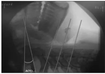
We determined the atlanto-occipital distance (AOD), refer-ence lines, common line and angles in accordance with liter-ature studies (Figure 1) (6, 8).
C0 (Mc Gregor’s) line: Line extends from the upper surface of the posterior edge of the hard palate to the most inferior point of the occipital bone.
C1 line: Line passing through the anterior and posterior arches of the atlas.
C2, C3 and C4 lines: Lines parallel to the inferior endplates of the vertebrae.
AOD: Vertical distance between the most inferior point of the occipital bone and the C1 reference line measured in
mil-limetre (mm).
The lower horizontal edge of the radiograph was used as a common reference line.
Angles between C0 C1, C0 C2, C0 C3, C0 C4, C1 C2 and C2 C3 lines were measured using a goniometer. All angles were cal-culated from the vertebrae minus the next vertebrae.
Angles between C0 C1, C0 C2, C0 C3, C0 C4 = C0 to common line angle minus C1, C2, C3 and C4 to common line angle, respectively.
Angle between C1 C2 = C1 to common line angle minus C2 to common line angle.
Angle between C2 C3 = C2 to common line angle minus C3 to common line angle.
Statistical analysis
Using previously published data for the C1 C2 angle from Ru-dolph et al. (6), we calculated the sample size as 22 patients for 80% power to detect a 10% difference in the C1 C2 angle. We used the Statistical Package of Social Science of Windows 16 (SPSS Inc.; Chicago, IL, USA) software. Values were given as mean ± standard deviation or median (interquartile range). For categorical data, we used chi-square test. For continuous data, normality was checked using the Kolmogorov-Smirnov test. If the distribution was found to be normal, parametric tests (Paired sample t-test) were used for the analysis; other-wise, nonparametric tests (Wilcoxon-Signed Ranks test) were used. Distribution was not normal for the measurement of C0 C1 using the Macintosh laryngoscope, C0 C4 in a neutral position, C2 C3 in a neutral position and C2 C3 angles using the LMA CTrach. A value of p<0.05 was considered as statis-tically significant.
Results
The demographic characteristics of patients are summarised in Table 1. Macroglossia was not detected in any patient. All patients’ mandibular protrusions were ‘A’ except one. All pa-tients were easily ventilated, only one of them needed Guedel airway insertion. Eighteen patients were intubated
success-46
Table 1. Demographic variables of patients. Values are mean (SD) or number (n)
Age (years) 39.4 (12.9)
Gender (Male/Female) (n) 4/18
ASA I/II 16/6
Mallampati I/II 17/5
Teeth morphology full/lack 17/5
Sternomental distance (cm) 14.6 (2.2)
Interincisor distance (cm) 4.4 (0.5)
Weight (kg) 74.8 (18.3)
Tiromental distance (cm) 8.1 (1)
Cormack-Lehane grade (I/II) 20/2
Figure 1. Fluoroscopic image of the upper C-spine and the skull base. This image shows the AOD and standard of the reference lines AOD: atlanto-occipital distance
fully at the first attempt, but 4 patients were intubated at the second or third attempt.
The mean AOD was measured as 20.4 mm in the neutral po-sition, which decreased significantly during the laryngoscopy procedure using all the three devices (Table 2).
The mean angle value measured between C0 C1 lines was 8.8 degrees in the neutral position and decreased to 5, 5.6 and 7.3 degrees when Macintosh blade, LMA CTrach and C-MAC videolaryngoscope, respectively, were inserted, and laryngoscopy was simulated.
The mean angle values in neutral position between C0 C2, C0 C3, C0 C4 and C1 C2 lines were measured as 19.9, 21, 20 and 25.8 degrees, respectively. All angles between C0 C2, C0 C3, C0 C4 and C1 C2 lines were significantly increased from the neu-tral position when the Macintosh blade, LMA CTrach and C-MAC videolaryngoscope were inserted, and laryngoscopy was simulated (p<0.001, p=0.012 for Macintosh laryngo-scope and p=0.010 for the LMA CTrach in the C0 C4 angle). The p values, measured values of angles in the neutral posi-tion and values of angles during laryngoscopy using the LMA CTrach, C-MAC videolaryngoscope and Macintosh laryngo-scope are outlined in Table 2.
When the LMA CTrach was compared to Macintosh laryn-goscopy, the AOD and the measured values of angles between C0 C2, C0 C3, C0 C4 and C1 C2 lines were significantly lower. The p values are summarised in Table 3.
When the LMA CTrach was compared to C-MAC video-laryngoscopy, the measured values of angles between C0 C1, C0 C2, C0 C3, C0 C4, C1 C2 and C2 C3 lines were significantly lower. The p values are summarised in Table 3.
When C-MAC was compared to Macintosh laryngoscope, no significant difference was observed in the angles measured between C0 C1, C0 C2, C0 C3, C0 C4, C1 C2 and C2 C3 lines.
The change in AOD was not significant (Table 3).
Discussion
This study showed a remarkable movement of the C-spine during laryngoscopy, which is proved by the evaluation of a significant change of certain angles from the neutral po-sition and the decrease in AOD. The changes in the angles were observed using the LMA CTrach, Macintosh blade and C-MAC videolaryngoscope. Intubation attempt using the LMA CTrach resulted in lesser C-spine movements in cer-tain angles when compared to the conventional laryngoscopy using the Macintosh blade and C-MAC videolaryngoscopy. Similar studies have reported that the C-spine motion mostly exceeds between C0 and C4 cervical segments with different types of airway devices, such as the conventional laryngo-scope with Macintosh and Miller blades, Bonfils rigid fibre-optic intubation device (Karl Storz Endoscopy-America, Inc. CA, USA), fibreoptic bronchoscope, GlideScope (Verathon Medical, Bothell, WA, USA) and Trachlight stylet (Laerdal Medical Limited, Kent, UK) (6, 8-12). Regarding these stud-
47
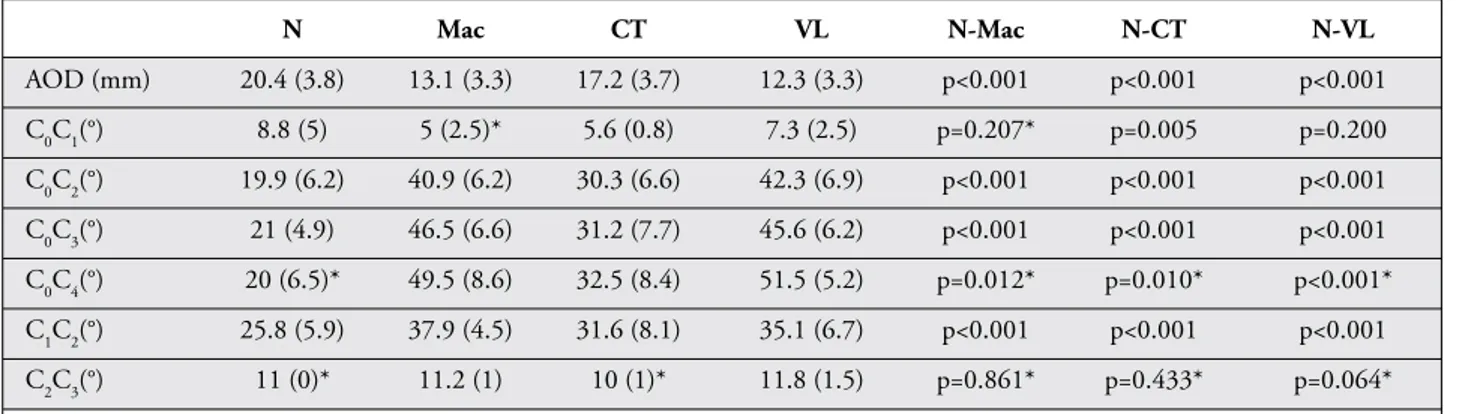
Table 2. AOD (mm) and C-spine motion measurements of patients using the Macintosh laryngoscope, LMA CTrach orC-MAC videolaryngoscope. Values are mean (SD) or median (interquartile range)
N Mac CT VL N-Mac N-CT N-VL AOD (mm) 20.4 (3.8) 13.1 (3.3) 17.2 (3.7) 12.3 (3.3) p<0.001 p<0.001 p<0.001 C0C1(°) 8.8 (5) 5 (2.5)* 5.6 (0.8) 7.3 (2.5) p=0.207* p=0.005 p=0.200 C0C2(°) 19.9 (6.2) 40.9 (6.2) 30.3 (6.6) 42.3 (6.9) p<0.001 p<0.001 p<0.001 C0C3(°) 21 (4.9) 46.5 (6.6) 31.2 (7.7) 45.6 (6.2) p<0.001 p<0.001 p<0.001 C0C4(°) 20 (6.5)* 49.5 (8.6) 32.5 (8.4) 51.5 (5.2) p=0.012* p=0.010* p<0.001* C1C2(°) 25.8 (5.9) 37.9 (4.5) 31.6 (8.1) 35.1 (6.7) p<0.001 p<0.001 p<0.001 C2C3(°) 11 (0)* 11.2 (1) 10 (1)* 11.8 (1.5) p=0.861* p=0.433* p=0.064*
*p values of AOD and the angles compared with the head in a neutral position and in laryngoscopy position using the LMA CTrach, Macintosh or C-MAC videolaryngoscope.
*Wilcoxon-Signed Ranks test was used. Values are median (interquartile range).
AOD: atlanto-occipital distance; N: neutral; Mac: Macintosh; CT: CTrach; VL: videolaryngoscope; SD: standard deviation.
Table 3. Comparison of p values of AOD and all angles using the LMA CTrach, Macintosh and C-MAC videolaryngoscope CT-Mac CT-VL Mac-VL AOD (mm) p=0.003 p=0.100 p=0.100 C0C1 (°) p=0.714* p=0.040 p=0.105* C0C2 (°) p<0.001 p<0.001 p=0.800 C0C3 (°) p<0.001 p<0.001 p=0.600 C0C4 (°) p<0.001 p<0.001 p=0.700 C1C2 (°) p=0.004 p=0.020 p=0.100 C2C3 (°) p=0.336* p=0.003* p=0.100
*Wilcoxon-Signed Ranks test was used. CT: LMA CTrach; VL: videolaryngoscope; AOD: atlanto-occipital distance
ies, our major aim was to investigate the C-spine motion be-tween C0 and C4 cervical segments. Although
videolaryngo-scopes improve the laryngeal view and have an easy learning curve, no advantages over Macintosh laryngoscopy in terms of the C-spine motion and intubation procedure time have been observed (13-17). Consistent with these studies, our study revealed that the C-MAC videolaryngoscope has no su-periority over Macintosh laryngoscope regarding the C-spine motion. However, the degree of C-spine movements with videolaryngoscopes remains controversial. GlideScope (Ver-athon Medical) videolaryngoscopy resulted in reduced move-ments of the C-spine in patients with unsecured C-spines compared to conventional Macintosh laryngoscope in a study that used video motion analysis (18). Truview videolaryngo-scope (Truphatek International, Netanya, Israel) was associat-ed with an improvassociat-ed laryngeal view and less movement at the C-spine (19). In a fluoroscopic study of a comparison of Air-Way Scope (Pentax Corporation, Tokyo, Japan), McCoy la-ryngoscope and Macintosh lala-ryngoscope, the AirWay Scope (Pentax Corporation) produced lesser movement of the up-per C-spine in patients with a normal C-spine (20). In a cine-fluoroscopy study, the use of a GlideScope videolaryngoscope (Verathon Medical) produced better glottic visualisation but did not significantly decrease the movement of the nonpatho-logic C-spine when compared with direct laryngoscopy (14). Videolaryngoscopy devices have a great variety. In an emer-gency situation, it is recommended to use the videolaryngos-copy device that proper experience has been acquired. As the patients included in the study did not have expected difficult airways, we preferred the C-MAC videolaryngoscope with the Macintosh blade because we had a good experience with C-MAC. Many of these devices have various sizes, angles of blades and different laryngoscopy techniques (some allowing direct view of glottis versus others allowing an indirect view with increased angles). Relevant differences in C-spine move-ments may occur between different blades of videolaryngo-scopes in an intubation scenario.
Fibreoptic intubation seems to be the preferred airway device over other intubation techniques in case of cervical instabil-ity (9, 21, 22). Although intubation with fibreoptic bron-choscope resulted in the lowest cervical motion compared to other intubation devices, fibreoptic intubation is not always possible without the use of head tilt-chin lift or jaw thrust manoeuvres, which causes the mostly intense motion in the unstable cervical segment and thereby great caution should be taken while performing these manoeuvres. Other disad-vantages of fibreoptic intubation include longer intubation times and the requirement of a skilled operator (21-23). The LMA CTrach resulted in lesser C-spine movements com-pared to laryngoscopy in our study. It has additional advan-tages, as noted by previous studies, such as easy insertion, high first intubation success rates and the ability of oxygen-ation and ventiloxygen-ation throughout the procedure (24-26). The intubating laryngeal mask produces segmental movement of the C-spine when applied with manual in-line stabilisation
in patients with C-spine pathology (27). However, supraglot-tic devices cause C-spine movements lesser or same as that by conventional laryngoscopes (23, 28). Many authors have studied about the fibreoptic intubation, ILMA and Mac-intosh laryngoscope. Endotracheal intubation with ILMA caused lesser spine movements when cervical extension angles were evaluated (21-24).
The LMA CTrach has a different design and technique of use when compared to the conventional laryngoscopy and videolaryngoscopy. In our opinion, an operator who is expe-rienced in ILMA could easily use the LMA CTrach. Supra-glottic airway devices are suggested in case of a ‘can’t ventilate, can’t intubate’ scenario, and ILMA is one of the alternative difficult intubation approaches in difficult intubation guide-lines when an intubation attempt using a conventional laryn-goscopic device fails (29). The LMA CTrach has a remarkable advantage over a standard ILMA with regard to providing a view of glottis. One disadvantage of the LMA CTrach is being a special device, which may not be available in small medical centres and emergency clinics, although it is success-fully used in many anaesthesiology clinics around the world. Limitations of our study were that we studied only healthy adult patients undergoing elective procedures, which did not really represent a true C-spine injury. Moreover, there was the inability to assess cervical movements in the coronal plane as our fluoroscopy images were captured only in the sagit-tal plane. Another limitation is that only laryngoscopy was performed with the LMA CTrach, an endotracheal tube was not inserted. Passing the endotracheal tube through the LMA CTrach may need additional manoeuvres.
Conclusion
The LMA CTrach resulted in lesser C-spine movements during endotracheal intubation of healthy surgical patients compared to Macintosh laryngoscope and C-MAC videolaryngoscope. The most important advantage of the LMA CTrach is the abil-ity of oxygenation and ventilation throughout the procedure. It may be considered one of the first-line intubating tools and may be helpful for a less traumatic endotracheal intubation for adult patients with suspected C-spine injury.
Ethics Committee Approval: Ethics committee approval was re-ceived for this study from the ethics committee of Kocaeli Universi-ty School of Medicine (2008/42, İAEK: 7/1)
Informed Consent: Written informed consent was obtained from patients who participated in this study.
Peer-review: Externally peer-reviewed.
Author Contributions: Concept – T.Ş., Z.İ.A.; Design – T.Ş., Z.İ.A., G.A.; Supervision – T.Ş., M.S., K.T.; Resources – T.Ş., Z.İ.A., G.A.; Materials – T.Ş., K.T.; Data Collection and/or Pro-cessing – T.Ş., Z.İ.A.; Analysis and/or Interpretation – T.Ş., Z.İ.A., G.A., O.B., D.B., M.S., K.T.; Literature Search – Z.İ.A., O.B., D.B.; Writing Manuscript – T.Ş., Z.İ.A., O.B.; Critical Review – T.Ş., G.A., M.S.
Conflict of Interest: No conflict of interest was declared by the authors. Financial Disclosure: The authors declared that this study has re-ceived no financial support.
Etik Komite Onayı: Bu çalışma için etik komite onayı Kocaeli Üniversitesi Tıp Fakültesi’nden (2008/42, İAEK: 7/1) alınmıştır. Hasta Onamı: Yazılı hasta onamı bu çalışmaya katılan hastalardan alınmıştır.
Hakem Değerlendirmesi: Dış bağımsız.
Yazar Katkıları: Fikir – T.Ş., Z.İ.A.; Tasarım – T.Ş., Z.İ.A., G.A.; Denetleme – T.Ş., M.S., K.T.; Kaynaklar – T.Ş., Z.İ.A., G.A.; Mal-zemeler – T.Ş., K.T.; Veri Toplanması ve/veya İşlemesi – T.Ş., Z.İ.A.; Analiz ve/veya Yorum – T.Ş., Z.İ.A., G.A., O.B., D.B., M.S., K.T.; Literatür Taraması – Z.İ.A., O.B., D.B.; Yazıyı Yazan – T.Ş., Z.İ.A., O.B.; Eleştirel İnceleme – T.Ş., G.A., M.S.
Çıkar Çatışması: Yazarlar çıkar çatışması bildirmemişlerdir. Finansal Destek: Yazarlar bu çalışma için finansal destek al-madıklarını beyan etmişlerdir.
References
1. Hastings RH, Marks JD. Airway management for trauma pa-tients with potential cervical spine injuries. Anesth Analg 1991; 73: 471-82. [CrossRef]
2. Ball PA. Critical care of spinal cord injury. Spine (Phila Pa 1976) 2001; 26: 27-30. [CrossRef]
3. Ghafoor AU, Martin TW, Gopalakrishnan S, Viswamitra S. Caring for the patients with cervical spine injuries: what have we learned? J Clin Anesth 2005; 17: 640-9. [CrossRef]
4. Liu EH, Goy RW, Chen FG. The LMA CTrach, a new lary-ngeal mask airway for endotracheal intubation under vision: evaluation in 100 patients. Br J Anaesth 2006; 96: 396-400.
[CrossRef]
5. Tripathi DC, Jha PS, Trivedi LP, Doshi SM, Modia B. LMA C Trach aided endotracheal intubation in simulated cases of cervical spine injury: A series of 30 cases. Saudi J Anaesth 2013; 7: 165-9. [CrossRef]
6. Rudolph C, Schneider JP, Wallenborn J, Schaffranietz L. Mo-vement of the upper cervical spine during laryngoscopy: a com-parison of the Bonfils intubation fiberscope and the Macintosh laryngoscope. Anaesthesia 2005; 60: 668-72. [CrossRef]
7. McElwain J, Laffey JG. Comparison of the C‐MAC, Airtraq and Macintosh laryngoscopes in patients undergoing tracheal intubation with cervical spine immobilization. Br J Anaesth 2011; 107: 258-64. [CrossRef]
8. Wong DM, Prabhu A, Chakraborty S, Tan G, Massicotte EM, Cooper R. Cervical spine motion during flexible bronchoscopy compared with the Lo-Pro GlideScope. Br J Anaesth 2009; 102: 424-30. [CrossRef]
9. Brimacombe J, Keller C, Kunzel KH, Gaber O, Boehler M, Puhringer F. Cervical spine motion during airway manage-ment: a cinefluoroscopic study of posteriorly destabilized third cervical vertebrae in human cadavers. Anesth Analg 2000; 91: 1274-8. [CrossRef]
10. Houde BJ, Williams SR, Cadrin-Chenevert A, Guilbert F, Drolet P. A comparison cervical spine motion during orotra-cheal intubation with the Trachlight or the flexible fiberoptic
bronchoscope. Anesth Analg 2009; 108: 1638-43. [CrossRef]
11. Urakami Y, Takenaka I, Nakamura M, Fukuyama H, Aoyama K, Kadoya T. The reliability of the Bellhouse test for evaluating extension capacity of the occipitoatlantoaxial complex. Anesth Analg 2002; 95: 1437-41. [CrossRef]
12. LeGrand SA, Hindman BJ, Dexter F, Weeks JB, Todd MM. Craniocervical motion during direct laryngoscopy and orotra-cheal intubation with the Macintosh and Miller blades: an in vivo cinefluoroscopic study. Anesthesiology 2007; 107: 884-91.
[CrossRef]
13. Turkstra TP, Craen RA, Pelz DM, Gelb AW. Cervical spine motion: a fluoroscopic comparison during intubation with the lighted stylet, GlideScope and Macintosh laryngoscope. Anesth Analg 2005; 101: 910-5. [CrossRef]
14. Robitaille A, Williams SR, Tremblay MH, Guilbert F, Theriault M, Drolet P. Cervical spine motion during tracheal intubation with manual in-line stabilization: direct laryngoscopy versus GlideScope videolaryngoscopy. Anesth Analg 2008; 106: 935-41. [CrossRef]
15. Savoldelli GL, Schiffer E, Abegg C, Baeriswyl V, Clergue F, Wa-ebar JL. Learning curves of the Glidescope, the McGrath and the Airtraq laryngoscopes: a manikin study. Eur J Anaesthesiol 2009; 26: 554-8. [CrossRef]
16. Stroumpoulis K, Pagoulatou A, Violari M, Ikonomou I, Ka-lantzi N, Kastrinaki K, et al. Videolaryngoscopy in the manage-ment of the difficult airway: a comparison with the Macintosh blade. Eur J Anaesthesiol 2009; 26: 218-22. [CrossRef]
17. Wasem S, Lazarus M, Hain J, Festl J, Kranke P, Roewer N, et al. Comparison of the Airtraq and the Macintosh laryngoscope for double-lumen tube intubation. Eur J Anaesthesiol 2013; 30: 180-6. [CrossRef]
18. Kill C, Risse J, Wallot P, Seidl P, Steinfeldt T, Wulf H. Videolar-yngoscopy with Glidescope reduces cervical spine movement in patients with unsecured cervical spine. J Emerg Med 2013; 44: 750-6. [CrossRef]
19. Bharadwaj A, Khurana G, Jindal P. Cervical Spine Movement and Ease of Intubation Using Truview or McCoy Laryngoscope in Difficult Intubation. Spine (Phila Pa 1976) 2016; 41: 987-93. [CrossRef]
20. Maruyama K, Yamada T, Kawakami R, Kamata T, Yokochi M, Hara K. Upper cervical spine movement during intubation: fluoroscopic comparison of the AirWay Scope, McCoy laryn-goscope, and Macintosh laryngoscope. Br J Anaesth 2008; 100: 120-4. [CrossRef]
21. Gercek E, Wahlen BM, Rommens PM. In vivo ultrasound re-al-time motion of the cervical spine during intubation under manual in–line stabilization: a comparison of intubation met-hods. Eur J Anaesthesiol 2008; 25: 29-36. [CrossRef]
22. Sahin A, Salman MA, Erden IA, Aypar U. Upper cervical spine vertebrae movement during intubating laryngeal mask, fibre-optic and direct laryngoscopy: a video-fluoroscopic study. Eur J Anaesthesiol 2004; 21: 819-23. [CrossRef]
23. Wahlen BM, Gercek E. Three-dimensional cervical spine mo-vement during intubation using the Macintosh and Bullard lar-yngoscopes, the Bonfils fibrescope and the intubating laryngeal mask airway. Eur J Anaesthesiol 2004; 21: 907-13. [CrossRef]
24. Waltl B, Melischek M, Schuschnig C, Kabon B, Erlacher W, Nasel C, et al. Tracheal intubation and cervical spine excursion: direct laryngoscopy vs. intubating laryngeal mask. Anaesthesia
25. Lui EH, Goy RW, Lim Y, Chen FG. Success of tracheal intuba-tion with intubating laryngeal mask airways: a randomized trial of the LMA Fastrach and LMA CTrach. Anesthesiology 2008; 108: 621-6. [CrossRef]
26. Malik MA, Subramaniam R, Churasia S, Maharaj CH, Har-te BH, Laffey G. Tracheal intubation in patients with cervical spine immobilization: a comparison of the AirwayScope, LMA CTrach and the Macintosh Laryngoscopes. Br J Anaesth 2009; 102: 654-61. [CrossRef]
27. Kihara S, Watanabe S, Brimacombe J, Taguchi N, Yaguchi Y, Yamasaki Y. Segmental cervical spine movement with the
in-tubating laryngeal mask during manual in-line stabilization in patients with cervical pathology undergoing cervical spine sur-gery. Anesth Analg 2000; 91: 195-200. [CrossRef]
28. Kılıç T, Goksu E, Durmaz D, Yıldız G. Upper cervical spine movement during intubation with different airway devices. Am J Emerg Med 2013; 31: 1034-6. [CrossRef]
29. Apfelbaum JL, Hagberg CA, Caplan RA, Blitt CD, Connis RT, Nickinovich DG, et al. Practice guidelines for management of the difficult airway: an updated report by the American Society of Anesthesiologists Task Force on Management of the Diffi-cult Airway. Anesthesiology 2013; 118: 251-70. [CrossRef]