Images in cardio-thoracic surgery
Dyspnea due to giant goiter
Fatih Alper
a,*, Mecit Kantarci
a, Ibrahim Can Kurkcuoglu
b, Ahmet A. Balik
ca
Department of Radiology, School of Medicine, Atatu¨rk University, 25240 Erzurum, Turkey
b
Department of Thoracic Surgery, School of Medicine, Atatu¨rk University, 25240 Erzurum, Turkey
cDepartment of General Surgery, School of Medicine, Atatu¨rk University, 25240 Erzurum, Turkey
Received 2 April 2003; received in revised form 10 April 2003; accepted 15 April 2003
Keywords: Goiter; Dyspnea; Thoracic wall
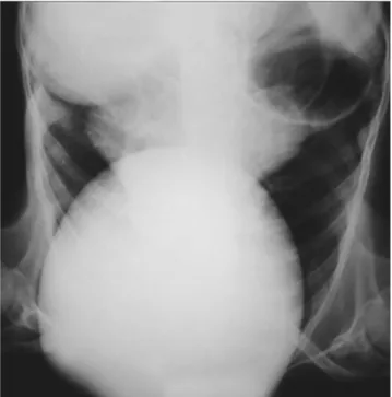
A 53-year-old man was admitted with limitation of cervical
movements, progressive dyspnea, and a slow-growing and
painful mass originating from both lobes of the thyroid
gland (
Fig. 1
). On physical examination, the dimensions of
the mass were 20 £ 25 £ 30 cm. The goiter appeared as a
cervical mass with a deviation of the trachea (
Fig. 2
). Via a
cervical incision subtotal thyroidectomy was performed
and a tissue weighing 1700 g was removed. After the
removal of the huge mass, cutaneous deformation was
corrected with resection. The histopathologic examination
revealed adenomatous multinodular goiter. The
postopera-tive course was uneventful and the patient was discharged
home 8 days after surgery. He is doing well at the 6-month
follow-up visit.
European Journal of Cardio-thoracic Surgery 24 (2003) 302
www.elsevier.com/locate/ejcts
1010-7940/03/$ - see front matter q 2003 Elsevier Science B.V. All rights reserved. doi:10.1016/S1010-7940(03)00286-0
Fig. 1. Chest radiograph demonstrating a huge mass.
Fig. 2. Lateral chest X-ray with cervical mass. * Corresponding author. Tel.: 316-6333x2266; fax:
þ90-442-316-6340.
E-mail address: [email protected] (F. Alper).