An Extraordinary Cause of Asymmetric Right Upper Lung
Edema: Mitral Regurgitation
Asimetrik Sağ Üst Pulmoner Ödemin Olağandıșı Nedeni: Mitral Yetersizlik
Ömer Faruk Keskin
1, Uygar Çağdaș Yüksel
1, Serkan Asil
1, Veysel Özgür Barıș
11 Gülhane Research and Education Hospital, Ankara, Turkey
In asymmetric pulmonary edema and consolidation patterns, pneumonia is always the primary diagnosis; however, mitral regurgitation should be considered in asymmetric pulmonary edema, where right upper pulmonary involvement predominates. Asymmetric right upper pulmonary edema may be caused from the jet flow directed towards the upper right pulmonary vein and increased hydrostatic pressure. So in these cases; investigation of mitral insufficiency by bedside transthoracic echocardiography is recommended.
Key Words: Asymmetric pulmonary edema, Mitral Regurgitation
Konsolidasyon ve asimetrik pulmoner ödemin en sık nedeni pnömoni olmasına rağmen sağ üst pulmoner tutulumun ön planda olduğu asimetrik pulmoner ödem tablolarında mitral yetersizlik göz önünde bulundurulmalıdır. Sağ üst pulmoner vene doğru yönelen jet akıșı ve artmıș hidrostatik basınç asimetrik pulmoner ödeme yol açabilir. Bu durumlarda yatakbașı transtorasik ekokardiyografi mitral yetersizliğinin gösterilmesinde önerilmektedir.
Anahtar Sözcükler: Asimetrik pulmoner ödem, Mitral yetersizlik
A 71 year old woman presented to the emergency department with progressive dyspnea, orthopnea, and dry cough. Her medical history was notable for hypertension and paroxysmal atrial fibrillation for which she was taking dabigatran 2x150 mg and nifedipine 1x60 mg. Physical examination revealed sinus tachycardia at 105 bpm, a blood pressure of 170/ 90 mm Hg, respiratory rate 24 breaths/min, and a grade 3/6 pansystolic murmur that was most prominent over the 5th intercostal space. Fine crackles were detected in the lung examination, which were more pronounced in the right lung upper zone. The patient’s O2 saturation was 70% in room air and 88% with 5 lt/min nasal O2 support. Laboratory examination was normal except Pro BNP which was 1400 mg/dl.
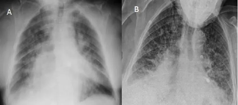
A chest radiograph obtained at the time of admission showed cardiomegaly and alveolar edema in the upper lobes of both lungs which was more pronounced on the right side than on the left with bilateral pulmonary vascular congestion (Figure 1).
Electrocardiography showed sinus tachycardia and ST depressions in the
inferolateral derivations. Transthoracic echocardiography revealed normal left ventricular systolic function, moderate-severe eccentric mitral regurgitation, moderate tricuspit regurgitation and a systolic pulmonary artery pressure of 60 mmHg.A high resolution computed tomographic scan of the chest showed asymmetric pulmonary edema, predominantly in the right upper lobe (Figure 2).
Laboratory tests and her history ruled out conditions with the same radiologic appearance, namely lobar pneumonia,
AnkaraÜniversitesiTıpFakültesiMecmuası 2018, 71 (1)
DOI: 10.1501/Tıpfak_000001001
DAHİLİ TIP BİLİMLERİ/ MEDICAL SCIENCES
Olgu Sunumu / Case Report
Received: Feb. 07, 2018 Accepted: April 04, 2018 Corresponding Author:
Veysel Özgür Barıș
E-mail: [email protected] GSM: +90 (505) 454 50 54
University of Health Science, Dıșkapı Yıldırım Beyazıt Research Gülhane Research and Education Hospital, Ankara,Turkey,
Ankara Üniversitesi Tıp Fakültesi Mecmuası 2018, 71 (1)
An Extraordinary Cause of Asymmetric Right Upper Lung Edema: Mitral Regurgitation
74
bronchial obstruction, veno-occlusive disease, unilateral aspiration, and pulmonary contusion .Since she had moderate to severe mitral regurgation, asymmetric right upper lung edema was considered as the primary diagnosis. The patient was put on diuretics and antihypertensive agents. After stabilization of the patient, transesophageal echocardiography was performed to investigate the mitral valve. Transesophageal echocardiography revealed moderate
eccentric mitral regurgitation with annular calcification. The reduction of the severity of mitral regurgitation was attributed to stabilization of the patient's volume status.
Discussion
Cardiogenic pulmonary edema, as the non-cardiogenic variety, can be both interstitial and alveolar, depending on the degree of alteration of the intravascular homeostasis. Initially,
chest radiographs of cardiogenic edema show peribronchial cuffing, Kerley lines, and subpleural fluid that is most pronounced centrally (1). Pulmonary edema associated with mitral valve insufficiency is usually bilateral; however, isolated or predominantly right upper lobe involvement can also be seen (2,3). Schnyder et al reported that asymmetric pulmonary edema was seen 9% in mitral regurgitation patients (4). Asymmetric pulmonary
Figure 1: A: Chest radiography shows cardiomegaly and alveolar edema in the upper lobes. B: Chest radiography of patients after the diuretic treatment.
Figure 2: A:Isolated right upper pulmonary consolidation. B: Bilateral pulmonary edema in the middle zones espicially right lung.C: Bilateral pulmonary edema in the lower zones espicially right lung
Journal of Ankara University Faculty of Medicine 2018, 71 (1)
Ömer Faruk Keskin, Uygar Çağdaș Yüksel, Serkan Asil, Veysel Özgür Barıș 75
edema due to mitral insufficiency has been reported to occur more frequently in posterior leaflet involvement (5,6). Increased intravascular hydrostatic pressure of mitral valve insufficiency has been found to result in consequent pulmonary edema (7). It is thought that the cause of right upper pulmonary edema may be the jet flow directed towards the upper right pulmonary vein with increased
hydrostatic pressure. Roach et al reported that the jet of regurgitant flow in a patient with a flail posterior valve leaflet is directed specifically toward the right pulmonary vein (5). In our patient we considered that the right upper lobe edema is the result of an asymmetric increase in the hydrostatic pressure of the right upper lung caused by asymmetric blood flow from the left atrium.
Conclusion
In asymmetric pulmonary edema and consolidation patterns, pneumonia is always the primary diagnosis. Mitral regurgitation, however, should also be considered in asymmetric pulmonary edema, where right upper pulmonary
involvement predominates. Investigation of mitral insufficiency by
bedside transthoracic echocardiography is recommended in all patients.
REFERENCES
1. Ponikowski P, Voors AA, Anker SD, et al. 2016 ESC Guidelines for the diagnosis and treatment of acute and chronic heart failure: The Task Force for the diagnosis and treatment of acute and chronic heart failure of the European Society of Cardiology (ESC). Developed with the special contribution of the Heart Failure Association (HFA) of the ESC. Eur J Heart Fail 2016;18:891-975.
2. Diana A, Guglielmini C, Pivetta M, et al. Radiographic features of cardiogenic pulmonary edema in dogs with mitral regurgitation: 61 cases (1998-2007). J Am Vet Med Assoc 2009;235:1058-1063.
3. Raman S, Pipavath S. Images in clinical medicine. Asymmetric edema of the upper lung due to mitral valvular dysfunction. N Engl J Med 2009;361:e6.
4. Schnyder PA, Sarraj AM, Duvoisin BE, et al. Pulmonary edema associated with mitral regurgitation: prevalence of predominant involvement of the right upper lobe. AJR Am J Roentgenol 1993;161:33-36.
5. Roach JM, Stajduhar KC, Torrington KG. Right upper lobe pulmonary edema caused by acute mitral regurgitation. Diagnosis by transesophageal echocardiography. Chest
1993;103:1286-1288.
6. Miyatake K, Nimura Y, Sakakibara H, et al. Localisation and direction of mitral regurgitant flow in mitral orifice studied with combined use of ultrasonic pulsed Doppler technique and two dimensional echocardiography. Br Heart J 1982;48:449-458.
7. Young AL, Langston CS, Schiffman RL, et al. Mitral valve regurgitation causing right upper lobe pulmonary edema. Tex Heart Inst J 2001;28:53-56.