ABSTRACT
Introduction: Although bioresorbable scaffolds (BRS) are considered a new paradigm in stent technology, operators are often discouraged from implanting BRS because of increased strut thickness, reduced radial force, requirement for pre- and postdilatation of the lesions, and concerns about the risk of stent thrombosis. We compared procedure and fluoroscopy duration, cumulative radiation dose, and contrast agent volume among patients undergoing BRS or drug-eluting stent (DES) implantation.
Patients and Methods: One hundred thirty-four patients with a total of 165 coronary lesions, including 64 patients (78 lesions) with BRS and 70 patients (87 lesions) with DES, were selected. Clinical and procedural characteristics and angiographic features were calculated. Procedure and fluoroscopy time, volume of contrast medium, and cumulative radiation dose (Gy) were compared.
Results: The number of predilated and postdilated lesions was higher in the BRS group than in the DES group, although baseline lesion morphologies were similar. Stent diameters were comparable between the two groups. Larger postdilatation balloon sizes were chosen in the BRS group. Mean procedure time (45.4 ± 16.1 minutes vs. 38.3 ± 15.1 minutes; p= 0.010), volume of contrast medium (207.7 ± 80.7 mL vs. 154.7 ± 74.6 mL; p= 0.001), fluoroscopy duration (15.9 ± 6.6 minutes vs. 13.1 ± 6.6 minutes; p= 0.014), and radiation dose (1.80 ± 1.08 Gy vs. 1.44 ± 0.91 Gy, p= 0.037) were significantly higher in the BRS group than in the DES group. Conclusion: BRS implantation leads to prolonged fluoroscopy, longer procedure duration, greater contrast volume, and higher radiation exposure compared with DES procedures.
Key Words: Coronary artery disease; percutaneous coronary intervention
Eriyebilen Stentlerin Prosedür Üzerine Olan Etkilerinin İlaç Kaplı Stentlerle Karşılaştırılması
ÖZET
Giriş: Artmış strut kalınlığı, azalmış stentin açılma kuvveti, stent takılmadan önce ve sonrasında balon yapma ihtiyacı, stent trombozu riskinde artış gibi birçok faktör operatörleri yenilikçi bir teknoloji olarak sunulan eriyebilen stentlerin (ES) kullanılmasında endişeye itmiştir. Bu çalışmada ES ile ilaç salınımlı stent (İSS) kullanılan hastalardaki işlem süresi ve floroskopi zamanı, opak miktarı ve radyasyon dozu karşılaştırılması hedeflenmiştir.
Hastalar ve Yöntem: Çalışmada koroner arter hastalığı tanısı alan 134 hastadaki 165 lezyon ele alınmış olup, bunlardan ES takılan 64 hastadaki 78 lezyon ile İSS takılan 70 hastadaki 87 lezyon dahil edilmiştir. İşlemle ilgili, klinik ve anjiyografik özellikler her grup için hesaplanmıştır. Her iki gruptaki prosedür ve floroskopi zamanı, kontrast miktarı ve toplam radyasyon dozu (Gy) karşılaştırılmıştır.
Bulgular: Başlangıç lezyon özellikleri her iki grupta benzer saptanmasına rağmen balon ile pre- ve post-dilatasyon anlamlı olarak ES takılanlarda daha yüksek saptandı. Stent çapları iki grupta da benzer iken ES grubunda postdilatasyon için daha yüksek çaplı balonlar tercih edildi. Ortalama işlem süresi (sırasıyla 45.4 ± 16.1 dakika ve 38.3 ± 15.1 dakika, p= 0.01), kontrast volümü (sırasıyla 207.7 ± 80.7 mL ve 154.7 ± 74.6 mL; p= 0.001), floroskopi zamanı (sırasıyla 15.9 ± 6.6 dakika ve 13.1 ± 6.6 dakika, p= 0.014), radyasyon dozu (sırasıyla 1.8 ± 1.08 Gy ve 1.44 ± 0.91 Gy, p= 0.037) ES grubunda İSS grubuna kıyasla anlamlı olarak daha yüksek bulunmuştur.
Sonuç: ES kullanımı İSS ile karşılaştırıldığında uzamış floro ve işlem süresi, artmış opak yükü ve radyasyon dozu ile ilişkilidir.
Anahtar Kelimeler: Koroner arter hastalığı; perkütan koroner girişim Beytullah Çakal1
1 University of Istanbul Medipol, Faculty of Medicine, Department of Cardiology, Istanbul, Turkey
Impact of Bioresorbable Scaffold Use on
Procedural Features Compared with
New-Generation Drug-Eluting Stents
Beytullah Çakal
E-mail: [email protected] Submitted: 17.10.2019 Accepted: 21.02.2020
Available Online Date: 30.04.2020
© Copyright 2020 by Koşuyolu Heart Journal. Available on-line at
www.kosuyoluheartjournal.com Correspondence
Cite this article as: Çakal B. Impact of bioresorbable scaffold use on procedural features compared with new-generation drug-eluting stents. Koşuyolu Heart J 2020;23(1):38-45.
INTRODUCTION
Drug- eluting stents (DES), which inhibit cell proliferation as compared with bare metal stents (BMS), were introduced in 1999, followed by second-generation DES, which significantly reduce restenosis and target lesion revascularization (TLR) and are as-sociated with a lower rate of stent thrombosis(1). Despite these
positive effects, the use of metallic DES has several disadvan-tages, such as vessel caging, diminished vasomotion, jailing side branches, preclusion of further bypass grafting, and foreign-body response that stimulates inflammation due to the implantation of permanent metallic material(2,3).
To overcome the limitations of DES, bioresorbable scaffolds (BRS) represent a new era of stent technology that elicits resorp-tion of foreign material, promoting vasomotor tone of the vessel and providing regulation of coronary blood flow(4,5). Advances in
the development of BRS began with the introduction of the Ab-sorb scaffold (Abbott Vascular, Santa Clara, CA, USA) and con-tinued with at least 22 BRS devices, some of which are still under development(6).
Although BRS appear to be appealing alternatives to metallic stents, factors such as increased strut thickness, concerns regard-ing late or very late stent thrombosis, target-vessel revasculariza-tion, weaker radial strength, inferior deliverability, and suboptimal visualization under fluoroscopy have discouraged operators from using these devices(7).
The aforementioned features and specific technical imple-mentation of BRS make these devices technically more demand-ing than DES deployment. The purpose of this study was to inves-tigate procedure and fluoroscopy duration, cumulative radiation dose, and volume of contrast agent in patients treated with BRS compared with new-generation DES.
PATIENTS and METHODS
Ethics committee approval was received for this study from the Istanbul Medipol University Non-Interventional Clinical Researches Ethics Committee (10840098-604.01.01-E.199).
Patients
In this single-center retrospective study, we included a total of 134 patients with 165 lesions. Sixty-four patients with 78 lesions underwent single or multivessel percutaneous coronary intervention with an everolimus-eluting BRS device (n= 19; Absorb BRS; Abbott Vascular, Santa Clara, CA, USA) or the novolimus-eluting BRS device (n= 59; Elixir Medical Corpo-ration) from February 2017 until January 2018, and 70 patients with a total of 87 lesions were treated with new-generation DES (Xience Pro, Abbott Vascular) from May 2018 until December 2018 at the Cardiology Clinic of Istanbul Medipol University. The inclusion criteria were stable coronary artery disease or
unstable angina or non-ST elevation myocardial infaction and age ≥ 18 years. Major exclusion criteria were defined as acute ST-segment elevation myocardial infarction, hemodynamically unstable arrhythmias, left ventricular ejection fraction of 30%, lesions in arterial or saphenous vein grafts, or restenotic lesions in either BRS- or DES-implanted patients. The choice to im-plant a BRS or DES was left to the decision of the operator.
Baseline clinical and procedural characteristics were record-ed for each case. Procrecord-edural time was definrecord-ed as the time from the onset of guided catheter angiography to final angiography of the target vessel. Total air kerma at the interventional reference point (Ka, r, Gy) was the procedural cumulative air kerma (X-ray energy delivered to air) at the interventional reference point, which assessed the hazard at that specific location and was re-corded for each procedure. Total fluoroscopy time was defined as the total length of time of fluoroscopic radiation exposure as measured during the period of coronary intervention.
Angiography Procedure and BRS Implantation
The recommended technique for BRS included adequate lesion preparation (P), appropriate sizing (S), and post dilata-tion (P), known as the PSP protocol. The goal was to achieve a final stenosis diameter of < 10% with a +0.5 mm noncompliant balloon inflated to high pressure (> 16 atm)(8).
The American College of Cardiology/American Heart As-sociation (ACC/AHA) classification was used to define the le-sions(9). The size of the guide catheter was either 6 or 7 Fr.
Lesion preparation using dedicated devices before stenting and after dilatation were left to the discretion of the operator.
Scaffold sizing was based on the visual evaluation of the ves-sel. The implantation of a scaffold was performed with a gradual increase in pressure of 1 atm every five seconds without exceed-ing the rated burst pressure. At this point, the balloon was de-flated rapidly, and the same balloon was inde-flated again and kept at a nominal pressure for 15 to 30 seconds. Finally, a further an-giogram was obtained to evaluate BRS expansion. Postdilatation was performed with noncompliant balloons with either the same size of BRS or a BRS that was 0.25- to 0.5 mm larger.
Quantitative coronary angiography (QCA) was performed using standard techniques with automated edge detection al-gorithms (CAAS 5.7.1, Pie Medical Imaging, Masstricht, The Netherlands) in the hospital’s angiographic analysis center. Reference vessel diameter (RVD), minimal lumen diameter (MLD), lesion length, stenosis percentage, minimal lumen di-ameter after balloon (BminLD), final minimum lumen didi-ameter (FminLD), and acute gain were measured. An upstream 300 mg loading dose of oral aspirin was followed by 100 mg aspirin daily in patients not receiving chronic aspirin treatment. Up-stream loading doses of clopidogrel 600 mg, prasugrel 60 mg,
or ticagrelor 180 mg were followed by a daily maintenance dose of clopidogrel 75 mg or prasugrel 10 mg or a twice-daily dose of ticagrelor 90 mg for 12 months in thienopyridine-naive patients.
All patients were anticoagulated with unfractionated hepa-rin duhepa-ring the procedure to achieve an activated clotting time of 250 seconds.
Statistical Analysis
SPSS 23.0 statistical software (SPSS Inc., Chicago, IL, USA) was used for statistical analysis. Continuous variables were expressed as mean ± standard deviation (SD), and cat-egorical variables were expressed as numbers and percentag-es. The Kolmogorov-Smirnov test was used to test normality of the distribution of continuous variables. Group means for continuous variables were compared using Student’s t-test or Mann-Whitney U test, as appropriate. The association between categorical variables was compared using the chi-square test. Lesion morphology, predilatation use, and postdilatation use were compared between the BRS and DES groups. The results were demonstrated with a separate box-plot graphic. The ac-cepted level of statistical significance was 0.05 for all com-parisons.
RESULTS
Baseline Patient Characteristics
Baseline patient characteristics are shown in Table 1. Sixty four patients were treated by BRS, and 70 patients were treated by DES. Patients treated with BRS tended to be younger com-pared with patients treated with DES (57.4 ± 9.4 vs. 61.1 ± 11.0 years, respectively; p= 0.038). Most of the patients were male (89% in the BRS group, 80% in the DES group). Among the patients in the BRS group, 78% had hypertension, 37% had diabetes, 39% had prior myocardial infarction (MI), and 56% had stable angina. In the DES group, 56% had hypertension, 39% had diabetes, 37% had prior MI, and 69% had stable an-gina. The radial approach was the most preferred approach in both groups (80% vs. 83%, p= 0.667; BRS vs. DES groups, respectively). The mean left ventricular ejection fractions of the BRS- and DES-implanted patients were 52.9% ± 7.1% and 53.5% ± 8.2%, respectively. Laboratory findings in both groups mainly showed statistically similar results.
Lesion Characteristics
A total of 175 lesions in 134 patients were treated (Table 2). Approximately half of the BRS and DES were implanted in
Table 1. Demographic characteristics of the study population according to the stent/scaffold type
Variable BRS (patients, n= 64) DES (patients, n= 70) p
Age (years) 57.4 ± 9.4 61.1 ± 11.0 0.038 Gender (male), n (%) 57 (89%) 56 (80%) 0.149 Hypertension, n (%) 50 (78%) 56 (80%) 0.574 Diabetes, n (%) 24 (37%) 27 (39%) 0.898 Hyperlipidemia, n (%) 39 (61%) 51 (73%) 0.142 Smoking, n (%) 40 (63%) 35 (50%) 0.145 Family history, n (%) 26 (41%) 22 (31%) 0.267 MI history, n (%) 25 (39%) 26 (37%) 0.819 Access site Femoral Radial 13 (20 %) 51 (80 %) 12 (17 %) 58 (83 %) 0.667 Clinical presentation Stable angina, n (%) USAP/NSTEMI, n (%) 36 (56%) 28 (44%) 48 (69%) 22 (31%) 0.141 Heart failure, n (%) 6 (9%) 7 (10%) 0.903
Ejection fraction (EF) (%) 52.9 ± 7.1 53.5 ± 8.2 0.654
Hemoglobin (g/dL) 13.03 ± 2.36 13.55 ± 1.80 0.154
Platelets 248.5 ± 115.6 242.3 ± 63.4 0.703
Creatinine 0.95 ± 0.21 0.98 ± 0.59 0.619
the left anterior descending artery as the target vessel [left ante-rior descending artery: n= 43 (55%), 50 (57%); left circumflex artery: n= 14 (18%), 18 (21%); and right coronary artery: n= 21 (27%), 19 (22%) in the BRS vs. DES groups, respectively].
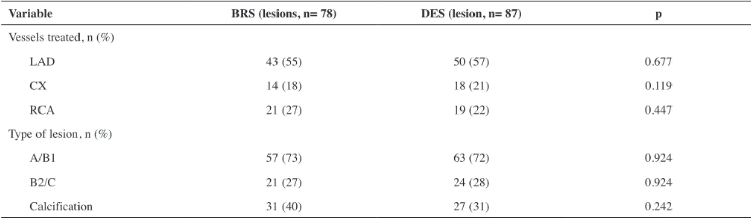
More than 70% of lesions in both groups were categorized as types A and B1 per the ACC/AHA classification (73% in the BRS group and 72% in the DES group, p= 0.924). Calcification was recorded in 31 lesions (40%) in the BRS group and in 27 lesions (31%) in the DES group (p= 0.242).
Procedural Characteristics
The main procedural characteristics determined by QCA analyses are shown in Table 3. The mean RVD was similar in
both groups (3.1 ± 0.4 mm in the BRS group, 3.1 ± 0.4 mm in the DES group, p= 0.256). The DES group had a lower MLD (0.75 ± 0.41 mm vs. 0.98 ± 0.46 mm, p= 0.001), higher steno-sis percentage (75.1% ± 12.4% vs. 69.4% ± 12.3%, p= 0.004), and longer lesion length (21.3 ± 4.4 mm vs. 24.5 ± 7.4 mm, p= 0.001). Larger stent sizes were chosen in BRS-treated patients (3.09 ± 0.4 mm vs. 2.89 ± 0.4 mm; p= 0.001) despite similar reference vessel diameters. Longer stent lengths were favored in the DES group (24.9 ± 5.3 mm vs. 30.3 ± 8.9 mm; p< 0.001).
Balloon predilation and postdilatation were performed more frequently in the BRS group than in the DES group (100% vs. 75%; p< 0.001; 98% vs. 84%; p= 0.004,
respec-Table 2. Angiographic characteristics of the lesions according to stent/scaffold type
Variable BRS (lesions, n= 78) DES (lesion, n= 87) p
Vessels treated, n (%) LAD CX RCA 43 (55) 14 (18) 21 (27) 50 (57) 18 (21) 19 (22) 0.677 0.119 0.447 Type of lesion, n (%) A/B1 B2/C Calcification 57 (73) 21 (27) 31 (40) 63 (72) 24 (28) 27 (31) 0.924 0.924 0.242
DES: Drug-eluting stent, BRS: Bioresorbable scaffold, LAD: Left anterior descending artery, CX: Left circumflex artery, RCA: Right coronary artery.
Table 3. Procedural characteristics measured by QCA
Variable BRS DES p value
Reference vessel diameter (mm) 3.1 ± 0.4 3.1 ± 0.4 0.256
Minimum lesion diameter (mm) 0.98 ± 0.46 0.75 ± 0.41 0.001
Stenosis percentage (%) 69.4 ± 12.3 75.1 ± 12.4 0.004
Lesion length (mm) 21.3 ± 4.4 24.5 ± 7.4 0.001
Predilatation, n (%)
Predilatation balloon size (mm) BminLD (mm) 78 (100) 2.8 ± 0.5 2.2 ± 0.5 65 (75) 2.5 ± 0.5 2.0 ± 0.5 < 0.001 < 0.001 0.045 Acute gain (mm) 2.0 ± 0.5 2.2 ± 0.5 0.017
Stent number per lesion 1.36 ± 0.56 1.41 ± 0.49 0.505
Stent diameter (mm) 3.09 ± 0.4 2.89 ± 0.4 0.001
Stent length (mm) 24.9 ± 5.3 30.3 ± 8.9 < 0.001
Postdilatation, n (%) 77 (98) 73 (84) 0.004
Postdilatation balloon size (mm) 3.3 ± 0.4 3.1 ± 0.4 0.036
FminLD (mm) 2.98 ± 0.40 2.97 ± 0.43 0.871
tively), although both groups had similar lesion morphologies (Figure 1). Predilatation and postdilatation balloon sizes were also significantly higher in BRS-treated patients (2.8 ± 0.5 mm vs. 2.5 ± 0.5 mm; p< 0.001; 3.3 ± 0.4 mm vs. 3.1 ± 0.4 mm, p< 0.036; pre- and postdilatation, respectively).
Increased predilatation balloon calibers provided higher BminLD following predilatation in the BRS group as com-pared with the DES group (2.2 ± 0.5 mm vs. 2.0 ± 0.5 mm, p< 0.045, respectively). Of note, even longer lesion lengths in the DES group were present, and the average number of stents per lesion was similar in both groups (1.36 ± 0.56 in BRS patients and 1.41 ± 0.49 in the DES group).
The total procedure time, radiation dose, fluoroscopy time, and amount of contrast agent were all significantly greater in patients treated with BRS compared with those treated with DES, as shown in Table 4 (Figure 2).
DISCUSSION
In this study, BRS interventions appeared to have a long-er procedure and fluoroscopy duration as well as increased amount of contrast agents and radiation dose compared with new-generation DES. The unique feature of the current study was that procedural aspects of BRS implantation focusing on procedure and fluoroscopy time, contrast volume, and amount
of radiation exposure were investigated and compared with DES procedures.
Most evidence on BRS comes from studies that have used the Absorb BRS(10-12). DESolve BRS is made from a
poly-l-lac-tide polymer backbone similar to that of the Absorb BRS elut-ing anti-inflammatory novolimus, offerelut-ing advantages such as a larger limit of expansion, with diminished strut fracture risk and self-correction of minor malapposition(13). Some technical
issues regarding the implantation technique have been reached by consensus and are applicable to both BRS methods used in our study(14,15). The consensus suggested the use of the PSP
protocol for BRS procedures, as data from studies advocate that rates of scaffold thrombosis could be reduced significantly if this approach is followed(8,16). Previously published large-
scale, randomized clinical trials and cohort studies reported variable rates of pre dilatation and post dilatation; interestingly, the rate of post dilatation was on average < 50%, with low intra-coronary imaging(17,18). Contrary to previously published data,
we found that predilatation and postdilatation rates were higher in the BRS group compared with the DES group (predilata-tion 100% vs. 75% and postdilata(predilata-tion 98% vs. 84%). Our mean BRS length was longer than in previous studies(17). It might be
speculated that longer lesion lengths might have affected the operator’s choice for higher pre- and postdilatation rates in the BRS group. However, even though lesion lengths were longer
Figure 1. Comparison of lesion characteristics and pre- and postdilatation rates in BRS and DES groups. Percentage of lesion classification according to the ACC/AHA definition between the DES and BRS groups (A); comparison of predilatation (B) and postdilatation rates (C) in both groups.
A
B
C
Lesion type (A/B1) Lesion type (B2/C) Number of Lesions Number of Lesions Number of Lesions Predilatation (-)
Predilatation (+) Post-dilatation (-)Post-dilatation (+)
p= 0.924 p< 0.001 p= 0.004
BRS DES BRS DES BRS DES
60 40 20 0 80 60 40 20 0 80 60 40 20 0
Table 4. Procedural variables
Variable BRS (patients, n= 64) DES (patients, n= 70) p
Fluoroscopy time (min) 15.9 ± 6.6 13.1 ± 6.6 0.014
Procedure time (min) 45.4 ± 16.1 38.3 ± 15.1 0.010
Contrast volume (mL) 207.7 ± 80.7 154.7 ± 74.6 0.001
in the DES group, the average number of stents per lesion did not differ between the groups because of the available longer stent sizes in the DES group.
Operators have become more cautious when it comes to using BRS because of the above-mentioned drawbacks. In ad-dition, increased strut thickness and inferior deliverability have made BRS implantation more challenging. However, despite these challenging and technically demanding issues, data on the duration of operation and fluoroscopy, contrast volume, and radiation dose during BRS deployment are scarce.
Sato et al. compared 96 patients treated with BRS and 96 matched controls treated with DES, both of whom had pre-dominantly complex lesions(19). They reported that primary
endpoints including procedure time, total contrast medium ad-ministered, and fluoroscopy time were higher in the BRS group (p< 0.001, p= 0.02, and p< 0.001, respectively). Our data show that these outcomes can also translate into similar results even
for patients with noncomplex lesion subsets. Wiebe et al. and Ozel et al. shared their data from a relatively small number of patients with chronic total occlusions regarding the amount of contrast volume and procedure and fluoroscopy time, but they did not conduct a comparison between BRS and DES groups(20,21). Thus, this paper, which aimed to evaluate
con-trast load, radiation exposure, and duration of procedure and fluoroscopy, is of clinical importance for both operators and patients as it highlights the increased dose of radiation and con-trast volume and longer procedural and fluoroscopy times in BRS-treated patients as compared with DES-treated patients.
In the current study, lesions in the DES group were longer than those in the BRS group. Hence, operators chose to implant significantly longer stents in the DES group. Unlike DES, com-mercially available scaffold sizes are also restricted. Although it might be expected that long stents selected for longer lesions with significantly most severe stenosis and lower predilatation
Figure 2. Association between procedural features in the study groups. Fluoroscopy time (A), procedure time (B), contrast volume (C), and radiation dose (D) of the BRS and DES procedures.
35 30 25 20 15 10 5 0 110 100 90 80 70 60 50 40 30 20 10 0 4.00 3.50 3.00 2.50 2.00 1.50 1.00 0.50 0.00 BRS BRS BRS BRS DES DES DES DES p= 0.014 Fluor o Time (min) Contrast Volume (mL)
Radiation Dose (GY)
Pr ocedur e T ime (min) p= 0.001 p= 0.037 p= 0.01 500 450 400 350 300 250 200 150 100 50 0
A
B
C
D
rates would increase the procedure times in DES group, there are several factors that make BRS deployment more demand-ing. Moreover, poor visualization of the scaffold markers as well as slow inflation time during BRS implantation also con-tributed additional time and excessive opaque use.
Because of this study’s single-center design and retrospec-tive nature, the current study is subject to selection and recall biases. Despite the relatively small sample size and the use of varied types of stents in the DES and BRS group, we believe that our study has sufficient power to reveal the observed dif-ferences in the procedural aspects between the BRS and DES groups.
Another limitation to our study was the lack of intravascu-lar imaging (intravascuintravascu-lar ultrasound/optical coherence tomog-raphy) for BRS and DES procedures to test radiation exposure, procedure time, and opaque load. However, Sato et al. did not find an association between intravascular imaging and longer procedural times(19). Finally, we were unable to establish a
re-lationship between BRS use and clinical outcomes, because long-term results were not evaluated in the current study.
CONCLUSION
BRS implantation results in a longer procedure and fluor-oscopy duration. Excessive radiation exposure and opaque volume were observed in BRS-implanted patients. Awareness of ways to minimize the likelihood of contrast nephropathy as well as inappropriate excessive radiation use is of paramount importance during BRS procedures. Randomized trials are warranted to investigate radiation dose, procedural and fluoros-copy times of BRS devices, and the impact of these parameters on clinical outcomes.
ACKNOWLEDGEMENT
The author thanks Ibrahim Oğuz Karaca, Bilal Boztosun, and Hacı Murat Güneş for their invaluable help during the ex-ecution of this project.
Ethics Committee Approval: Ethics committee approval was received for this study from the Istanbul Medipol University Non-Interventional Clin-ical Researches Ethics Committee (10840098-604.01.01-E.199).
Informed Consent: Written informed consent was obtained from pa-tients who participated in this study.
Peer-review: Externally peer-reviewed.
Author Contributions: Concept/Design – BÇ; Analysis/Interpretation – BÇ; Data Collection – BÇ; Writing – BÇ; Critical Revision – BÇ; Final Approval – BÇ; Statistical Analysis – BÇ; Obtained Funding – BÇ; Overall Responsibility - BÇ
Conflict of Interest: The authors have no conflict of interest to declare. Financial Disclosure: The authors declared that this study has received no financial support.
REFERENCES
1. Sousa J, A Costa M, Abizaid AS, Feres F, Pinto I, C Seixas A, et al. Lack of neointimal proliferation after implantation of sirolimus-coated stents in human coronary arteries: A quantitative coronary angiography and three-dimensional intravascular ultrasound study. Circulation 2001;103(2):192-5.
2. Joner M, Finn AV, Farb A, Mont EK, Kolodgie FD, Ladich E, et al. Pathol-ogy of drug-eluting stents in humans: delayed healing and late thrombotic risk. J Am Coll Cardiol 2006;48(1):193-202.
3. Serruys PW, Garcia-Garcia HM, Onuma Y. From metallic cages to tran-sient bioresorbable scaffolds: change in paradigm of coronary revasculari-zation in the upcoming decade? Eur Heart J 2012;33(1):16-25b. 4. Ormiston JA, Serruys PW, Regar E, Dudek D, Thuesen L, Webster MW, et
al. A bioabsorbable everolimus-eluting coronary stent system for patients with single de-novo coronary artery lesions (ABSORB): a prospective open-label trial. Lancet 2008;371(9616):899-907.
5. Sotomi Y, Onuma Y, Collet C, Tenekecioglu E, Virmani R, Kleiman NS, et al. Bioresorbable scaffold: the emerging reality and future directions. Circulation Research 2017;120(8):1341-52.
6. Capodanno D. Bioresorbable scaffolds in coronary intervention: unmet needs and evolution. Korean Circulation Journal 2018;48(1):24-35. 7. Jinnouchi H, Torii S, Sakamoto A, Kolodgie FD, Virmani R, Finn AV.
Fully bioresorbable vascular scaffolds: lessons learned and future direc-tions. Nat Rev Cardiol 2019;16(5):286-304.
8. Ortega-Paz L, Capodanno D, Gori T, Nef H, Latib A, Caramanno G, et al. Predilation, sizing and post-dilation scoring in patients undergoing everolimus-eluting bioresorbable scaffold implantation for prediction of cardiac adverse events: development and internal validation of the PSP score. EuroIntervention 2017;12(17):2110-7.
9. Bonow RO, Masoudi FA, Rumsfeld JS, Delong E, Estes NA 3rd, Goff DC Jr, et al. ACC/AHA classification of care metrics: performance meas-ures and quality metrics: a report of the American College of Cardiology/ American Heart Association Task Force on Performance Measures. J Am Coll Cardiol 2008;52(24):2113-7.
10. Tan HC, Ananthakrishna R. Bioresorbable vascular scaffolds in rou-tine clinical practice: should we wait longer? JACC Cardiovasc Interv 2016;9(16):1664-6.
11. Ellis SG, Kereiakes DJ, Metzger DC, Caputo RP, Rizik DG, Teirstein PS, et al. Everolimus-eluting bioresorbable scaffolds for coronary artery dis-ease. N Engl J Med 2015;373(20):1905-15.
12. Kereiakes DJ, Ellis SG, Metzger C, Caputo RP, Rizik DG, Teirstein PS, et al. 3-year clinical outcomes with everolimus-eluting bioresorb-able coronary scaffolds: The ABSORB III Trial. J Am Coll Cardiol 2017;70(23):2852-62.
13. Verheye S, Ormiston JA, Stewart J, Webster M, Sanidas E, Costa R, et al. A next-generation bioresorbable coronary scaffold system: from bench to first clinical evaluation: 6- and 12-month clinical and multimodality imag-ing results. JACC Cardiovasc Interv 2014;7(1):89-99.
14. Rizik DG, Hermiller JB, Kereiakes DJ. The ABSORB bioresorbable vascular scaffold: A novel, fully resorbable drug-eluting stent: Current concepts and overview of clinical evidence. Catheter Cardiovasc Interv 2015;86(4):664-77.
15. Tamburino C, Latib A, van Geuns RJ, Sabate M, Mehilli J, Gori T, et al. Contemporary practice and technical aspects in coronary interven-tion with bioresorbable scaffolds: a European perspective. EuroIntervent 2015;11(1):45-52.
16. Stone GW, Abizaid A, Onuma Y, Seth A, Gao R, Ormiston J, et al. Ef-fect of technique on outcomes following bioresorbable vascular scaf-fold implantation: analysis from the ABSORB trials. J Am Coll Cardiol 2017;70(23):2863-74.
17. Ali ZA, Serruys PW, Kimura T, Gao R, Ellis SG, Kereiakes DJ, et al. 2-year outcomes with the Absorb bioresorbable scaffold for treatment of coronary artery disease: a systematic review and meta-analysis of seven randomised trials with an individual patient data substudy. Lancet 2017;390(10096):760-72.
18. Yamaji K, Raber L, Windecker S. What determines long-term outcomes using fully bioresorbable scaffolds-the device, the operator or the lesion? EuroIntervent 2017;12(14):1684-7.
19. Sato K, Latib A, Panoulas VF, Kawamoto H, Naganuma T, Miyazaki T, et al. Procedural feasibility and clinical outcomes in propensity-matched pa-tients treated with bioresorbable scaffolds vs new-generation drug-eluting stents. Can J Cardiol 2015;31(3):328-34.
20. Wiebe J, Liebetrau C, Dorr O, Most A, Weipert K, Rixe J, et al. Feasibil-ity of everolimus-eluting bioresorbable vascular scaffolds in patients with chronic total occlusion. Int J Cardiol 2015;179:90-4.
21. Ozel E, Tastan A, Ozturk A, Ozcan EE, Kilicaslan B, Ozdogan O. Proce-dural and one-year clinical outcomes of bioresorbable vascular scaffolds for the treatment of chronic total occlusions: a single-centre experience. Cardiovasc J Afr 2016;27(6):345-9.