Erectile Dysfunction Is Positively Correlated with Mean Platelet Volume
and Platelet Count, but Not with Eosinophil Count in Peripheral Blood
Alper Otunctemur,1 Muammer Bozkurt,1 Hüseyin Beşiroğlu,1 Emre Can Polat,2* Levent Ozcan,3 Emin Ozbek1 Purpose: Increased eosinophil count (EC), mean platelet volume (MPV), and platelet count (PC) are important in vascular disorders which are main factors resulting in endothelial dysfunction. We aimed to investigate the associ-ation between MPV, and EC, with erectile dysfunction (ED).Materials and Methods: Two hundred thirty participants (130 patients with ED, and 100 healthy controls) were enrolled in this study. A detailed psychosexual history obtained, and physical, and laboratory examination were performed. International Index of Erectile Function (IIEF)-5 questionnaire was used to evaluate the erectile status objectively. IIEF-5 score was applied to all patients, and IIEF-5 score under 22 was considered as ED. The MPV, PC, and EC were compared between the two groups.
Results: The mean age of the patients with ED and control group was 55.62 ± 8.90 years and 54.19 ± 4.10 years, respectively. MPV and PC levels were significantly higher in ED group (8.51 ± 1.00 fL and 8.16 ± 0.94 fL; 244.59 ± 57.3 cells/μL and 230.17 ± 48.44 cells/μL, respectively (P < .05). EC and white blood cell count were not signif-icantly different between study and control groups.
Conclusions: In our study a relationship was found between elevated MPV, and PC with ED. MPV and PC may be used as a biomarker in patients with ED.
Keywords: erectile dysfunction; etiology; risk factors; blood platelets; platelet count; eosinophils; leukocyte count. INTRODUCTION
E
rectile dysfunction (ED) is defined as a difficulty in initiating or maintaining penile erection adequate for sexual inter course. Penile erection is the result of a complex interaction between psychological, neu-ral, vascular, and endocrine factors. One of the largest current studies of ED, the Massachusetts Male Aging Study, found that ED may be present in up to half of the male population between 40 and 70 years old.(1)This condition has been estimated to affect 150 million individuals worldwide,(2) and data from The
Enhanc-ing Neuro ImagEnhanc-ing Genetics through Meta-Analysis (ENIGMA) Consortium study in 2004 suggested that the condition is prevalent in approximately 17% of all European men are affected.(3)
Several epidemiological studies have reported that ED is a marker of cardiovascular disease (CVD).(4-6) A 2011
meta-analysis of 12 prospective cohort studies provid-ed strong evidence that ED is indeprovid-ed significantly and independently associated with an increased risk of not only CVD but also coronary heart disease, stroke, and all-cause mortality.(7) Clearly, ED is now regarded as a
major health problem for the increasingly healthy aging population.
In the etiology of ED, generally, organic and
psycho-genic factors come together. However, if the penis is considered as a specialized vascular bed, it is well-known that vascular reasons dominate in the etiolo-gy of ED.(8) During the last 20 years, many new facts
about the basic physiology and pathology of ED have been determined, especially at the molecular level. Di-abetes mellitus, atherosclerosis, coronary disease, and hypertension contribute to the development of ED via endothelial dysfunction and peripheral artery disease.(9)
It has also been hypothesized that ED is an early mes-senger of CVD.(10)
The mean platelet volume (MPV) (expressed as fem to litre, fL) is one of the leading indicator in platelet func-tion reflecting the platelet producfunc-tion rate and platelet stimulation. Elevated MPVs are reported in CVDs.(11)
The MPV, the most commonly used measure of platelet size, is a potential marker of platelet reactivity. Larger platelets are metabolically and enzymatically more ac-tive and have greater prothrombotic potential. Elevated MPV is associated with other markers of platelet ac-tivity, including increased platelet aggregation, throm-boxane synthesis, and increased expression of adhesion molecules.(12) Furthermore, a higher MPV may take
place in vascular pathologies and increase the risk of CVD, suggesting a common mechanism by which these factors may increase the risk of CVD and ED. An
as-1 Department of Urology, Okmeydani Training and Research Hospital, Istanbul, Turkey. 2 Department of Urology, Istanbul Medipol University, Istanbul, Turkey.
3 Department of Urology, Derince Training and Research Hospital, Kocaeli, Turkey.
*Correspondence: Department of Urology, Istanbul Medipol University, Istanbul, Turkey. Tel: +90 532 71496 04. E-mail: [email protected].
sociation between ED and ischemic heart disease has been suggested as a consequence of vascular lesions of the penile arteries.(13) There is a relationship between
vascular dysfunction with eosinophilia. It is known that eosinophils play an important role inendothelial dys-function, vasoconstriction, inflammation, and thrombo-sis.(14) Eosinophils stimulate the activation and
aggre-gation of platelets. Moreover, they ease the formation of thrombosis via inhibition of thrombomodulin.(15,16)
Perhaps, increased eosinophil count (EC, expressed as cells/μL) could lead to ED through endothelial dysfunc-tion.
These pathophysiological approach to the issue let us consider that ED might be associated with increased platelet count (PC, expressed as cells/μL) and volume as well as EC. In this study, we aimed to investigate the association between the MPV, PC, and EC with ED, in comparison with a control group.
MATERIALS AND METHODS
We have conducted a prospective study in participants who visited Okmeydani Training and Research Hospi-tal. A total of 230 patients were evaluated for ED and they were divided into two groups: 130 patients suf-fering from ED for > 1 year were classified as a study group and 100 patients without ED who were sexually active and married were classified as a control group. Local ethics committee approval had been obtained before the beginning of the study. All patients had a complete detailed and careful history taken, with spe-cial attention to the sexual history, including details to differentiate between psychogenic and organic ED; a complete physical examination, including genital and neurological examination; blood glucose assay, urine analysis, complete blood count (CBC), and kidney and liver function. Erectile function was assessed using the five-item version of the International Index of Erectile Function questionnaire (IIEF-5), a validated, self-ad-ministered questionnaire. The score of 22–25 indicate normal erectile function, while scores < 22 indicate ED. According to the IIEF-5 score, ED was classified as severe (5–7), moderate (8–11), mild-to-moderate (12–16), or
mild (17–21). Exclusion criteria included the followings:
the patients using anti-platelet or anticoagulant drugs, patients with congestive heart failure (ejection fraction < 50%), pulmonary hypertension, coroner artery dis-ease, stroke, known peripheral atherosclerotic disdis-ease, surgical coronary intervention, percutaneous coronary angioplasty and/or stenting, stable and unstable angina pectoris, impaired renal function (creatinine > 1.4 mg/ dL), unstable endocrine or metabolic diseases, patients with Peyronie’s disease, acute/chronic hepatic or hepa-tobiliary disease, and malignancy. Patients who have undergone radical prostatectomy and/or pelvic sur-gery, history of pelvic trauma and taking beta-blocker, spironolactone, corticosteroids, antioxidant vitamins, and alcohol were also excluded from the study. Fur-thermore patients who had a recent history of an acute infection and/or high body temperature (> 38°C), an in-flammatory, or an allergic disease were excluded from the study. Blood samples of all patients were taken from an antecubital vein following an overnight fasting state. Fasting blood glucose, MPV, PC, EC, white blood cell (WBC) total cholesterol, high-density lipoprotein cho-lesterol (HDL-C), low-density lipoprotein chocho-lesterol (LDL-C), and triglyceride (TG) levels were measured in the hospital’s chemistry laboratory.
Statistical Analysis
Statistical analyses were performed by the Statistical Package for the Social Science (SPSS Inc, Chicago, Illi-nois, USA) version 15.0. The quantitative demographic values were evaluated by student’s t-test or Mann Whit-ney U test whether the parameters were suitable for nor-mal distribution or not. If the parameters are qualitative, chi-square test was used. Kruskal-Wallis test was con-ducted to evaluate the difference between subgroups of the patients stratified for age. Logistic regression anal-yses were conducted to estimate the risk ratios. Pearson correlation test was performed to determine possible correlation between MPV, and PC with IIEF-5 score. All tests were performed using a 2-tailed analysis, and a
P value of < .05 was considered statistically significant.
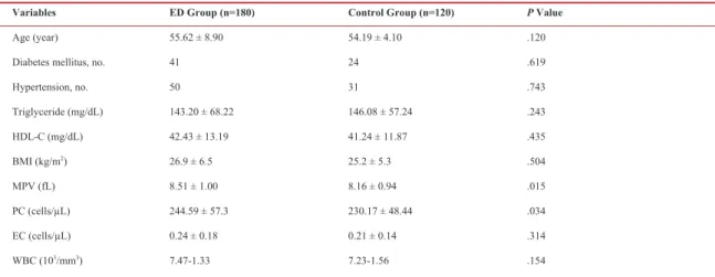
RESULTS Table 1. Clinical characteristic of study patients.
Variables ED Group (n=180) Control Group (n=120) P Value
Age (year) 55.62 ± 8.90 54.19 ± 4.10 .120
Diabetes mellitus, no. 41 24 .619
Hypertension, no. 50 31 .743 Triglyceride (mg/dL) 143.20 ± 68.22 146.08 ± 57.24 .243 HDL-C (mg/dL) 42.43 ± 13.19 41.24 ± 11.87 .435 BMI (kg/m2) 26.9 ± 6.5 25.2 ± 5.3 .504 MPV (fL) 8.51 ± 1.00 8.16 ± 0.94 .015 PC (cells/μL) 244.59 ± 57.3 230.17 ± 48.44 .034 EC (cells/μL) 0.24 ± 0.18 0.21 ± 0.14 .314 WBC (103/mm3) 7.47-1.33 7.23-1.56 .154
Abbreviations: ED, erectile dysfunction; HDL, high-density lipoprotein; BMI, body mass index; MPV, mean platelet volume; PC, platelet count; EC, eosinophil count; WBC, white blood cells.
A total of 230 patients between the ages of 40 and 65 years were analyzed and divided into two groups: 130 patients with ED and 100 patients without ED (control group). The baseline characteristics of the patients are demonstrated in Table 1. The mean age was 55.62 ± 8.90 years in patients with ED and 54.19 ± 4. 10 years in control group. There was no significant differences be-tween two groups with respect to age, body mass index (BMI), frequencies of diabetes mellitus, hypertension, smoking and levels of fasting blood glucose, Total-C, LDL-C, HDL-C, TG, and WBC (P > .05 for all). We compared the MPV, PC, EC values between groups. It seems that the patients with ED have higher MPV and PC levels than control group. MPV level was 8.51 ± 1.00 fL in patients with ED and 8.16 ± 0.94 fL in con-trol group. PC level was 244.59 ± 57.3 μL in patients with ED and 230.17 ± 48.44 μLin control group. There was statistically significant difference for MPV and PC levels between the patients with ED and control group (P < .05) (Table 1). In logistic regression analyses the parameters assumed to be related to ED were examined. The patients were stratified into three groups as 40-50 years, 50-60 years, and 60-70 years. Patients with > 70 years old and < 40 years old were not evaluated as sepa-rate group, since the frequencies of them were not suit-able for a robust statistical analysis. Although MPV and PC were not statistically different between each group, IIEF-5 score was statistically different. The details are demonstrated in Table 2.
We also evaluated the groups in terms of ED severity which was classified as mild, mild to moderate, moder-ate and severe. When examining the distribution of the patient frequencies into the subgroups stratified by ED severity, there was statistically significant difference between three groups (P = .02). The details are shown in Table 3.
In logistic regression analyses the parameters assumed to be related to ED were evaluated. The parameters were adjusted for age, diabetes mellitus, hypertension, dyslipidemia, and alcohol consumption. Table 4 rep-resents the 95% confidence interval (CI) and adjusted odds ratios (ORs) for the associations between certain relevant associated risk factors and ED. Patients with
higher PC (OR = 1.005; 95% CI: 1.003-1.010) and MPV (OR = 1.256; 95% CI: 1.088-1.4) had increased risk for development of ED. The EC was not correlated-with ED in logistic regression model.
DISCUSSION
ED is one of the most prevalent urological disorders resulting from variable organic and psychologic de-rangements. Vascular pathologies take great part in or-ganic causes via the impairment of endothelial function which is crucial in erection physiology. The presence of a number of common risk factors, the presence of sev-eral known pathophysiologic links, and a number of ret-rospective association studies have reinforced the idea that the link between ED and CAD is important and real. This idea is defined that ED and coronary artery disease (CAD) are different manifestations of a com-mon underlying vascular pathology. ED may be the ear-ly clinical manifestation of a generalized vascular dis-ease and carries an independent risk for cardiovascular events.(17,18) Many patients present with underlying
sys-temic CVD and their first symptom can be ED.(19) One
study of 132 men correlated angiographic results with ED symptoms and scores on the IIEF-5; 58% reported experiencing ED before the diagnosis of CVD.(20)
Pro-spective angiographic study showed that almost one in five men presented with erectile function abnormalities of vascular origin had angiographically documented si-lent CAD.(21) In the light of this information, young men
with ED may be ideal candidates for cardiovascular risk factor screening and medical intervention.
ED precedes other manifestations of systemic athero-sclerosis, such as CAD and cerebrovascular disease, may be partially explained by blood vessel size.(22) The
penile arteries are typically 1 to 2 mm in diameter, whereas the coronary arteries are 3 to 4 mm in diameter and the carotid arteries, 5 to 7 mm in diameter. There-fore, an atherosclerotic plaque of a given size should occlude and hemodynamically affect a penile artery earlier than a coronary or carotid artery. Ultimately, small arteries such as the pudendal and penile arteries begin to degenerate, and end-organ ischemia results. Table 2. The study parameters stratified by age (years).
Variables 40-50 (n = 31) 50-60 (n = 66) 60-70 (n = 75) P Value
Platelet count (cells/μL) 237.99 ± 37.5 240.05 ± 61.04 247.82 ± 56.32 .530
Mean platelet volume (fL) 8.24 ± 0.68 8.54 ± 1.08 8.60 ± 1.09 .553
IIEF-5 score 15.12 (7-22) 16.19 (7-22) 13.16(6-21) .001
Abbreviation: IIEF, international index of erectile function. Data are presented as mean ± SD.
Abbreviation: ED, erectile dysfunction.
ED severity, no. 40-50 (n = 31) 50-60 (n = 66) 60-70 (n = 75) P Value
Mild 6 35 17
Mild to moderate 16 15 33 .02
Moderate 7 10 14
Severe 2 6 11
(23) Pathophysiologic link between ED and CAD is
en-dothelial dysfunction. Many patients with ED exhibit evidence of inflammation and endothelial dysfunction independent of their CAD status.(24,25)
Endothelial dysfunction is the key event in the patho-physiology of ED and, importantly, men with penile vascular.(24) Endothelial dysfunction can carry a
height-ened risk of future CAD events because it results in dysregulated intimal proliferation, inappropriate va-soconstriction, and a pro-inflammatory environment that causes plaque destabilization.(26) The fact that ED
and atherosclerotic vascular diseases share such a large number of common risk factors has led to the clinical consensus that most cases of organic ED are probably part of the spectrum of atherosclerotic vascular disease.
(10)
There are many studies about the relationship between MPV and some thrombotic and cardiac disorders.(27,28)
It has been demonstrated that MPV is correlated with platelet function and activation.(29,30) Small platelets
have lower functional capabilities than larger ones.(30)
There are many evidences suggesting the important role of MPV as a marker of inflammation, disease activity and efficacy of anti- inflammatory treatment in several chronic inflammatory disorders. Therefore, MPV has been used as an indicator of platelet function for inflam-matory diseases.(29-31) Due to vascular causes, we
inves-tigated the relationship between ED and MPV, and PC. In a study by Ciftci and colleagues(32) on 90 cases, PC
and MPV values were increased in patients with vascu-logenic ED. In our study, we found that the MPV and PC values were significantly higher in patients with ED than in the controls.
Eosinophils activate coagulation system and platelets, and they also cause vasospasm such as coronary artery spasm. Also eosinophil granule proteins are involved in vascular injury, and eosinophils may also affect cardio-vascular system through inflammatory cell infiltration.
(33) Recent studies showed that eosinophils were
associ-ated with stent thrombosis, stent restenosis, and acute coronary syndromes. Umemoto and colleagues reported that peripheral ECs were significantly higher in patients with severe coronary spasm than that in patients with no spasm. They also speculated that EC could predict va-sospastic angina pectoris.(33) Eosinophils are equipped
with several granule-associated molecules that play a role in the occurrence of thrombosis and vascular in-jury. Eosinophils generate an increased tendency to thrombosis through leukocyte, platelet stimulation, and release of tissue factor.(34-36) All these effects contribute
to procoagulation through preventing the activation of thrombin and formation of endorsing fibrin. Sakai and colleagues demonstrated that large thrombus has great-er EC both in thrombi and in pgreat-eriphgreat-eral blood.(37) They
also speculated that thrombus growth might be
facilitat-ed in patients with higher EC in the peripheral blood.(37)
The powerful vasoconstrictor and procoagulant effects of eosinophils, made us hypothesize that there might be a correlation between EC and ED. Increased EC in patients with ED might be due to vasoconstriction and thrombosis.
Study addressing the relationship between EC and ED has not been conducted yet. Thrombotic and vascular effects of EC and CAD with common etiology of ED are known. In light of this, we might hypothesize that ED could be associated with higher MPV and PC. How-ever, there was no significant relationship between ED and EC in our study. Further studies with larger popu-lation are needed to yield more reliable results in this issue.
CONCLUSIONS
There is a relationship between elevated MPV, and PC with ED, through endothelial dysfunction. Whereas there is no statistically significant relationship between ED and EC. In the light of this data, MPV and PC val-ues may be used as a biomarker in patients with ED. CONFLICT OF INTEREST
None declared. REFERENCES
1. Feldman HA, Goldstein I, Hatzichristou DG, Krane RJ, McKinlay JB. Impotence and its medical and psychosocial correlates: results of the Massachusetts Male Aging Study. J Urol. 1994;151:54–61.
2. Aytaç IA, McKinlay JB, Krane RJ. The likely worldwide increase in erectile dysfunction between 1995 and 2025 and some possible policy consequences. BJU Int. 1999;84:50–6.
3. de Boer BJ, Bots ML, Lycklama a Nijeholt AA, et al. Erectile dysfunction in primary care: prevalence and patient characteristics. The ENIGMA study. Int J Impot Res. 2004;16:358–64.
4. Inman BA, Sauver JL, Jacobson DJ, et al. A population-based, longitudinal study of erectile dysfunction and future coronary artery disease. Mayo Clin Proc. 2009;84:108–13.
5. Clark NG, Fox KM, Grandy S. SHIELD Study Group. Symptoms of diabetes and their association with the risk and presence of diabetes: findings from the Study to Help Improve Early evaluation and management of risk factors Leading to Diabetes (SHIELD). Diabetes Care. 2007;30:2868–73.
Table 4. Multivariate logistic regression analysis for the risk factors for erectile dysfunction adjusted for age, diabetes mellitus, hypertension, alcohol consumption, and dyslipidemia.
Variables OR CI (95%) P Value Wald
Mean platelet volume 1.256 1.088-1.484 .014 7.083
Platelet count 1.005 1.003-1.010 .026 4.236
Eosinophil count 0.937 0.821-0.993 .165 0.031
6. Chung SD, Chen YK, Lin HC, Lin HC. Increased risk of stroke among men with erectile dysfunction: a nationwide population-based study. J Sex Med. 2011;8:240–6.
7. Dong JY, Zhang YH, Qin LQ. Erectile dysfunction and risk of cardiovascular disease: meta-analysis of prospective cohort studies. J Am Coll Cardiol. 2011;58:1378–85.
8. Simonsen U, García-Sacristán A, Prieto D. Penile arteries and erection. J Vasc Res. 2002;39:283–303.
9. Gratzke C, Angulo J, Chitaley K, et al. Anatomy, physiology, and pathophysiology of erectile dysfunction. J Sex Med. 2010;7:445– 75.
10. Jackson G, Rosen RC, Kloner RA, Kostis JB. The second Princeton consensus on sexual dysfunction and cardiac risk: new guidelines for sexual medicine. J Sex Med. 2006;3:28– 36.
11. Pizzulli L, Yang A, Martin JF, Lüderitz B. Changes in platelet size and count in unstable angina compared to stable angina or non-cardiac chest pain. Eur Heart J. 1998;19:80–4.
12. Kamath S, Blann AD, Lip GY. Platelet activation: assessment and quantification. Eur Heart J. 2001;22:1561–71.
13. Virag R, Bouilly P, Frydman D. About arterial risk factors and impotence. The Lancet. 1985;1:1109–10.
14. Wang J, Mahmud SA, Thompson JA, Geng JG, Key NS, Slungaard A. The principal eosinophil peroxidase product. HOSCN. Is a uniquely potent phagocyte oxidant inducer of endothelial cell tissue factor activity: a potential mechanism for thrombosis in eosinophilic inflammatory states? Blood. 2006;107:558-65.
15. Rohrbach MS, Wheatley CL, Slifman NR, Gleich GJ. Activation of platelets by eosinophil granule proteins. J Exp Med. 1990;172:1271-4.
16. Olsen EG, Spry CJ. Relation between eosinophils and endomyocardial disease. Prog Cardiovasc Dis. 1985;27:241-54.
17. Thompson IM, Tangen CM, Goodman PJ, Probstfield JL, Moinpour CM, Coltman CA. Erectile dysfunction and subsequent cardiovascular disease. JAMA 2005;294:2996–3002.
18. Ponholzer A, Temml C, Obermayr R, Wehrberger C, Madersbacher S. Is erectile dysfunction an indicator for increased risk of coronary heart disease and stroke? Eur Urol. 2005;48:512–8.
19. Roumeguère T, Wespes E, Carpentier
Y, Hoffmann P, Schulman CC. Erectile dysfunction is associated with a high prevalence of hyperlipidemia and coronary heart disease risk. Eur Urol. 2003;44:355-9.
20. Solomon H, Man JW, Jackson G. Erectile
dysfunction and the cardiovascular patient:
endothelial dysfunction is the common denominator. Heart. 2003;89:251-3.
21. Vlachopoulos C, Rokkas K, Ioakeimidis N, et al. Prevalence of asymptomatic coronary artery disease in men with vasculogenic erectile dysfunction: a prospective angiographic study. Eur Urol. 2005;48:996-1002.
22. Montorsi P, Ravagnani PM, Galli S, et al. The artery size hypothesis: a macrovascular link between erectile dysfunction and coronary artery disease. Am J Cardiol. 2005;96:19M-23M.
23. O’Rourke MF, Hashimoto J. Mechanical
factors in arterial aging: a clinical perspective. J Am Coll Cardiol. 2007;50:1-13.
24. Elesber AA, Solomon H, Lennon RJ, et
al. Coronary endothelial dysfunction is associated with erectile dysfunction and elevated asymmetric dimethylarginine in patients with early atherosclerosis. Eur Heart J. 2006;27:824-31.
25. Vlachopoulos C, Aznaouridis K, Ioakeimidis N, et al. Unfavourable endothelial and inflammatory state in erectile dysfunction patients with or without coronary artery disease. Eur Heart J. 2006;27:2640-8.
26. Rodriguez JJ, Al Dashti R, Schwarz ER.
Linking erectile dysfunction and coronary artery disease. Int J Impot Res. 2005;17(suppl 1):S12-S8.
27. Gasparyan AY, Ayvazyan L, Mikhailidis
DP, Kitas GD. Mean platelet volume: a link between thrombosis and inflammation? Curr Pharm Des. 2011;17:47-58.
28. Yazici S, Yazici M, Erer B, et al. The platelet indices in patients with rheumatoid arthritis: mean platelet volume reflects disease activity. Platelets. 2010;21:122-5.
29. Martin JF, Trowbridge EA, Salmon G,
Plumb J. The biological significance of platelet volume: Its relationship to bleeding time, thromboxane B2 production and megakaryocyte nuclear DNA concentration. Thromb Res. 1983;32:443-60.
30. Thompson CB, Eaton KA, Princiotta SM,
Rushin CA, Valeri CR. Size dependent platelet subpopulations: Relationship of platelet volume to ultrastructure, enzymatic activity, and function. Br J Haematol. 1982;50:509-19.
31. Gasparyan AY, Sandoo A,
Stavropoulos-Kalinoglou A, Kitas GD. Mean platelet volume in patients with rheumatoid arthritis: the effect of anti- TNF-α therapy. Rheumatol Int. 2010;30:1125-9.
32. Ciftci H,Yeni E, Demir M, Yagmur I,et al. Can the Mean Platelet Volume Be a Risk Factor for Vasculogenic Erectile Dysfunction? World J Mens Health. 2013;31:215-9.
33. Umemoto S, Suzuki N, Fujii K, et al.
Eosinophil counts and plasma fibrinogen in patients with vasospastic angina pectoris. Am J Cardiol. 2000;85:715-9.
34. Moosbauer C, Morgenstern E, Cuvelier SL, et al. Eosinophils area major intravascular location for tissue factor storage and exposure. Blood. 2007;109:995-1002.
35. Levi M, Hack CE, de Boer JP, Brandjes DP, Buller HR, ten Cate JW. Reduction of contact activation related fibrinolytic activity in factor XII deficient patients. Further evidence for the role of the contact system on fibrinolysis in vivo. J Clin Invest. 1991;88:1155-60.
36. Rodeghiero F, Castaman G, Ruggeri M, Cazzavillan M, Ferracin G, Dini E. Fibrinolytic studies in 13 unrelated families with factor XII deficiency. Haematologica. 1991;76:28-32.
37. Sakai T, Inoue S, Matsuyama TA, et al. Eosinophils may be involved in thrombus growth in acute coronary syndrome. Int Heart J. 2009;50:267-77.