ABSTRACT
A fi fty years old female patient with the history of Laser in situ keratomileusis (LASIK) presented at emergency room complaining of insertion of a hair clip piece into her left eye. On slit-lamp examination, there was a foreign body impacted in inferotemporal paracentral cornea extending into the lens via penetrating iris. Two and a half months after the corneal fi xation and traumatic cataract extraction, iridoplasty and secondary intraocular lens (IOL) implantation were applied. The stable refraction was – 0.50 – 0.75/160 degrees at the fi rst year of trauma. In traumatic aphakic eyes with the history of refractive surgery choosing appropriate IOL calculation formula may become more diffi cult because routinely used methods to calculate IOL power do not guarantee the same accuracy compared with naive eyes. Haigis-L formula might be a preferable method to calculate IOL power for these eyes.
Key words: Refractive surgery; intraocular lenses; traumatic aphakia; penetrating eye injuries.
ÖZ
Geçirilmiş Laser in situ keratomileusis (LASIK) öyküsü olan 50 yaşında kadın hasta sol gözüne saç tokası parçası gelme şikayeti ile acil servise başvurdu. Biyomikroskopik incelemesinde alt temporal parasantral korneadan irisi penetre ederek lense uzanan yabancı cisim izlenmekteydi. Korneal sütürasyon ve travmatik katarakt ekstraksiyonundan 2,5 ay sonra hastaya iridoplasti ve sekonder göziçi lens implantasyonu uygulandı. Travmanın birinci yılında stabil refraksiyon -0.50 -0.75/160 derece düzeyinde idi. Refraktif cerrahi öyküsü olan travmatik afakik hastalarda uygun göziçi lens formülasyonunun seçilmesi rutinde kullanılan diğer yöntemlerin naif gözlerdeki doğruluğu garanti etmemesi nedeniyle zordur. Haigis-L formülünün bu gözler için tercih edilebilir bir yöntem olabileceği düşünülmek-tedir.
Anahtar kelimeler: Refraktif cerrahi, göziçi lensleri, travmatik afaki, penetran göz yaralanmaları.
Intraocular Lens Power Calculation in Traumatic Aphakic
Patient Following Myopic LASIK Operation: Case Report
Miyopik LASIK Cerrahi Öyküsü Olan Travmatik Afakik Gözde Göziçi Lens
Gücünün Hesaplanması: Olgu Sunumu
Ceyhun ARICI1, Rengin YILDIRIM2, Funda DİKKAYA3, Cansu YÜKSEL-ELGİN4, Umut ONUR5
1- Uz. Dr., Istanbul Üniversitesi Cerrahpaşa Tıp Fakültesi, Göz Hastalıkları, İstanbul- TÜRKİYE
2- Prof. Dr., Istanbul Üniversitesi Cerrahpaşa Tıp Fakültesi, Göz Hastalıkları, İstanbul- TÜRKİYE
3- Uz. Dr. Medipol Üniversitesi, Medipol Mega Hastanesi, Göz Hastalıkları, İstanbul- TÜRKİYE
4- Uz. Dr., Sarıyer Devlet Hastanesi, Göz Hastalıkları, İstanbul- TÜRKİYE 5- Uz. Dr., Bakırköy Dr.Sadi Konuk Eğitim ve Araştırma Hastanesi, Göz
Hastalıkları, İstanbul- TÜRKİYE
Geliş Tarihi - Received: 03.08.2016 Kabul Tarihi - Accepted: 28.10.2016
Glo-Kat 2017; 12: 222-225
Yazışma Adresi / Correspondence Adress:
Funda DİKKAYA Medipol Üniversitesi, Medipol Mega Hastanesi, Göz Hastalıkları, İstanbul- TÜRKİYE
Phone: +90 503 364 3086
E-mail: [email protected] 222
INTRODUCTION
In traumatic aphakic eyes with the history of refractive surgery keeping the refraction at emmetropic levels may become more diffi cult because routinely used methods to calculate intraocular lens (IOL) power do not guarantee the same accuracy compared with naive eyes. Numerous meth-ods have been described to prevent this refraction error in
patients with previous refractive surgery but an appropriate IOL calculation formula for a trauma induced aphakic eye could not be obtained in the literature.
Thus, the aim of this paper was to report outcomes using the Haigis-L formula to calculate IOL power in a traumatic aphakic eye with repaired corneal perforation that had pre-vious LASIK.
CASE REPORT
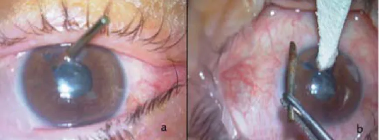
A fi fty years old Caucasian female patient presented with complaining of insertion of a metal hair clip piece into her left eye. On examination, visual acuity was limited to fi n-ger counting. On slit-lamp examination, there was a metal foreign body impacted in inferotemporal paracentral cornea extending into the lens via penetrating iris at 4 o’clock po-sition, and traumatic cataract (Figure 1a). Besides the trau-matic fi ndings, a LASIK scar was also observed. A compre-hensive fundoscopic examination could not be achieved due to the traumatic cataract.
The patient underwent immediate operation, foreign body was extracted (Figure 1b) and the corneal wound was closed. Next, the perforated anterior capsule was stained with trypan blue, capsulorhexis was performed and lens was aspirated. It was noted that posterior capsule had not been ruptured. (Figure 2a).
Corneal sutures were removed 2 months after primary re-pair and iris reparation and secondary IOL implantation was planned (Figure 2b).
Axial length (AL), anterior chamber depth (ACD) and kera-tometry measurements were taken using with the IOLMaster 500 partial coherence interferometer (Carl Zeiss Meditec, Dublin, CA). Preoperative measurements were recorded as follows: AL: 23.80mm, K1: 40.00D/8.43 mm @ 140° , K2: 41.46 D/ 8.14 mm @ 50°, ACD: 3.93 mm.
However, an appropriate IOL calculation formula for a trauma induced aphakic eye could not be obtained in the literature, Haigis-L algorithm within the partial coherence interferometer’s software was chosen to calculate the IOL power preoperatively.
In the operation, clear corneal incision (3.00 mm) was per-formed in the steep corneal axis (50°) to decrease corneal astigmatism. Then, +26.0 D Alcon Acrysof MA60AC
(Al-223
Glo-Kat 2017; 12: 222-225 Arıcı et al.
Figure 1: Photographies from operating microscope. a : Preoperatively b: Just after removing of hair clip.
Figure 2: Slitlamp photographies with diffuse illumination postoperative corneal fi xation and traumatic cataract extraction a: 1 week
con Laboratories, Inc., Texas, USA) three pieces foldable IOL was implanted into the capsular bag based on adequate capsular support.
Then traumatic iris rupture was fi xed using PC9 suture with the technique of Siepser sliding knot (Figure 3a). The sta-ble refraction postoperatively was – 0.50 – 0.75/160 degrees and BCVA was 10/10 at the fi rst year of trauma (Figure 3b). CONCLUSION
After corneal refractive surgery, routinely used methods to calculate IOL power do not guarantee the same accuracy compared with naive eyes. There are three main reasons for this challenges; incorrect measurement of the corneal radi-us due to inability to measure the anterior corneal curvature correctly, changed refractive index and incorrect estimation of the effective lens position in IOL calculation formulas. Combination of these errors result in postoperative hyper-opic refraction.1
To prevent or minimize this refraction error numerous new methods have been developed to calculate IOL power. Clin-ical history method 2 had been considered as gold standart to
calculate IOL power after refractive surgery but nowadays it has been replaced by new methods that do not require his-torical data.3 At present there are two most popular software packages for post-myopic refractive eyes; the Holladay IOL Consultant Program and the ASCRS IOL Power Calculator. ASCRS IOL Power Calculator system contains Wang/Koch/ Maloney method,Shammas No-History with Regression Analysis, the Haigis-L, ASCRS-Average, ASCRS-Mini-mum and ASCRS-MaxiASCRS-Mini-mum formula.4These methods cal-culate the corneal power from standard keratometry using a conversion formula obtained by regression analysis. There are two benchmark standards which are proposed by the British National Health Service for IOL calculating
for-mulas in virgin cornea cataract surgeries, that is, 55% of the eyes should be within 0.50 D of the target spherical equiva-lent and 85% within 1.00 D.5
The mean absolute error (MAE) is an another index which is used for evaluating accuracy of IOL formulation methods, is defi ned as the absolute difference between the intended formula-derived spherical equivalent (SE) refractive target and the actual postoperative SE.
Haigis L formula which does not need historical data, uses linear regression analysis to correct corneal power which obtained from the IOLMaster.6 We preferred using Haigis –L formula in this case, because it is easy to achieve de-pending on the availability on the IOL Master and it had acceptable refractive outcome in the studies.3,6,7
Haigis described the Haigis-L formula for the calculation of IOL power after refractive laser surgery in a study of 187 eyes with a mean AL of 27.02 ±2.01 mm. 6 In this study,
the MAE was 0.37 D and the percentages of correct refrac-tion predicrefrac-tions within ±1.00 and ±0.50 D were 84.0% and 61.0%, respectively which fulfi ll the criteria of British Na-tional Health Service.6
With the Haigis-L formula, Chee-Wai Wong et al.8 found that the overall predictability for achieving ±0.50 D and ±1.00 D of the target refraction were 45.2% and 71.0%, re-spectively and MAE of 0.77 ± 0.74 D in a study of 62 eyes with ALs greater than 25.0 mm. This results still fall short of the benchmark standards proposed by the British National Health Service but when compared with other modern IOL power calculation formulas, had the highest proportion ac-ceptable accuracy to predict and achieve target outcomes. According to these studies the Haigis-L formulahas accept-able accuracy to predict IOL power after phacoemulsifi ca-tion in eyes that had previous LASIK. In the literature there
Figure 3: Slitlamp photographies with diffuse illumination postoperative secondary IOL implantation and iridoplasty. a: , 1 day
is no study which shows accuracy of Haigis-L method in traumatic aphakic patients following myopic LASIK opera-tion, also there is not any other study which shows the most suitable method for IOL power calculation in these eyes. In our case we preferred Haigis-L method and patient had ac-ceptable refraction like – 0.50 – 0.75/160° at the fi rst year of trauma.
This case provides the fi rst evaluation of the accuracy of Haigis L methods for IOL power prediction in traumatic aphakic patients following myopic LASIK operation and re-fractive result showed that it might be a preferable method for these eyes.
REFERENCES / KAYNAKLAR
1- Utine CA, Çakır H, Çakır M, Perente I. Intraocular le ns power cal-culation after keratorefractive surgery. T Oft Gaz. 2007; 37: 427-32.
2- Holladay JT. Determining the power of an intraocular lens to achieve a postoperative correction of -1.00D. Refract Corneal Surg. 1989; 5: 202-3.
3- Wang L, Hill WE, Koch DD. Evaluation of intraocular lens pow-er prediction methods using the Ampow-erican Society of Cataract and Refractive Surgeons Post-Keratorefractive Intraocular Lens Power Calculator. J Cataract Refract Surg. 2010; 36: 1466-73.
4- Yang R, Yeh A, George MR, Rahman M, Boerman H, Wang M. Comparison of intraocular lens power calculation methods after myopic laser refractive surgery without previous refractive surgery data. J Cataract Refract Surg. 2013; 39: 1327–35.
5- Gale RP, Saldana M, Johnston RL, Zuberbuhler B, McKibbin M. Benchmark standards for refractive outcome after NHS cataract surgery. Eye. 2009; 23: 149–52.
6- Haigis W. Intraocular lens calculation after refractive surgery for myopia: Haigis-L formula. J Cataract Refract Surg. 2008; 34: 1658–63.
7- McCarthy M, Gavanski GM, Paton KE, Holland SP. Intraocular lens power calculations after myopic laser refractive surgery: a comparison of methods in 173 eyes. Ophthalmology. 2011; 118: 940-4.
8- Wong CH, Yuen L, Tseng P, Han DCY. Outcomes of the Haigis-L formula for calculating intraocular lens power in Asian eyes after refractive surgery. J Cataract Refract Surg. 2015; 41: 607–12.
225