66
Case Report / Vaka Sunumu Cardiovascular Surgery / Kalp Damar Cerrahisi
Medeniyet Medical Journal 32(1):66-69, 2017 doi:10.5222/MMJ.2017.1008
ISSN 2149-2042 e-ISSN 2149-4606
Epicardial permanent pacemaker implantation via coronary
sinus in a patient with mechanical tricuspid valve prosthesis
Mekanik triküspid kapak protezi olan bir hastada koroner sinüs yolu ile
kalıcı pacemaker implantasyonu
Tolga Demİr1, Filiz KızılırmaK2, Nurşen TaNrıKulu3, Fethi KılıçarslaN2, Ergun Demİrsoy1
received: 23.01.2016 accepted: 20.02.2016
1Kolan International Hospital, Department of Cardiovascular Surgery, Istanbul, Turkey 2Medipol University Hospital, Department of Cardiology, Istanbul, Turkey
3Kolan International Hospital, Department of Anesthesiology, Istanbul, Turkey
yazışma adresi: Tolga Demir, Kolan International Hospital, Department of Cardiovascular Surgery, Istanbul e-mail: [email protected]
ıNTroDuCTıoN
Complete atrioventricular block (CAVB) following tricuspid valve surgery is an important complication which requires permanent pacemaker implantation. However, it is not possible to pass the pacing lead through the mechanical tricuspid valve. In these pati-ents epicardial pacemaker implantation via coronary sinus (CS) can be preferred as an alternative method to surgical epicardial pacemaker implantation1-4. In this manuscript, we present a patient who had CAVB following mechanical tricuspid valve replacement and treated with epicardial pacing via CS.
Case rePorT
A 58-year-old female patient who underwent mit-ral valve replacement 15 years ago, presented with shortness of breath. Her electrocardiogram (ECG) revealed atrial fibrillation, echocardiographic exami-nation revealed normal LV systolic function, normal mitral mechanic prosthetic valve, severe tricuspid re-gurgitation and enlargement of the right heart cham-bers (Figure 1a). Surgical treatment was recommen-ded for severe tricuspid regurgitation. A mechanical prosthesis was implanted. During the surgery, CAVB developed after removal of the aortic cross clamp
aBsTraCT
Pacemaker implantation in patients with tricuspid prosthesis is a challenging situation. It is not possible to pass the pacing lead through tricuspid valve in these patients. Therefore, ventricular pacing can be achieved either epicardial or endocardial left vent-ricular stimulation. For epicardial pacing, lead implantation can be done transvenously via coronary sinus or surgically. In this paper, we present a patient who had complete atrioventricular block (CAVB) following tricuspid valve prosthesis implantation and treated with epicardial pacing via coronary sinus. In pati-ents who develop CAVB after tricuspid valve replacement, can be treated with permanent pacemaker implantation via CS. This technique enables a minimally invasive approach and effective stimulus in patients with inaccessible right ventricle because of the presence of prosthetic tricuspid valve.
Key words: Mechanical tricuspid valve prosthesis, coronary si-nus, permanent pacemaker
ÖZ
Triküspid protezi olan hastalarında pacemaker implantasyonu zor bir durumdur. Bu hastalarda, pacemaker elektrodunu triküspit kapaktan geçirmek olası değildir. Bu nedenle, ventriküler uyarı, ancak epikardiyal veya endokardiyal sol ventriküler stimülasyon ile elde edilebilir. Epikardiyal uyarı için, elektrodun implantas-yonu, tranasvenöz yol ile koroner sinüse veya cerrahi yöntemle yapılabilir. Bu makalede, triküspid kapak replasmanı sonrasın-da gelişen tam kalp bloğunun, koroner sinüs yoluyla epikardiyal stimülasyon elde edilerek tedavi edildiği bir hastayı sunuyoruz. Triküspid kapak replasmanı sonrası tam kalp bloğu gelişen hasta-lar koroner sinüs yolu ile implante edilen kalıcı kalp pili ile tedavi edilebilirler. Bu teknik, hem minimal invaziv bir yaklaşım hem de protez triküspid kapak nedeniyle sağ ventriküllerine erişilemeyen hastalarda etkili bir uyarı sağlar.
Anahtar kelimeler: Mekanik triküspid kapak protezi; koroner si-nüs; kalıcı pacemaker
67
T. Demir et al., Epicardial permanent pacemaker implantation via coronary sinus in a patient with mechanical tricuspid valve prosthesis
and the patient was paced by epicardial temporary pacing leads. On the 7th postoperative day, perma-nent pacemaker implantation was decided due to the persistence of CAVB. (Figure 1b). Due to the pre-sence of a mechanical tricuspid valve, epicardial pa-cemaker implantation via CS was planned.
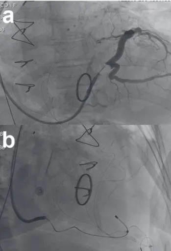
A subcutaneous pocket was created at the left pec-toral region. Following venous access with modified Seldinger technique the J wire was advanced along inferior vena cava. CS was cannulated and occlusive CS angiogram was performed (Figure 2a). The middle cardiac vein (MCV) was targeted. A 0.014” guidewire was positioned into the MCV and CS pacing lead was advanced over it (Figure 2b). The lead was stable at the apical segment of the MCV. However, at this posi-tion because of diaphragmatic stimulaposi-tion we could not implant the lead to MCV. Then, the posterolate-ral branch was targeted. A 5 F coronary angiography catheter was introduced through the CS sheath and posterolateral branch was cannulated. A 0.014 gui-dewire was advanced through proximal tortous
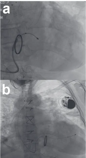
seg-ment. This could be possible with the support of CS sheath and 5-F coronary angiography catheter (Figure 3a). After several attempts we managed to place LV lead in apical region of LV over the guidewire. In this position, pacing threshold, impedance and R wave amplitude were within acceptable ranges and there was no diaphragmatic stimulation (threshold: 0,5 V, Impedance: 780 ohms, R wave: 14 mV). The lead was connected to pacemaker generator (Medtronic RE-LIA) (Figure 3b). Postprocedural ECG revealed pace-maker rhythm with right bundle branch morphology. The patient was discharged two days after implan-tation without any complication. At the follow- up exam, fluoroscopy revealed stable lead position and all pacemaker functions were normal with acceptab-le interrogation measurements.
Figure 1. Preoperative 12-lead electrocardiogram (eCG) sho-wing atrial fibrillation (a). Postoperative eCG shosho-wing complete atrioventricular block with atrial fibrillation.
Figure 2. Coronary sinus (Cs) angiogram (a), Cs pacing lead in the middle cardiac vein (b).
68
Med Med J 32(1):66-69, 2017
DısCussıoN
CAVB following tricuspid prosthesis valve surgery is a common complication mostly related to valve sutu-res on the septal leaflet near the AV node. A limited number of studies have shown that the need of pa-cemakers after a tricuspid valve surgery during the early perioperative phase is higher (in 13% to 28% of the cases), than other valve interventions5.
Pace-maker implantation in these patients is troublesome since it is impossible to reach right ventricle. Epicar-dial pacing (either surgically or via CS) or endocarEpicar-dial LV pacing are the alternative options. Due to pree-xisting mitral valve prosthesis, LV endocardial pacing was not possible in our patient. Also because of the history of two cardiac surgeries she was not a good candidate for surgical epicardial pacing. Therefore we decided to perform pacemaker implantation via CS. However, if pacing via CS could not be achieved, we have planned surgical epicardial pacing as an al-ternative treatment option.
Several case reports have been published on epicar-dial permanent pacemaker implantation via CS in patients with tricuspid valve prostheses3,4. In parallel with the frequent use of cardiac resynchronization therapy, the number of pacemacer implantations increased, and permanent pacing via CS have beca-me a viable option in these patients. However, vari-ations in CS anatomy, diaphragmatic stimulation and inability to detect appropriate pacing threshold may sometimes make CS lead implantation impossible. In our case, we had to change position of the lead due to diaphragmatic stimulation in MCV. In patients who underwent surgery for tricuspid valve disease CS dis-tortion and dilation along with dilated right heart chambers may further complicate lead implantation in CS.
Lead dislodgement is an important issue for CS lead implantation. Pacemaker dislocation in a pacema-ker dependent patient with total AV block may lead to pacemaker dysfunction which can result in mor-tal consequences. Therefore, lead stability is crucial in these patients. In our patient, although the tor-tuosity of the targeted side branch made the imp-lantation difficult, it has provided a good stability. At the follow up exam, functions of the pacemaker were normal. There are leads with active fixation capability which are developed to increase CS lead stability. However, we did not prefer those leads because it would be difficult to advance leads into posterolateral branch of CS due to angled and tor-tuous anatomy.
Figure 3. Coronary sinus (Cs) pacing lead advanced through pos-terolateral brunch with support of Cs sheath and 5 F coronary angiography catheter (a), Cs pacing lead positioned into poste-rolateral brunch and connected to pacemaker generator (b).
69
T. Demir et al., Epicardial permanent pacemaker implantation via coronary sinus in a patient with mechanical tricuspid valve prosthesis
Permanent pacemaker implantation in the right ventricle causes left bundle branch block in ECG after the procedure. In these patients, right bundle branch morphology after permanent pacemaker implantati-on may be a sign for lead malpositiimplantati-on. However, it is an expected and normal ECG finding in patients un-dergoing permanent pacemaker implantations via CS lead.
In conclusion, patients who develop CAVB after tri-cuspid valve replacement, can be treated with per-manent pacemaker implantation via CS. This tech-nique enables a minimally invasive approach and effective stimulus in patients with inaccessible right ventricle due to the presence of prosthetic tricuspid valve.
reFereNCes
1. Grimard C, Clémenty N, Fauchier L, Babuty D. Ventricular pa-cing through coronary sinus in patients with tricuspid prost-hesis. Ann Thorac Surg 2010;89:e51-2.
http://dx.doi.org/10.1016/j.athoracsur.2010.03.084 2. Demir AD, Sen N, Erbay AR, Atak R. An effective and safe
al-ternative to epicardial pacemaker placement for permanent pacemaker implantation in a patient with mechanical tricus-pid valve: stimulation of the left ventricle through the coro-nary sinus. Turk Kardiyol Dern Ars 2011;39:244-7.
http://dx.doi.org/10.5543/tkda.2011.01322
3. Herre JM, Bullaboy CA, Derkac WM, Dow MT. Permanent transvenous dual-chamber pacing using the coronary sinus in a patient with a mechanical prosthetic tricuspid valve.
Pe-diatr Cardiol 2004;25:65-6.
http://dx.doi.org/10.1007/s00246-003-0493-4
4. Vijayakumar M, Kamath P, Pai PG. Permanent pacing in a pa-tient with tricuspid prosthesis-widening therapeutic use of coronary sinus. Indian Heart J 2013;65:611-3.
http://dx.doi.org/10.1016/j.ihj.2013.08.012
5. Jokinen JJ, Turpeinen AK, Pitkänen O, Hippeläinen MJ, Harti-kainen JE. Pacemaker therapy after tricuspid valve operati-ons: implications on mortality, morbidity, and quality of life.
Ann Thorac Surg 2009;87:1806-14.