Charge storage and release onto Au and Ag nanoparticles in
aqueous medium as probed by optical spectroscopy
Ilknur Tunca, Hikmet Sezenb, Haci Osman Guvencc, Sefik Suzer*
Bilkent University, Chemistry Department, 06800 Ankara, Turkey a
Present Address: Turk Hava Kurumu Universitesi, Ankara, Turkey b
Present Address: Karlsruhe Institute of Technology, Karlsruhe, Germany c
Present Address: Heidelberg University, Heidelberg, Germany *
Author for correspondence: Sefik Suzer, email: [email protected] Received 18 Mar 2013; Accepted 22 Apr 2013; Available Online 22 Apr 2013
1. Introduction
Gold and silver nanoparticles have captured the interest of many researchers because of their unique properties which do not exist in bulk metals nor in molecules. Particularly, strong and well-defined surface plasmon resonance (SPR) band of the nanoparticles develop in the visible region after a certain size [1]. Hence, photochemical properties can be followed by the corresponding spectral changes in these bands and for utilization in certain technological applications like sensing, photodynamic therapy, etc [2-5].
The position of the surface plasmon band depends strongly on size, shape and environment. Silver and gold nanoparticles smaller than 3 nm in diameter do not exhibit a significant absorption, but particles with 3-50 nm size show strong SPR bands around 390-420 and 520-560 nm for silver and gold respectively, which shift to larger wavelengths with increasing size [6-10]. Shape-dependency of the position of SPR band is also strongly related with the interaction between the incoming light and the particles [11]. Additionally, increase in the refractive index of the medium leads to a red-shift [12]. Composition-dependence in Au-Ag alloys is another factor for tuning the SPR band, such that, the absorption band shifts between 380 and 530 nm as a function of the alloy content, as first reported by Link et al. [13], and recently by us [14].
Optical properties of small metal nanoparticles are dominated by the collective oscillation of conduction electrons (surface plasmon) resulting from the interaction with electromagnetic radiation and the Mie theory provides a description of this interaction of the electromagnetic radiation
for spherically symmetric particles in a dielectric medium. Accordingly, the total extinction coefficient of small metallic particles is given as the summation of all oscillations contributing to the absorption and scattering of the interacting electromagnetic radiation [11]. However, the derivation of Mie theory is based on zero electrical charge density ( = 0), and is not extendable to charged clusters. On the other hand, the Drude model [1,15] offers an explanation for predicting the shifts in the absorption spectrum of metallic nanoparticles, which strongly depend on 2 factors; (i) dielectric constant of the medium, and (ii) density of the electrons. Accordingly, the bulk plasmon frequency of metal clusters is proportional to electron density on the particles. Thus, stored additional electrons on the nanoparticles are expected to lead to a blue-shift in the spectrum [12,16,17].
Addition of electron donor species such as borohydride ions to metal nanoparticles solution in aqueous medium also provides an increase in the electron density around the particles, which is also evidenced by the blue shift of the corresponding SPR band. Along these lines, we have recently reported on the charge storage capacities of alloys and core-shell-structured Ag-Au nanoparticles with different compositions, where we presented data showing that the extent of the blue shift as a result of electron storage increases quasi-linearly with the mole fraction of silver in the nanoparticles. The trend is very similar to reported ones previously for both the frequency and the extinction coefficient of the plasmon band shifts of the neutral nanoparticles [13,15]. Accordingly, Au(core)@Ag(shell) nanoparticles exhibit considerably larger optical shifts than that of Au-Ag nanoalloys with the same composition due to the enhanced Ag content on the surface of the particles [14]. Abstract
Gold and silver nanoparticles in aqueous solutions can store negative or positive charges when, respectively, NaBH4, KI are introduced into the same media. The charge storage can be followed by the spectral shifts in the corresponding surface plasmon resonance (SPR) bands of these nanoparticles. In a similar way, the kinetics of these two processes can be monitored by the same shifts. Accordingly, we show that although Au nanoparticles exhibit smaller spectral shifts upon both negative and positive charge storage, when compared with Ag nanoparticles, their kinetics are faster towards reduction (electron storage) and comparable towards oxidation. Hence, if not the spectral shifts, the kinetics of the electron storage process can be correlated to the larger electron affinity of Au nanoparticles, when compared with those of Ag. The similarity of the kinetics towards oxidation must also be related to the small difference between the ionization potentials of Au and Ag nanoparticles.
These negatively charged nanoparticles are not stable and loose the stored electrons back to oxygen dissolved in the aqueous medium, as evidenced by the gradual red-shift back to their initial positions in time. Alternatively, strong electron acceptor agents such as methyl viologen, or thionine preferentially capture the electrons and/or prevent completely the electron storage on the metal nanoparticles. As an experimental demonstration, Henglein et al. reported a red-shift in the SPR band of colloidal silver particles due to the oxidation of surface atoms by the nucleophilic reagents chemisorbed [18,19]. Later, they reported on the changes of the SPR band of colloidal Ag by addition of mixed KI and NaSH solutions. Red-shift and damping in the SPR band were reported as a result of surface atoms complexation [20].
Control of the charging/discharging abilities of metal nanoparticles are expected to provide control over different photocatalytic, luminescent and sensing properties of these particles, and optical absorption provides and easy and convenient technique for assessing these properties. As an extension of our previous work [14], we now provide a kinetic investigation on charge storage and releasing abilities of Au and Ag nanoparticles by following the shifts of the respective absorption bands after sequential addition of electron donor and acceptor species, for furthering our basic understanding of the factors contributing to and/or governing these important physicochemical processes.
2. Experimental Details
HAuCl4, AgNO3, trisodium citrate dihydrate, NaBH4,
Thionine and KI were purchased from Aldrich and used without further treatment. Milli-Q grade water was used in all preparations. Gold colloidal nanoparticles were synthesized following the protocol by Turckevich et. al. [21,22]. A 100 mL solution of 5.0x10⁻⁴ M HAuCl4 was allowed to boil, at
which point 5.0 mL of 1.0 % (w/v) sodium citrate was added dropwise with stirring. Following the addition of sodium citrate, the solution began to darken and turn bluish-gray or purple. After approximately 5 min. the reaction was complete and the final color of the solution was a deep wine red. Similarly colloidal silver nanoparticles were prepared by
rapidly adding 1.0 mL of 0.01 M AgNO3 to 99 mL of solution
containing 1.0 mM NaBH4 and 0.30 mM sodium citrate under
vigorous stirring at room temperature.
Electron storing and releasing (optical) properties of Au and Ag nanoparticles were characterized by a Ocean Optics (HR4000 Model) UV-Vis spectrophotometer. To study the kinetics of charge injection to the nanoparticles, freshly prepared 4 mM, ice-cool aqueous borohydride, thionine or KI solutions were introduced drop-wise, and changes in the absorption spectra were recorded in time.
3. Results
3.1. Reduction
3.1.1. Spectral shifts due to electron storage
Negative charge storage of Au and Ag nanoparticles is based on oxidation of borohydride upon addition to aqueous solutions of Au and Ag nanoparticles. Generated electrons are injected to the nanoparticles, leading them to be cathodically polarized. The representative reaction is given as follows:
BH4- + Mn + 3H2O BO33- + Mn8- + 10H+
Electron addition to molecules generally leads to a
red-shift in absorption band of these molecules. However,
electron injection into metallic nanoparticles causes a
blue-shift of the corresponding SPR band, along with an increase in
the extinction coefficient. Using the Mie theory, numerous workers have shown that the plasmon band position as well as its extinction coefficient are both related to the electron density (n), the electron effective mass (meff), and the dielectric
function (ε2) of the medium through the relation [11]:
2 max 0 eff
1
2ne
m
Hence storage of additional electrons on the nanoparticles is expected to lead to a blue-shift in the spectrum.
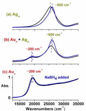
Figure 1 shows the response of separate aqueous solutions of Au and Ag nanoparticles, as well as a mixture with approximately equal concentration and size, before and after addition of NaBH4. To follow the changes in the
frequency of the SPR band, we have used the energy/frequency scale (in cm-1) to display the spectra, rather than the conventional wavelength scale (in nm). The extent of the blue-shift as a result of charge storage of the Ag nanoparticles is always larger (ca. 600 cm-1) than that of Au nanoparticles (ca. 200 cm-1), both in separate and mixed solutions, as was also discussed in our previous paper [14].
Figure 1. Optical spectra of ca. 20 nm Au and Ag nanoparticles
separately (a and c) and together (b) in aqueous medium, before (black) and after (blue) introduction of NaBH4.
Relevant data are given in Table 1. Note that oxygen dissolved in water competes with this reaction and the blue shifted spectra return back to its original position within several minutes.
3.1.2. Spectral shifts due to electron release
On the other hand, thionine is an organic dye molecule, which is widely used in following charge transfer and/or electron donor-acceptor reactions, since it exists as a monovalent cation in near-neutral pH values, and captures electrons strongly via the reaction: [16,17]
Thio+ + 2H+ + 2e - → ThioH2+
Accordingly, when introduced to the medium with negatively charged nanoparticles it leads to the corresponding
discharging process and the SPR bands start shifting back to their original positions, as also shown in Figure 2.
3.1.3. Kinetics of electron storage and release
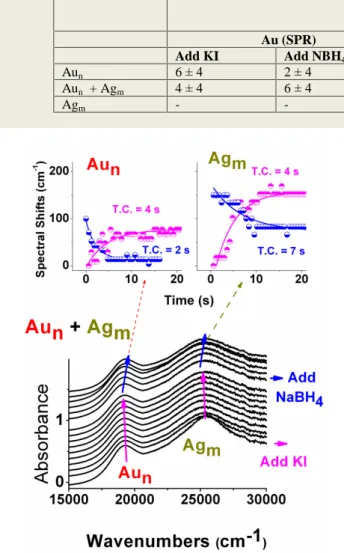
To study the kinetics of charge storing and releasing processes of Au and Ag nanoparticles, time resolved response of the particles are measured by recording the UV-Vis-NIR spectra at 0.6 second intervals, upon addition of NaBH4 and
later on thionine respectively, as shown in Figure 2. Exponential fits to the time data reveals relative time constants (T.C.) of these two processes for Au and Ag nanoparticles separately, as well as in their mixture, which are indicated in the inset of Figure 2, and are collected in Table 1. As was reported in our previous paper, and also stressed above, Ag nanoparticles exhibit a larger spectral shift upon electron addition when compared to Au nanoparticles, both separately, as well as in the same solution [14]. However, the situation is the opposite when the time constants are considered. When present in the same solution, the reduction time constant of Au nanoparticles is 12 s, whereas that of Ag is 20 s. In separate solutions, as well as together Au nanoparticles always exhibit faster kinetics towards both electron capture and release.
3.2. Oxidation
As a next step, the result of oxidation of the nanoparticles by complexation with KI is studied. The position of the maximum of the plasmon band changes upon chemisorption of I- anions, due to oxidation of surface atoms of the Au/Ag nanoparticles. The proposed structure is Ag+ I-
for silver complex, existence of which is monitored by the red-shift on the SPR band with respect to that of bare Ag nanoparticles [19]. These anodically polarized particles are then exposed to NaBH4 to reverse the process. The spectral
changes, as well as the kinetics of these two processes are given in Figure 3, and pertinent data related also with the derived time constants (T.C.) are collected in Table 2. Close examination of the data reveals that, although the extent of the spectral shifts are again larger for Ag nanoparticles, no significant differences are seen between the time constants of oxidation of Au and Ag nanoparticles by complexation of I-, nor during the back-reduction with NaBH4.
4. Discussion
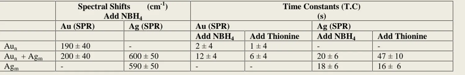
As mentioned in the introduction section several parameters can contribute to the spectral shifts observed like shape, size, and environment. Unless special care is taken to foster the growth in certain directions, most chemical routes, including the citrate reduction method that we use, yield Table 1. Spectral Shifts Observed and Kinetic Data (Time Constants) of Electron Storage (by addition of NaBH4) and Release (by addition of Thionine) to Au and Ag nanoparticles separately and together in aqueous medium.
Spectral Shifts (cm-1) Add NBH4
Time Constants (T.C) (s)
Au (SPR) Ag (SPR) Au (SPR) Ag (SPR)
Add NBH4 Add Thionine Add NBH4 Add Thionine
Aun 190 ± 40 - 2 ± 4 1 ± 4 - -
Aun + Agm 200 ± 40 600 ± 50 12 ± 4 6 ± 4 20 ± 6 47 ± 10
Agm - 590 ± 50 - - 18 ± 6 16 ± 6
Figure 2. A sequence of spectra recorded in time to the mixture
solution of Figure 1 (b) following introduction of NaBH4, and later of Thionine respectively. The inset displays the time variation of the positions of the corresponding SPR bands of Au and Ag nanoparticles. Exponential fits to the data for driving the time constants of the processes upon addition of NaBH4 and Thionine are also depicted in the inset (see also Table 1).
spherical nanoparticles. Hence, shape can be ruled out. Only the nanoparticles larger than 3 nm develop strong SPR band, and an optimum and reproducible size of 15-20 nm is obtained by our procedure judged by the reproducible position of the SPR band within ± 5 nm. Furthermore most chemical treatments result in enlarging the size of the particles, through aggregation, resulting a permanent red shift in the SPR band. Similarly, when the particles are forced to approach each other, by coating on a solid surface, or by evaporation of the solvent, etc., additional bands arise, which are, however, also strongly red-shifted from the SPR band. The change in the refractive index of the medium also causes shifts in the SPR band. For aqueous solution this shift is also towards higher wavelength (red-shift) since water has the lowest refractive index, as recently reported by Templeton et al. where 8 nm red-shift was reported when the refractive index was increased from 1.33 (water) to 1.55 [23]. As can be seen from this discussion, all these changes are expected to cause; (i) red,
and (ii) permanent shifts in the corresponding SPR band,
therefore they cannot explain our experimental observations that; (i) blue, and temporary shifts upon addition of NaBH4 to
the aqueous solutions containing our Ag, and Au nanoparticles either separately or together in the same vessel. Furthermore, experiments carried out at different dilution conditions yielded similar shifts. Therefore, we are left to correlate these shifts to partial charge accumulation on the particles, allowed by their certain electrochemical properties, like the double-layer capacitance, or the allowed Fermi-Level difference from water, etc [23, 24].
If we are also to attempt to control photocatalytic, luminescent and sensing properties of charged nanoparticles we need to better understand the physical and chemical parameters affecting the various processes involved, when deriving information from experimental observations, in this particular case, spectral shifts. We can start by asking the following questions:
Question #1: Can we correlate the charging capacity of nanoclusters by the spectral shifts observed in the optical spectra? If so, which has a higher capacity, Au or Ag?
Question #2: Are the spectral shifts related with simple electron affinities (E.A.) and ionization potentials (I.P.) of these particles?
Question #3: Are the kinetics of these processes related with the same parameters (E.A. and I.P.)?
It was shown by Link et. al. that although the theoretical effective masses and the electron densities are very similar for gold and silver, both the positions and the extinction coefficients of the two are very different [13]. Accordingly, the extinction coefficient of 20 nm pure silver nanoparticles is approximately 5 times larger than that of pure gold nanoparticles. Furthermore, by using experimentally derived optical parameters from thin films of gold-silver alloys, they were also able to calculate the optical spectra of
gold-silver nanoalloys, which closely matched the
experimental data [13, 25]. This is also evidenced by our Figure 1, where the SPR band of Ag is much more intense than that of Au. In our previous paper, we used this finding that, when compared to gold, silver either has a larger electron density or a smaller effective mass to cause both a larger shift in the position of the frequency of the plasmon band and also in its extinction coefficient, to guide us in understanding, qualitatively, the spectral shifts we observed. Accordingly, we compared the optical shifts observed upon charge injection onto Au-Ag nanoalloys vs. core-shell nanoparticles, both with a nominal 25% Au content. As a result of charge storage upon NaBH4 addition, the plasmon bands of the alloy and the
Au@Ag are blue-shifted by 350 cm-1 and 1000 cm-1, respectively. Such a large difference is undoubtedly related with the different Ag and Au allocation within these nanoparticles. Although conduction electrons of the Ag and Table 2. Kinetic Data (Time Constants) of Oxidation (by addition of KI) and Back-Reduction (by addition of NBH4) to Au and Ag nanoparticles separately and together in aqueous medium.
Time Constants (T.C) (s)
Au (SPR) Ag (SPR)
Add KI Add NBH4 Add KI Add NBH4
Aun 6 ± 4 2 ± 4 - -
Aun + Agm 4 ± 4 6 ± 4 4 ± 4 3 ± 4
Agm - - 3 ± 4 3 ± 4
Figure 3. Recorded shifts in the SPR band of Au and Ag
nanoparticles of Figure 1, by sequential addition of KI and NaBH4 with respect to time. The inset displays the time variation of the positions of the corresponding SPR bands of Au and Ag nanoparticles. Exponential fits to the data for driving the time constants of the processes upon addition of KI and NaBH4 are also depicted in the inset (see also Table 2).
Au atoms move freely within the whole cluster in the alloy, those in the core-shell particles are restricted to their part, either nucleus or shell [11]. In the case of the alloy, both Au and Ag contribute to the plasmon absorption frequency. However, in the core-shell case it is the silver shell which contributes most, and in agreement with our assessment, causes a larger spectral shift. Hence, we, then, concluded our work by stating that the chemical nature of the nanoparticle is the dominating factor contributing to the extent of the spectral shifts in bimetallic systems, and silver is approximately 4-5 times more effective when compared to gold. As a consequence of these discussion we, now, come to the conclusion that the extent of the spectral shifts are not straightforwardly related with electron storage capacity of Au and Ag nanoparticles as raised in Question #1 above.
The gas-phase electron affinity of a single Au atom is 2.3 eV, which is significantly larger than that of Ag atom (1.3 eV). Moreover, those of small clusters of the same size are always higher for Au than Ag [26]. If one assumes that the electron affinities scale in the same way when extrapolated to the aqueous medium, one would expect Au nanoparticles to be a better electron capturer and display a larger spectral shift, when compared to those of Ag. Since the experimental observation is exactly in the opposite direction, in parallel to our answer to the Question #1, we can also state that simple electron affinity arguments, raised in Question #2, cannot be used to correlate the spectral shifts observed either.
The gas-phase ionization potentials of Au and Ag atoms are 9.2, and 7.6 eV respectively, again the small clusters follow more or less the same trend. Here, however, the relative difference between Au and Ag is not large, hence one should not expect a large difference between the oxidation behavior of their corresponding nanoparticles, in contrast with the larger spectral shift observed in Ag when compared to Au. In parallel to our answers stated above, we can also state that simple ionization potential arguments, raised in Question #2, cannot be used to correlate the spectral shifts observed.
On the other hand, if one correlates the kinetics of these processes, with the electron affinities and ionization potentials, the expected trend follows. The relatively larger electron affinity of Au nanoparticles renders to faster electron capture when compared with Ag, and not so-large a difference between their ionization potentials renders more or less similar kinetics towards oxidation. Hence, we finally postulate that kinetic data, rather than the spectral shifts, must be considered for assessing the charge/discharge capacities of nanoparticles in aqueous medium.
5. Conclusions
We have demonstrate that simple UV-Vis
spectroscopy, coupled with right electron donating/capturing molecules can be used to follow nanoparticles abilities for capturing and releasing of electrons in aqueous medium. In addition, we have raised and tried to answer questions towards
understanding why and how these nanoparticles perform these reactions, and whether or not they are related to simple electronic properties like ionization potentials and electron affinities. Naturally, we are hoping that we, as well as others, will take up from our findings and pursue both in experimental and theoretical directions to further understanding these issues.
References
1. P. Mulvaney, Langmuir 12 (1996) 788.
2. V. L. Colvin, M. C. Schlamp, and A. P. Alivisatos, Nature 370 (1994) 354.
3. R. Elghanian, J. J. Storhoff, R. C. Mucic, R. L. Letsinger, and C. A. Mirkin, Science 277 (1997) 1078.
4. M. Shim and P. Guyot-Sionnest, Nature 407 (2000) 981. 5. T. A. Taton, C. A. Mirkin, and R. L. Letsinger, Science 289
(2000) 1757.
6. A. Henglein, Langmuir 15 (1999) 6738.
7. A. Henglein and D. Meisel, Langmuir 14 (1998) 7392. 8. M. M. Alvarez, J. T. Khoury, T. G. Schaaff, M. N. Shafigullin,
I. Vezmar, and R. L. Whetten, J. Phys. Chem. B 101 (1997) 3706.
9. S. W. Chen, R. S. Ingram, M. J. Hostetler, J. J. Pietron, R. W. Murray, T. G. Schaaff, J. T. Khoury, M. M. Alvarez, and R. L. Whetten, Science 280 (1998) 2098.
10. C. A. Mirkin, R. L. Letsinger, R. C. Mucic, and J. J. Storhoff, Nature 382 (1996) 607.
11. U. Kreibig and M. Vollmer, Optical Properties of Metal Clusters, Springer, Berlin (1995).
12. P. V. Kamat, J. Phys. Chem. B 106 (2002) 7729.
13. S. Link, Z. L. Wang, and M. A. El-Sayed, J. Phys. Chem. B 103 (1999) 3529.
14. I. Tunc, H. O. Guvenc, H. Sezen, S. Suzer, M. A. Correa-Duatre, and L. M. Liz-Marzan, J. Nanosci. Nanotechnol. 8 (2008) 3003.
15. P. Mulvaney, J. Perez-Juste, M. Giersig, L. M. Liz-Marzan, and C. Pecharroman, Plasmonics 1 (2006) 61.
16. T. Hirakawa and P. V. Kamat, Langmuir 20 (2004) 5645.
17. T. Hirakawa and P. V. Kamat, J. Am. Chem. Soc. 127 (2005)
3928.
18. A. Henglein, T. Linnert, and P. Mulvaney, Ber. Bunsenges. Phys. Chem. 94 (1990) 1449.
19. T. Linnert, P. Mulvaney, and A. Henglein, J. Phys. Chem. 97 (1993) 679.
20. F. Strelow and A. Henglein, J. Phys. Chem. 99 (1995) 11834. 21. J. Turkevich, P. L. Stevenson, J. Hillier, Discuss. Faraday Soc.
11 (1951) 55.
22. B. V. Enustun, J. Turkevich, J. Am. Chem. Soc. 85 (1963) 3317.
23. A. C. Templeton, J. J. Pietron, R. C. Murray, and P. Mulvaney, J. Phys. Chem. B 104 (2000) 564.
24. G. Oldfield, T. UNG, and P. Mulvaney, Adv. Mater. 12 (2000) 1519.
25. M. P. Mallin and C. J. Murphy, Nano Lett. 2 (2002) 1235. 26. E. M. Fernandez, J. M. Soler, I. L. Garzon, and L. C. Balbas,
Phys. Rev. B. 70 (2004) 165473.
Cite this article as:
Ilknur Tunc et al.: Charge storage and release onto Au and Ag nanoparticles in aqueous medium as probed by optical