apoptotic markers in normozoospermic and
non-normozoospermic patients
Seda Karabulut1,2, Asuman Demiroğlu-Zergeroğlu3, Elif Yılmaz4, Pelin Kutlu5 andİlknur Keskin1,2
1
Istanbul Medipol University, International School of Medicine,İstanbul, Turkey;2Istanbul Medipol University, REMER (Regenerative and Restorative Medicine Research Center),İstanbul, Turkey;3Gebze Technical University, Department of Molecular Biology and Gynetics, Kocaeli, Turkey;4Medistate Hospital, IVF Unit,İstanbul, Turkey; and5Medicana Çamlıca Hospital, IVF Center, İstanbul, Turkey
Summary
The negative effects of cryopreservation on sperm parameters are well documented but little information is known about molecular basis of the process. The aim of the present study was to investigate the possible effects of sperm cryopreservation on main apoptotic signs including DNA fragmentation and caspase-3 activation and to determine if these effects vary according to sperm parameters. Sperm samples of 72 patients were cryopreserved. The patients were sub-grouped as normozoospermic or non-normozoospermic patients according to their semen parameters. DNA fragmentation rates and caspase-3 activation levels were analyzed before and after cryopreserva-tion in both groups. Mean DNA fragmentacryopreserva-tion rate was increased significantly from 23.98% in neat semen samples to 27.34% after cryopreservation (P= 0.03). DNA fragmentation rates were slightly higher in non-normozoospermic patients compared with the normozoospermic patients in both the neat semen and after cryopreservation (23.25 and 24.71% vs. 26.32 and 28.36%, respectively) although the difference obtained were not statistically significant. An increasing trend for caspase-3 activations (0.093 vs. 0.116) was observed after cryopreservation but the differences were not statistically significant. Caspase-3 activation was found to be slightly higher in non-normozoospermic patients both in the neat semen and after cryopreservation compared with the normozoospermic patients but the differences were not statistically significant. Caspase-3 expression was also shown using immunocytochemistry in both fresh ejaculated sperm and thawed sperm after cryopreservation but at different localizations. The cryopreservation process had detrimental effects on sperm quality but the quality of the sperm samples was not adversely effective for the apoptotic markers including DNA fragmentation and caspase-3 activation patterns. In fact, it was the cryopreservation process itself that adversely effected the above apoptotic markers and apoptosis. It was concluded therefore that sperm cell cryopreservation triggers apoptosis after thawing and this process adversely affects semen parameters.
Introduction
Cryopreservation of human semen is used as a method for preserving fertility in various cases of male factor infertility 1-2 (Cavalla et al., 2006; Anderson, 2008). Negative effects of cryo-preservation on fertilization capacity, motility and viability parameters of spermatozoa have been well documented in the literature (Yoshida et al., 1990; Nallella et al., 2004; Ozkavukcu et al., 2008). As a result, the use of frozen sperm cells in assisted reproductive techniques (ART) including intrauterine insemination (IUI), in vitro fertilization (IVF) and intracyto-plasmic sperm injection (ICSI), has been reported to reduce the outcomes of these treatments, therefore resulting in reduced fertilization rates, impaired developmental and implantation potential of embryos, and to an increase in abortion rates (Yildiz et al. 2007). There is limited knowledge of the molecular basis of the cryopreservation process and sperm damage. The supposed mechanisms underlying the negative effects of freezing include membrane damage and alterations in both membrane permeability, structure of cellular skeleton and mito-chondrial damage, plus DNA fragmentation, oxidative stress, capacitation and increase in sperm morphology (Royere et al., 1991; Parks & Graham, 1992; Cormier et al., 1997; Watson, 2000; Donnelly et al., 2001; Linfor & Mayers, 2002; Peña et al., 2003; Chohan et al., 2004; Ortega-Ferrusola et al., 2008; Ozkavukcu et al., 2008). Damage that arises as a result of the freezing procedure is called as‘cryodamage’ and may be due to intracellular and extracellular ice formation and osmotic stress during freezing and thawing processes (Mazur, 1984; Royere et al., 1991; Oehninger et al., 2000; Stanic et al., 2000; Schoolcraft et al., 2004; Yildiz et al., 2007; Said et al., 2010) which may cause apoptosis, a genetic process activated for self-suicide of the cell.
cambridge.org/zyg
Article
Cite this article:Karabulut S et al (2018) Effects of human sperm cryopreservation on apoptotic markers in normozoospermic and non-normozoospermic patients. Zygote 26: 308–313. doi:10.1017/S0967199418000254 Received: 16 November 2017
Revised: 3 June 2018 Accepted: 22 June 2018
First published online: 17 September 2018 Keywords:
Apoptosis; Caspase-3; Cryopreservation; DNA fragmentation; Sperm
Author for correspondence:
Seda Karabulut. Istanbul Medipol University, International School of Medicine, Department of Histology and Embryology, Kavacık mah, ekinciler cd. No. 19 Beykoz, Istanbul. Tel: +90 532 273 98 64. E-mail: [email protected]
Apoptosis is defined as ‘programmed cell death’ coded by intrinsic factors. It is activated to eliminate old and damaged cells in an organism. It is not possible to distinguish an apoptotic sperm cell during routine microinjection procedures. Therefore, it is impossible to eliminate the damaged sperm cell after cryopre-servation and this may cause impaired fertilization and embryo development, and reductions in embryo quality, pregnancy rates, implantation rates and also an increase in abortion rates.
Apoptosis is suggested to play an important role in the cryo-injury of sperm DNA, as the process of cryopreservation has been shown to increase the activation of specific aspartic acid-directed cysteine proteases (caspases) in both human (Paasch et al., 2004a, Wündrich et al., 2006; Karabulut et al., 2014) and bull sperma-tozoa (Martin et al., 2007). The exact apoptotic mechanisms involved in the aetiology of sperm cryopreservation, including those leading to cryo-injury of sperm DNA, have not been fully understood.
Signs of apoptosis include phosphatidylserine degradation products, changes in mitochondrial membrane potential, and particularly in DNA fragmentation and the caspase cascade as a result of caspase activation. Caspases, are known to play key roles in the cellular apoptotic cascade and eventual cell death (Thornberry & Lazebnik, 1998). Apoptosis markers such as DNA fragmentation and caspase activations are observed at the end of the apoptotic process and this change is irreversible after those signs are detected. Activated caspases attack target molecules such as nucleic acids, proteins and lipids that lead to different and diverse outcomes in the cells such as DNA fragmentation and lipid peroxidation (Hetts, 1998).
Caspase-3 is an executioner caspase and is activated after cleaving by the initiator caspases. The ICAD–CAD (Inhibitors of Caspase-Activated DNase–Caspase-Activated DNase) complex is one of the substrates of caspase-3, and is excised by activated caspase-3. CAD is an endonuclease and it is set released and activated, and degrades chromosomal DNA leading to DNA fragmentation (Karp, 2007).
The aim of the present study was to investigate the possible effects of sperm cryopreservation on main apoptotic signs including DNA fragmentation and caspase-3 activation, and to determine if these effects varied according to semen quality.
Methods Study design
Cryopreserved sperm samples of 72 patients who attended the Florence Nightingale Hospital, IVF Center between January 2016 and June 2016 because of different indications (medical treat-ments, n = 8, surgical treatments, n = 32 chemotherapy or radiotherapy, n = 16 cryoptozoospermia, n = 16) were included in the study. Samples from smokers, patients with a history of varicocele, and patients with genetic abnormalities were not included in the study. Semen analysis was performed according to World Health Organization (1999) criteria. Briefly, semen con-centration (million/ml), and total and progressive motile sperm rates (%) were assessed before and after cryopreservation. Normal morphology rate (%) were assessed according to Kruger’s strict criteria (Kruger et al., 1987). Semen analyses were performed by the same technician. DNA fragmentation rates and caspase-3 activation levels were analyzed before and after the cryopre-servation procedure.
Among the 72 participants, 39 were normozoospermic, and three had at least one abnormal sperm parameter including centration, motility and morphology (<20 million/ml sperm con-centration,<50% total motility) according to WHO-1999 criteria and 14% normal morphology according to Kruger’s strict criteria (Kruger et al., 1987): <70% normal morphology) (oligoastheno-teratozoospermic: 28%; e: 3%; astheno(oligoastheno-teratozoospermic: 2%).
Sperm cryopreservation and thawing
The liquid nitrogen vapour procedure was used to cryopreserve the samples. Sperm samples were collected in a sterile container following masturbation. Liquefaction semen analysis was per-formed according to WHO-1999 criteria. The cryoprotectant solution (TYB; Irvine Scientific, Santa Ana, CA, USA) was added (1:1 sample/cryoprotectant ratio) on the semen sample very slowly, and the samples were transferred cryotubes. The cryotubes were then placed 10–15 cm above the liquid nitrogen level and were exposed to liquid nitrogen vapour for 10 min until freezing, and then put directly into liquid nitrogen.
Frozen semen samples were thawed 7 days later by placing the cryotubes in a water bath at 30°C for 10 min until the solution melted completely. The samples were then left at room tem-perature for 20 min and washed with G-IVF (VitroLife, Sweden) medium. For this purpose, 1 ml of G-IVF is added to the solution which was then centrifuged at 1300 rpm for 10 min; the super-natant was discarded to get remove the cryoprotectant. This step was repeated two times and the pellet was used for examination.
Sperm DNA fragmentation assessment
The terminal deoxynucleotidyl transferase (TdT) dUTP Nick-End Labelling (TUNEL) assay was used to analyse DNA fragmentation in sperm cells. DNA fragmentation was analysed both before and after the cryopreservation procedure. In Situ Cell Death Detection Kit, Fluorescein, Roche, was used for TUNEL procedure, which was performed as recommended in the datasheet.
The samples were centrifuged at 2000 rpm and were separated from the seminal plasma. The pellets were suspended three times in 1 ml of distilled water, homogenized and centrifuged again. Following the addition of a hypotonic solution (KCl; 0.075 M) onto the pellets, they were centrifuged once more and incubated at 37ºC for 30 min. Next, 2 ml of fixative (2:1; methanol: acetic acid) was added on each pellet and the samples were kept at −20ºC for 15 min. Smears were prepared on slides, air dried, treated in 1 × phosphate-buffered saline (PBS) for 5 min, fol-lowed by washing in an ethyl alcohol series (70, 85 and 100%). The In Situ Cell Death Detection Kit, Fluorescein (Roche, Merck KGaA, Darmstadt, Germany) was used to prepare the stock solution, which was added onto the slides. Then slides were then covered to provide darkness, and were incubated at 37ºC for 45 min. Subsequently, they were washed three times in PBS for 1 min each, and left to dry at room temperature. Cells they were stained with DAPI, and incubated at 37ºC for 45 min covered with aluminium foil. Following the washing procedure with PBS for 1 min, samples were air dried, stained with DAPI again, and analyzed under a FITC–DAPI filter in a fluorescence microscope. Minimally, 200 sperm cells were analysed by observing at least 10 microscopic fields with a ×10 magnification. All sperm cells analyzed microscopically were chosen randomly from different parts of the slides. The sperm cells with DNA fragmentation were visualized as green, and those with no DNA fragmentation were visualized as blue.
Caspase-3 analysis
Caspase-3 expression level analysis
Caspase-3/CPP32 Colorimetric Assay Kit, Biovision was used to observe caspase-3 activation levels [optical density (OD) 405 nm]. The procedure was performed as recommended in the datasheet: 10µl of DTT (1 M) was added to 1 ml of 2 × Reaction Buffer to obtain a 10 mM solution. The sperm cells were added into the solution and counted to give a final suspension of 1 × 10⁷. Cryptozoospermic samples were counted to give a final suspension of 1 × 10⁶.
Results were multiplied by dilution rate (×10). Sperm counts were performed in replicates. The suspension was then re-suspended in chilling cell lysis buffer for 10 min on ice. Following centrifugation at 10,000 g for 1 min, the supernatant (cytosolic extract) was separated and kept at −80°C until analysis.
On the day of analysis, the frozen samples were thawed and protein concentration was determined as follows: samples were adjusted to have 200µg protein in 50 µl of Cell Lysis Buffer; 50 µl of 2 × Reaction Buffer (including 1 mM DTT) were added on this suspension which was taken into micro-wells. Subsequently, 5µl of 4 mM DEVD-PNA substrate was added to each well to give a 200µM final concentration and wells incubated at 37°C for 2 h. The absor-bance values (OD) of the samples were analyzed at 405 nm by ELISA.
Caspase-3 subcellular localization analysis
Immunohistochemistry (IHC) techiques were applied to detect the localization of caspase-3 expressions. Sperm smear slides were left to air dry, then fixed with 4% paraformaldehyde. After washing with PBS three times, a 3% hydrogen peroxide–methanol mixture was applied to the slides for 20 min. Antigen retrieval was performed using citrate buffer in a microwave. Slides were air dried, then washed with PBS three times. Blocking solution of Mouse Specific HRP (ABC) Detection IHC Kit (ab93677) (ABCA, Cambridge, UK) was applied according to the manufacturer’s protocol. Primer caspase-3 antibodies were prepared as a 1/100 dilution (sc56046) (Santa Cruz Biotechnology, Inc. Texas, USA) and applied on the slides overnight in a humidified chamber as recommended by the manufacturer. The day after, slides were washed and the streptavidin peroxidase and biotinylated goat anti-polyvalent solution of the IHC kit were applied as recommended by the manu-facturer. A DAB Substrate kit (ab64238) (ABCA, Cambridge, UK) was applied according to the manufacturer’s recommendations. Haema-toxylin was used to measure background staining. Then the slides were mounted with bio-mount medium. At least 100 sperm cells were analyzed by light microscopy (×100) and the results were calculated as percentage expression (%).
Statistical analysis
SPSS for Windows 16.0 software package (SPSS Inc., Chicago, IL, USA) was used for the statistical analysis. The Mann–Whitney
U-test was used to evaluate the differences between the groups. The results were evaluated in 95% confidence intervals and the statistical significance was defined as a P-value < 0.05.
Results
The mean age of the patients was 36.9± 18 years (range: 22–58 years) and mean baseline semen volume (ml) was 4.0± 2.2 ml. Semen characteristics of patients are given in Table 1. Sperm parameters (%) were investigated in 72 sperm samples prior to and after cryopreservation. Although there were no significant differences in sperm concentrations and normal morphology rate before and after cryopreservation, total motility, progressive motility and vitality rates decreased significantly after cryopre-servation (P = 0.032; P = 0.040; P = 0.041 respectively).
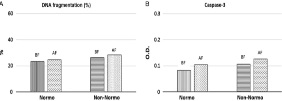
DNA fragmentation rates (%) were investigated in 72 sperm samples prior to and after cryopreservation and found to be increased significantly after cryopreservation (23.98± 8.07%; 27.34± 8.61% respectively) (P = 0.03). When the patients were sub-grouped as normozoospermic or non-normozoospermic, DNA fragmentation rates were found to be 23.25 and 24.71% in non-normozoospermic samples; and 26.32 and 28.36% in nor-mozoospermic samples respectively), although the differences obtained were not statistically significant (P = 0.07 and P = 0.07, respectively) (Fig. 1).
Caspase-3 activation levels (OD 405 nm) were measured in 72 sperm samples before and after cryopreservation. Mean caspase-3 activation values (OD 405 nm) of the patients were calculated as 0.093± 0.044 (OD 405 nm) before cryopreservation and 0.116± 0.105 (OD 405 nm) after cryopreservation. Differences were not statistically significant (P = 0.06). When the patients were grouped as normozoospermic and non-normozoospermic, caspase-3 activation (OD 405 nm) values were found to be 0.082 and 0.104 (OD 405 nm) in non- normozoospermic samples; 0.106 and 0.126 ((OD 405 nm) in normozoospermic samples respec-tively), but differences were not statistically significant (P = 0.07 and P = 0.08, respectively) (Fig. 1).
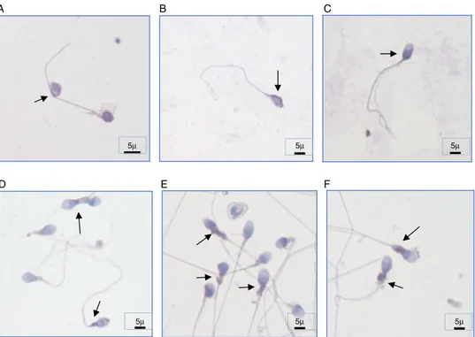
Subcellular localization of caspase-3 protein in human sperm was also investigated before and after cryopreservation. We observed that caspase-3 was expressed in both fresh ejaculated sperm and thawed sperm after cryopreservation and was localized along the entire human spermatozoa, including the acrosome region, the midpiece, and along the tail with variable immunos-taining intensity. Before cryopreservation, sperm samples showed prominent caspase-3 immunostaining at the most proximal part of the acrosome region (62% before cryopreservation and 32% after cryopreservation) and at the membrane of the head (48% before cryopreservation and 30% after cryopreservation). Whereas we observed a prominent staining at the midpiece part
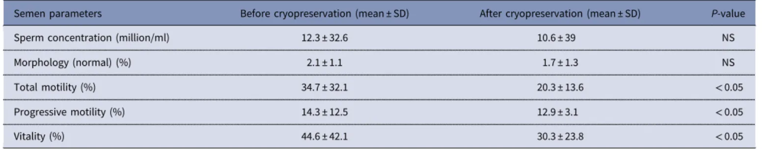
Table 1. Comparison of semen parameters before and after cryopreservation. Values are mean ± SD unless otherwise stated
Semen parameters Before cryopreservation (mean ± SD) After cryopreservation (mean ± SD) P-value
Sperm concentration (million/ml) 12.3 ± 32.6 10.6 ± 39 NS
Morphology (normal) (%) 2.1 ± 1.1 1.7 ± 1.3 NS
Total motility (%) 34.7 ± 32.1 20.3 ± 13.6 < 0.05
Progressive motility (%) 14.3 ± 12.5 12.9 ± 3.1 < 0.05
Vitality (%) 44.6 ± 42.1 30.3 ± 23.8 < 0.05
of the sperm (62% after cryopreservation and 32% before cryo-preservation) and at the cytoplasmic droplets of abnormal sperm cells (42% after cryopreservation and 28% before cryopreserva-tion) (Fig. 2). Interestingly, we observed weaker caspase-3 stain-ing in two particular spermatozoa regions: the postacrosomal region of the head (12% before cryopreservation and 15% after cryopreservation) and in the tail (8% before cryopreservation and 12% after cryopreservation) both before and after cryopreserva-tion. All of the cryopreserved sperm samples were used for intracytoplasmic sperm injection (ICSI) after thawing and resul-ted in an overall pregnancy rate of 32.6%, which was calcularesul-ted as 39.4% of the fresh ejaculate group.
Discussion
The mechanisms underlying the negative effects of the cryopre-servation process on sperm cells are not fully understood. DNA fragmentation and caspase activation are late signs of apoptosis and therefore used as markers to detect apoptosis in a cell. Caspase-3 has been implicated as an effector caspase associated with the initiation of the death cascade and is therefore an important marker of the cell’s entry point into the apoptotic signalling pathway. Caspase-3 is acti-vated by upstream caspase-8 and caspase-9 and, as it serves as a convergence point for different signalling pathways, it is well suited as a read-out in an apoptosis assay. Several studies have reported a relationship between caspase activation, phosphatidylserine externa-lization and DNA fragmentation and male infertility (Weng et al., 2002; Isachenko et al., 2004; Petyim & Choavaratana, 2006; Grunewald et al., 2009). It has also been demonstrated that the percentage of sperm cells with damaged DNA increased (>30–40%); sperm parameters (Sun et al., 1997; Zini et al., 2001), pregnancy rates (Evenson et al., 1999; Spanò et al., 2000) and fertilization rates fol-lowing IVF and ICSI decreased (Lopes et al., 1998; Host et al., 2000). Damaged sperm DNA led to embryos with damaged genetic material following ICSI cycles (Lopes et al., 1998; Ahmadi & Ng, 1999). As a result, sperm DNA fragmentation and its effects have attracted increased attention in studies, however effects of cryopreservation on the integrity of sperm DNA are controversial. Although, most studies have suggested that cryopreservation generates, as well as exacerbates, the extent of sperm DNA fragmentation in infertile patients (Hammadeh et al., 1999; Donnelly et al., 2001; de Paula et al., 2006; Thomson et al., 2009a,b,c), others have reported no effect of cryo-preservation on DNA integrity (Duru et al., 2001; Isachenko et al., 2004). Spanò and colleagues demonstrated that sperm DNA integrity was negatively affected by sperm cryopreservation procedures in 19 cases in which the SCSA test was used to analyze DNA content
(Spanò et al., 1999). Similar results were obtained by de Paula and colleagues in their study of 77 cases, in which the TUNEL assay was used (de Paula et al., 2006). In contrast, Isachenko and colleagues demonstrated using two different freezing methods (vitrification and slow freezing) that there was no effect of cryopreservation on DNA integrity. In the present study, we observed a significant increase in sperm DNA fragmentation rates in all of the patient samples after cryopreservation, regardless of the quality of the semen, when the slow freezing and TUNEL methods were used. Thia diversity may be due to the different cryopreservation techniques, DNA fragmentation assessment methods, and different sample sizes. The study of Petyim & Choavaratana (2006) justified this conclusion when they reported different DNA fragmentation rates in their study comparing two different cryopreservation. When we sub-grouped the patients as normozoospermic or non-normozoospermic, we observed that DNA fragmentation rates were similar between these groups with different sperm parameters before and after cryopreservation as it is the cryopreservation process itself that causes DNA fragmentation, not the quality of the semen sample.
The second parameter analyzed was caspase-3 activation status. Cryopreservation has been shown to increase the activation of cas-pases in humans in several studies (Paasch et al., 2004; Wündrich et al., 2006). It has been suggested that DNA fragmentation was partially a consequence of caspase-3 activation (Weng et al., 2002; Grunewald et al., 2009). However, Paasch et al. (2004) in their study of 84 samples observed no increase in sperm DNA fragmentation levels, although activation of caspases 3, 8 and 9 was observed fol-lowing cryopreservation. We showed that DNA fragmentation was increased in 252 samples but caspase-3 levels were not affected after cryopreservation. We observed that caspase-3 activation levels were similar before and after cryopreservation in 72 individuals. When the patients were grouped as normozoospermic or non-normo-zoospermic, we found no significant difference between these sub-groups. These results indicated that caspase-3 activation was neither affected by the cryopreservation process, nor by the quality of the sample. The only difference observed for caspase-3 was localization of this protein’s expression before and after cryopreservation. Although caspase-3 is expressed in both fresh ejaculated sperm and thawed sperm after cryopreservation and is localized along the entire human spermatozoa, at the acrosome, midpiece and along the tail with variable staining intensity. Sperm samples showed prominent immunostaining at the most proximal parts of the acrosome region and at the membrane of the head before cryopreservation but prominent staining at the midpiece part of the sperm and in the cytoplasmic droplets of abnormal sperm cells after cryopreservation. Caspase-3 immunostaining in the postacrosomal region of the head
Figure 1.DNA fragmentation rates (A) and caspase-3 activation levels (optical density, OD) (B), in normozoospermic or non-normozoospermic patients. DNA fragmentation rates were given as mean (%) and caspase-3 activation levels were given as mean OD. Abbreviations: BF, before freezing; AF, after freezing; Normo, normozoospermic patients; Non-normo: non-normozoospermic patients.
and in the sperm tail was weaker in both samples before and after cryopreservation.
According to our results, it may be concluded that the cryo-preservation procedure has a negative effect on these sperm para-meters, but when apoptotic signs are considered, there was no significant difference in sperm DNA fragmentation levels and caspase-3 activation levels. The values obtained for both apoptotic markers did not differ between the normozoospermic and non-normozoospermic subgroups, indicating that sperm sample quality had no effect on these apoptotic markers during cryopreservation, instead the cryopreservation procedure itself affected DNA frag-mentation levels negatively. The differences obtained for caspase-3 localization may be the result of different signalling pathways induced as a consequence of physical stress driven by the cryo-preservation process. Caspase-3 protein levels were higher in the midpiece region of the sperm after cryopreservation. Because mitochondria are mostly located in the midpiece region of the sperm cells these results may reflect the increasing activity of mitochondria to overcome stress produced by cryodamage and trigger the mitochondria-mediated intrinsic apoptotic pathway.
In conclusion, the results obtained in the present study may help to explain the causes of cryodamage observed after cryo-preservation and to help investigators to develop strategies to avoid or minimize the negative effects observed. Furthermore, the results may provide valuable information when comparing dif-ferent methods of sperm cryopreservation, DNA fragmentation and different sperm qualities. Further studies that observe more outcome apoptotic markers and with larger sample sizes should be conducted to analyse the effects of cryopreservation on dif-ferent sperm subpopulations.
Financial support. This research received no specific grant from any funding agency, or commercial or not-for-profit sectors.
Conflicts of interest. There are no conflicts of interest.
Ethical standards. All the patients were informed and gave written consent. The ethical committee of Istanbul Medipol University approved the study on 13 November 2017 and the study was given ethical number 10840098-604.01.01-E.42838.
References
Ahmadi A and Ng SC (1999) Fertilizing ability of DNA-damaged spermatozoa. J Exp Zool 284, 696–704.
Anderson RA(2008) Fertility preservation techniques: laboratory and clinical progress and current issues. Reproduction 136, 667–9.
Cavalla P, Rovei V, Masera S, Vercellino M, Massobrio M, Mutani R and Revelli A (2006) Fertility in patients with multiple sclerosis: current knowledge and future perspectives. Neurol Sci 27, 231–9.
Chohan KR, Griffin JT and Carrell DT (2004) Evaluation of chromatin integrity in human sperm using acridine orange staining with different fixatives and after cryopreservation. Andrologia 36, 321–6.
Cormier N, Sirard MA and Bailey JL (1997) Premature capacitation of bovine spermatozoa is initiated by cryopreservation. Asian J Androl 18, 461–8.
de Paula TS, Bertolla RP, Spaine DM, Cunha MA, Schor N and Cedenho AP(2006) Effect of cryopreservation on sperm apoptotic deoxyribonucleic acid fragmentation in patients with oligozoospermia. Fertil Steril 86, 597– 600.
Donnelly ET, McClure N and Lewis SE(2001) Cryopreservation of human semen and prepared sperm: effects on motility parameters and DNA integrity. Fertil Steril 76, 892–900.
Duru NK, Morshedi MS, Schuffner A and Oehninger A (2001) Cryopreservation-thawing of fractionated human spermatozoa is associated with membrane phosphatidylserine externalization and not DNA frag-mentation. J Androl 22, 646–51.
Evenson DP, Jost L.K, Marshall D, Zinaman M.J, Clegg E, Purvis K, de Angelis P and Claussen O.P (1999) Utility of the sperm chromatin structure assay as a diagnostic and prognostic tool in the human fertility clinic. Hum Reprod 14, 1039–49.
A B C
D E F
5µ 5µ 5µ
5µ 5µ 5µ
Figure 2.Subcellular localization of caspase-3 in human spermatozoa of different individuals. (A–C) Caspase-3 immunostaining regions before cryopreservation (prominent staining at the most proximal part of the acrosome region and at the membrane of the head). (D–F) Caspase-3 immunostaining regions after cryopreservation (prominent staining at the midpiece part of the sperm) ×100 magnification. Arrows indicate positive immunostaining regions. Scale bars are indicated (5µm).
Grunewald S, Sharma R, Paasch U, Glander HJ and Agarwal A (2009) Impact of caspase activation in human spermatozoa. Microsc Res Tech 72, 878–88.
Hammadeh ME, Askari AS, Georg T, Rosenbaum P and Schmidt W(1999) Effect of freeze-thawing procedure on chromatin stability, morphological alteration and membrane integrity of human spermatozoa in fertile and subfertile men. Int J Androl 22, 155–62.
Hetts S.W(1998) To die or not to die. JAMA 279, 300–7.
Host E, Lindenberg S and Smidt-Jensen S(2000) The role of DNA strand breaks in human spermatozoa used for IVF and ICSI. Acta Obstet Gynecol Scand 79, 559–63.
Isachenko E, Isachenko V, Katkov II, Rahimi G, Schöndorf T, Mallmann P, Dessole S and Nawroth F(2004) DNA integrity and motility of human spermatozoa after standard slow freezing versus cryoprotectant-free vitrification. Hum Reprod 4, 932–9.
Karabulut S, Demiroğlu-Zergeroğlu A, Yılmaz E, Sağır F and Delikara N (2014) p53 and mitogen-activated protein kinase pathway protein profiles in fresh and frozen spermatozoa. Andrologia 46, 1113–7.
Karp G(2007) Cell and Molecular Bıology: Concepts and Experiments, 5th edn, Canada: John Wiley & Sons Canada, Limited.
Kruger TF, Acosta AA, Simmons KF, Swanson RJ, Matta JF, Veeck LL, Morshedi M and Brugo S(1987) New method of evaluating sperm morphology with predictive value for human in vitro fertilization. Urology 30, 248–51.
Linfor JJ and Mayers SA(2002) Detection of DNA damage in response to cooling injury in equine spermatozoa using single-cell gel electrophoresis. J Androl 23, 107–13.
Lopes S, Sun JG, Jurisicova A, Meriano J and Casper RF (1998) Sperm deoxyribonucleic acid fragmentation is increased in poor-quality semen samples and correlates with failed fertilization in intracytoplasmic sperm injection. Fertil Steril 69, 528–32.
Martin G, Cagnon N, Sabido O, Sion B, Grizard G, Durand P and Levy R (2007) Kinetics of occurrence of some features of apoptosis during the cryopreservation process of bovine spermatozoa. Hum Reprod 22, 380–8. Mazur P(1984) Freezing of living cells: mechanism and implications. Am J
Physiol 16, 125–42.
Nallella KP, Sharma RK, Allamaneni SS, Aziz N and Agarwal A(2004) Cryopreservation of human spermatozoa: comparison of two cryopreserva-tion methods and three cryoprotectants. Fertil Steril 82, 913–8.
Oehninger S, Duru NK, Srisombut C and Morshedi M(2000) Assessment of sperm cryodamage and strategies to improve outcome. Mol Cell Endocrinol 169(1–2):3–10.
Ortega-Ferrusola C, Sotillo-Galán Y, Varela-Fernández E, Gallardo-Bolaños JM, Muriel A, González-Fernández L, Tapia JA and Peña FJ (2008) Detection of ‘apoptosis-like’ changes during the cryopreservation process in equine sperm. J Androl 29, 213–21.
Ozkavukcu S, Erdemli E, Isik A, Oztuna D and Karahuseyinoglu S(2008) Effects of cryopreservation on sperm parameters and ultrastructural morphology of human spermatozoa. J Assist Reprod Genet 25, 403–11. Paasch U, Grunewald S, Agarwal A and Glandera HJ(2004a) Activation
pattern of caspases in human spermatozoa. Fertil Steril 81, 802–9. Paasch U, Sharma RK, Gupta AK, Grunewald S, Mascha EJ, Thomas AJ Jr,
Glander HJ and Agarwal A (2004b) Cryopreservation and thawing is associated with varying extent of activation of apoptotic machinery in subsets of ejaculated human spermatozoa. Biol Reprod 71, 1828–37.
Parks JE and Graham JK(1992) Effects of cryopreservation procedures on sperm membranes. Theriogenology 38, 209–22.
Peña FJ, Johannisson A, Wallgren M and Rodríguez Martınez H (2003) Assessment of fresh and frozen–thawed boar semen using an annexin-V assay: a new method of evaluating sperm membrane integrity. Theriogenol-ogy 60, 677–89.
Petyim S and Choavaratana R (2006) Cryodamage on sperm chromatin according to different freezing methods, assessed by AO test. J Med Ass Thailand 89, 306–13.
Royere D, Hamamah S, Nicolle JC and Lansac J (1991) Chromatin alterations induced by freeze-thawing influence the fertilizing ability of human sperm. Int J Androl 14, 328–32.
Said TM, Gaglani A and Agarwal A(2010) Implication of apoptosis in sperm cryoinjury. Reprod BioMed Online 21, 456–62.
Schoolcraft WB, Moffatt O, Sakkas D, Seli E and Gardner D.K (2004) Extent of nuclear DNA damage in ejaculated spermatozoa impacts on blastocyst development after in vitro fertilization. Fertil Steril 82, 378–83. Spanò M, Bonde JP, Hjollund HI, Kolstad HA, Cordelli E and Leter G
(2000) Sperm chromatin damage impairs human fertility. The Danish First Pregnancy Planner Study Team. Fertil Steril 73, 43–50.
Spanò M, Cordelli E, Leter G, Lombardo F, Lenzi A and Gandini L(1999) Nuclear chromatin variations in human spermatozoa undergoing swim-up and cryopreservation evaluated by the flow cytometric sperm chromatin structure assay. Mol Hum Reprod 5, 29–37.
Stanic P, Tandara M, Sonicki Z, Simunic V, Radakovic B and Suchanek E (2000) Comparison of protective media and freezing techniques for cryopreservation of human semen. Eur J Obstet Gynecol Reprod Biol 1, 65– 70.
Sun J.G, Jurisicova A and Casper R.F(1997) Detection of deoxyribonucleic acid fragmentation in human sperm: correlation with fertilization in vitro. Biol Reprod 56, 602–7.
Thomson LK, Fleming SD, Aitken R.J, De Iuliis GN, Zieschang JA and Clark AM(2009a) Cryopreservation-induced human sperm DNA damage is predominantly mediated by oxidative stress rather than apoptosis. Hum Reprod 24, 2061–70.
Thomson LK, Fleming SD, Barone K, Zieschang J-A and Clark AM(2009b) The effect of repeated freezing and thawing on human sperm DNA fragmentation. Fertil Steril 93, 1147–56.
Thomson LK, Fleming SD, Schulke L, Barone K, Zieschang J-A and Clark AM(2009c) The DNA integrity of cryopreserved spermatozoa separated for use in ART is unaffected by the type of cryoprotectant used but is related to the DNA integrity of the fresh separated preparation. Fertil Steril 92, 991–1001.
Thornberry NA and Lazebnik Y (1998) Caspases: enemies within. Science 281, 1312–6.
Watson P.F(2000) The causes of reduced fertility with cryopreserved semen. Anim Reprod Sci 60, 481–92.
Weng SL, Taylor SL, Morshedi M, Schuffner A, Duran EH, Beebe S and Oehninger S(2002) Caspase activity and apoptotic markers in ejaculated human sperm. Mol Hum Reprod 8, 984–91.
World Health Organization (1999) WHO Laboratory Manual for the Examination and Processing of Human Semen, World Health Organization, Department of Reproductive Health and Research.
Wündrich K, Paasch U, Leicht M and Glander HJ (2006) Activation of caspases in human spermatozoa during cryopreservation- an immunoblot study. Cell Tissue Banking 7, 81–90.
Yildiz C, Ottaviani P, Law N, Ayearst R, Liu L and McKerlie C (2007) Effects of cryopreservation on sperm quality, nuclear DNA integrity, in vitro fertilization, and in vitro embryo development in the mouse. Reproduction 133, 585–95.
Yoshida H, Hoshiai H, Fukaya T, Ohi T, Tozawa H and Mandai Y(1990) Fertilizability of fresh and frozen human spermatozoa. Assist Reprod Tech Androl 1, 164–72.
Zini A, Bielecki R, Phang D and Zenzes MT(2001) Correlations between two markers of sperm DNA integrity, DNA denaturation and DNA fragmentation, in fertile and infertile men. Fertil Steril 75, 674–77.