Ocular morbidities of premature children with mild or no
retinopathy of prematurity
Murat Küçükevcilioğlu1, Fatih Mehmet Mutlu1, Serdar Ümit Sarıcı2
1Department of Ophthalmology, Gulhane Military Medical Academy and 2Department of Pediatrics, Ufuk University Faculty of Medicine, Ankara, Turkey.
E-mail: [email protected]
Received: 28 August 2014, Revised: 26 September 2014, Accepted: 10 November 2014
SUMMARY: Küçükevcilioğlu M, Mutlu FM, Sarıcı SÜ. Ocular morbidities of premature children with mild or no retinopathy of prematurity. Turk J Pediatr 2015; 57: 129-135.
This study reports the ocular and neurologic outcomes at 3 years of age of preterm children with mild or no retinopathy of prematurity (ROP). We were able to compile data from the medical records of 119 out of 585 (20.3%) children with mild and no ROP, who were screened between March 1999 and March 2012. There were 52 children with mild ROP and 67 with no ROP. In terms of ROP-related risk factors, the mild ROP and no ROP groups showed significant differences in birth weight (p<0.001) and gestational age (p<0.001), as well as in the presence of mechanical ventilation (p=0.04), respiratory distress syndrome (p=0.003), blood transfusion (p=0.006) and sepsis (p=0.024). However, both groups seemed identical in terms of the distribution of refractive errors, presence of strabismus, presence of unfavorable structural or functional outcome, and presence of neurologic sequelae. In particular, the high incidence of neurologic morbidity (100%) in subjects with very poor vision suggested a clear correlation.
Key words: childhood, morbidity, neurological, ocular, prematurity.
Prematurely born children are at risk for developing various ophthalmologic problems such as refractive errors, strabismus, reduced visual acuity and contrast sensitivity later in life1-11. The extent to which these ophthalmic
morbidities are attributable to retinopathy of prematurity (ROP) per se has not been clearly shown, however. Though the frequencies of these morbidities are much higher in children with severe ROP, prematurely born children with mild ROP or no ROP also have a greater risk than those born at term1-11.
Ophthalmological follow-up of these children is therefore warranted. Although there have been a few recommendations on the timing of follow-up examinations of prematurely born children, none of these have led to a worldwide consensus5,10. Based on their
findings from a prospective study, Hölmstrom et al.10 recommended the following: a first
examination at the age of 12 months; a second examination between 24 and 30 months of age; and a third examination between 42 and
48 months of age. They also suggested that follow-up could be intensified in children with severe ROP or adjusted according to the characteristics of the population. Interestingly, another report by Hölmstrom et al. 4 found that
retinoscopies performed at 2.5 years predicted strabismus, visual acuity, and refractive errors at a target age of 10 years.
We previously reported long-term follow-up outcomes of children with treated ROP who had been included in our screening program between March 1999 and March 201212. In the current report, we are presenting
ocular morbidities of children with mild or no ROP at 30–36 months of age who were screened during the same period. Based on the recommendations mentioned above, and as there has no large prospective study been done in Turkey regarding ophthalmic follow-up scheduling in prematurely born children, we arbitrarily chose the age of 30–36 months for retrospective chart review.
Material and Methods
Approval was obtained from the ethics committee of our institution for the present study. All efforts were made to comply with the guidelines of the Declaration of Helsinki Principles. Infants with a gestational age ≤ 34 weeks and/or a birth weight ≤ 1500 g were screened for ROP between 4 and 6 weeks after birth or at a postconceptional age of 31–33 weeks according to our protocol13. If possible,
follow-up examinations were performed at 3, 6, 9 and 12 months and on a yearly basis thereafter. In addition to general data concerning the neonatal period, we assembled information from the 30–36-month ophthalmic examinations of prematurely born children with a history of mild ROP (not reaching threshold ROP before 2005 and not reaching pre-threshold ROP thereafter) or no ROP (could be temporal avascular retina). Subjects with a history of severe ROP in one eye were excluded. We were also informed of the presence of any accompanying neurologic morbidity (hydrocephalus, periventricular leukomalacia, epilepsy, mental–motor retardation or cerebral palsy) by the pediatric service of our institution. Strabismus was diagnosed by the cover– uncover test and corneal light reflections. Refractive errors were determined by cycloplegic retinoscopy after instilling 1% cyclopentolate hydrochloride. Refractive values were converted to spherical equivalents and astigmatism was noted. Myopia ≤ -1 diopter (D), hypermetropia ≥ 2 D and astigmatism ≥ 1 D were considered significant. Slit lamp and dilated fundus examinations were also performed to document structural changes. Partial or total retinal detachment, pre-phthisis or phthisis bulbi, optic disk dragging, cataract blocking the view of the macula and total optic disk pallor were considered unfavorable structural outcomes. Visual acuity was tested with linear tumbling E cards and Snellen charts if possible. Otherwise, visual function was estimated by the presence or absence of normal fixation and presence or absence of nystagmus. Visual acuity ≤20/200 in Snellen-equivalent or uncentral/unsteady/ unmaintained fixation was considered an unfavorable functional outcome.
For refractive error and structural or functional outcome data, both eyes of the children were included. Data were analyzed using SPSS
version 16 (Statistical Package for the Social Sciences, SPSS Inc., Chicago, IL) for comparison of children with a history of mild ROP and those with no ROP.
Results
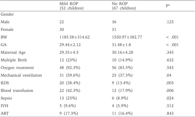
We were able to obtain ophthalmic records pertaining to the selected follow-up period for a total of 119 children (52 with mild ROP and 67 with no ROP). The mean age of follow-up was 32.64±2.63 months. There were 22 male (42%) and 30 female (58%) children in the mild ROP group and 36 male (53%) and 31 (47%) female children in the no ROP group. There was no significant difference in gender between two groups (p>0.05). In terms of ROP-related risk factors, the mild ROP and no ROP groups showed significant differences in birth weight (p<0.001) and gestational age (p<0.001), and in the presence of mechanical ventilation (p=0.04), respiratory distress syndrome (p=0.003), blood transfusion (p=0.006) and sepsis (p=0.024) (Table I). Both groups showed a similar distribution of refractive errors. The mean values of the spherical equivalents in the mild and no ROP groups were 1.8±1.36 D and 1.43±1.74 D, respectively. The mean values of the cylindric errors in the mild and no ROP groups were 0.21±0.52 D and 0.28±0.67 D, respectively (Table II). We found frequencies of 11.5% for myopia, 28.8% for hypermetropia and 16.3% for astigmatism in the mild ROP group, and corresponding frequencies of 6.7%, 22.3% and 9.7% in the no ROP group. Interestingly, we noted extreme refractive errors (myopia of < -5 D in two subjects, hypermetropia of > 5 D in two subjects and astigmatism of ≥ 3 D in two subjects) in the mild ROP group. However, in the present study, the frequencies and degree of refractive errors were similar in the mild ROP and no ROP groups (p> 0.05) (Figs. 1,2).
Strabismus was diagnosed in 8 children (15.3%) in the mild ROP group, of whom six were esotropic, with three having previously undergone surgical correction. In the no ROP group, strabismus was diagnosed in 13 children (19.4%), of whom nine were esotropic and three had previously undergone surgical correction. There were no significant differences in the rate of strabismus, presence of surgical
correction or angle of deviation between subjects with mild ROP and those with no ROP (p> 0.05) (Table II).
Visual acuity could be assessed with linear tumbling E cards and Snellen charts in 44 (36.9%) subjects who cooperated and were older than 30 months. Unfavorable functional outcome was observed in three eyes (2.8%) in the mild ROP group and two eyes (1.5%) in the no ROP group (p>0.05). We encountered unfavorable structural outcomes in in three eyes (2.8%) in the mild ROP group; two had total optic disk pallor, and one had aphakia associated with glaucoma. In the no ROP group, only one subject (0.75%) showed an unfavorable structural outcome, due to total optic disk pallor. There was no difference in the rate of unfavorable structural outcome between the mild and no ROP groups (p>0.05) (Table II). The rate of neurologic morbidity was 15.3% in the mild ROP and 10.4% in the no ROP groups (p>0.05). Of the eight subjects in the mild ROP group with such morbidities, there were three subjects with cerebral palsy; three with mental–motor retardation (MMR); and two with hydrocephaly who had undergone shunt
Fig. 1. Distribution of refractive status between eyes
with no ROP and eyes with mild ROP (no ROP, n=134; mild ROP, n=104)
Fig. 2. Distribution of astigmatism between eyes with no ROP and eyes with mild ROP (no ROP, n=134; mild ROP, n=104)
Mild ROP
(52 children) No ROP(67 children) P*
Gender Male 22 36 .125 Female 30 31 BW 1185.58±314.62 1550.97±382.77 < .001 GA 29.44±2.12 31.48±1.8 < .001 Maternal Age 29.35±4.5 30.16±4.28 .345 Multiple Birth 12 (23%) 10 (14.9%) .632 Oxygen treatment 48 (92.3%) 56 (83.5%) .543 Mechanical ventilation 31 (59.6%) 25 (37.3%) .04 RDS 20 (38.4%) 9 (13.4%) .003 Blood transfusion 22 (42.3%) 12 (17.9%) .006 Sepsis 13 (25%) 6 (8.9%) .024 IVH 5 (9.6%) 4 (5.9%) .512 ART 9 (17.3%) 11 (16.4%) .843
Table I. Perinatal Characteristics of Preterm Children Included in the Study
BW: Birth weight, GA: Gestational age, RDS: Respiratory distress syndrome, IVH: Intraventricular hemorrhage, ART: Assisted reproductive technology
surgery. In the no ROP group there were two subjects with cerebral palsy; two with MMR; one with periventricular leukomalacia; one with epilepsy; and one with hydrocephaly who had undergone shunt surgery. Interestingly, all four subjects with unfavorable functional or structural outcomes had one of these neurologic morbidities. Of these four, two had cerebral palsy with a history of intraventricular hemorrhage, and two had hydrocephalus that required shunt surgery.
Discussion
The current study indicates that mild ROP and no ROP groups are identical in terms of ocular and neurologic outcomes in early childhood. The distributions of refractive errors were quite similar in both groups, confirming
the observations of other authors4,14. A study
done in our country by Goktas et al.3 reported
a mean spherical equivalent of 1.1±3.1 D in eyes with no ROP and 1.2±1.0 D in eyes with ROP with no macular heterotopias. Although they also included eyes with treated ROP, and the mean age (37.6±1.1 months) was higher than ours (32.64±2.63 months), our figures (1.43±1.74 D in no ROP and 1.8±1.36 D in mild ROP) were comparable. Another study, by Ozdemir et al.8, reported a mean spherical
equivalent of 1.46±1.26 D in the right eyes and 1.22±0.91 D in the left eyes of 26 children between the ages of 5 and 7 with no history of ROP who had been born prematurely. All these results indicate a clear hyperopic tendency in premature children with mild or no ROP similar to that of term-born children. Mild ROP
(52 children/104 eyes)
No ROP
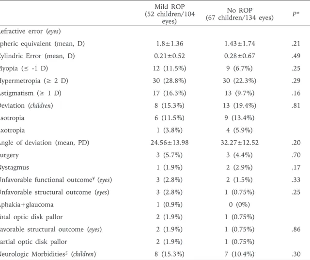
(67 children/134 eyes) P* Refractive error (eyes)
Spheric equivalent (mean, D) 1.8±1.36 1.43±1.74 .21
Cylindric Error (mean, D) 0.21±0.52 0.28±0.67 .49
Myopia (≤ -1 D) 12 (11.5%) 9 (6.7%) .25 Hypermetropia (≥ 2 D) 30 (28.8%) 30 (22.3%) .29 Astigmatism (≥ 1 D) 17 (16.3%) 13 (9.7%) .16 Deviation (children) 8 (15.3%) 13 (19.4%) .81 Esotropia 6 (11.5%) 9 (13.4%) Exotropia 1 (3.8%) 4 (5.9%)
Angle of deviation (mean, PD) 24.56±13.98 32.27±12.52 .20
Surgery 3 (5.7%) 3 (4.4%) .70
Nystagmus 1 (1.9%) 2 (2.9%) .17
Unfavorable functional outcome¥ (eyes) 3 (2.8%) 2 (1.5%) .33
Unfavorable structural outcome (eyes) 3 (2.8%) 1 (0.75%) .25
Aphakia+glaucoma 1 (0.9%) 0 (0%)
Total optic disk pallor 2 (1.9%) 1 (0.75%)
Favorable structural outcome (eyes) 2 (1.9%) 1 (0.75%) .86
Partial optic disk pallor 2 (1.9%) 1 (0.75%)
Neurologic Morbidities£ (children) 8 (15.3%) 7 (10.4%) .30
Table II. Ocular Outcomes and Associated Neurologic Morbidities of Preterm Children Studied
* T-test for continuous variables, and χ² for categorical variables ¥ Visual acuity ≤ 20/200
However, in our previous report we found a myopic tendency (-0.72±2.9 D) in children with treated ROP12. In the current study, the rate of
myopia was 11.5% in the mild ROP and 6.7% in the no ROP group, which falls in the 0-16% range previously reported for prematurely born children14-18. The rate was 15.9% in the study
by Goktas et al.3 and 0% in that by Ozdemir
et al.8. However, the rates we found definitely
exceed the rates given by large population-based screening studies for preschool children. In Turkey, studies done among school-aged children have shown the rate of myopia to vary considerably, from 3.2% to 32%, in different geographical regions19-21. Besides differences
in study design, this regional variation may be another reason for the disparate rates of myopia reported by different studies done in our country. Several studies have suggested that infants who develop any stage of ROP in the neonatal period are at greater risk for developing myopia or other ocular morbidities such as strabismus than are infants without ROP5,22,23. However, this was not the case in
our study. Therefore, our findings support the rationale for evaluating children with mild ROP and no ROP separately from those with treated ROP, which is confirmed by the findings of other authors7. While Ozdemir et al.8 reported
a lower rate of astigmatism (6.7%) than the 16.3% in the mild ROP group and 9.7% in the no ROP group found in our study; the mean cylindric error of 0.42±0.38 D in the right eye and 0.27±0.35 D in the left eye they reported was higher than the 0.21±0.52 D and 0.28±0.67 D found in the current study8. Such
variability is also evident in many other studies done in different countries. Some show that the rate of astigmatism may reach 15% in term-born subjects, a figure to which our rates are quite similar24-26. Tuppurainen et al.9 reported
a rate of 12.1% in prematurely born children at the age of 5, while Wang et al.1 reported an
astigmatism rate of 29.6% in preterm children with mild or no ROP at 2 years of age, and found that it did not change significantly up to 7 years of age in this particular group. We may state that the rates and severity of refractive errors in premature children with mild or no ROP do not seem to deviate widely from what is seen in term-born children.
The association between prematurity and strabismus is well established1,2,10,11. Strabismus
was present in 7.7% of the cases studied by Ozdemir et al.8, which included only premature
children without ROP, and in 22.1% of the cases investigated by Goktas et al.3, which also
included children with treated ROP. O’Connor et al.7 concluded that mild ROP confers no
additional risk for any of the ocular morbidities except strabismus. Interestingly, we found a higher rate of strabismus in the no ROP group than in the mild ROP group (19.4% versus 15.3%); however, the difference was not significant. Unlike the situation seen with regard to refractive error, the rate of strabismus in our cohort was much higher than that reported in large population-based screening studies in Turkey19-21. One reason could be
the high incidence of neurologic morbidity in our cohort. Of a total of 21 subjects with strabismus, 7 (33.3%) had neurologic sequelae. Similarly, Goktas et al.3 found that neurologic
morbidity was present in 33.8% of their subjects with strabismus. It is possible that noninclusion of many developmentally delayed children could account in part for the relatively low prevalence of strabismus detected in population-based studies.
The causes of blindness in premature children are reported to be severe ROP, congenital cataract and optic atrophy18,22, which is
consistent with our findings. As we did not include subjects with severe ROP, we had the opportunity to partly eliminate the factors causing very low vision in preterm children that are attributable to ROP per se. We detected unfavorable functional outcome, defined as ≤ 20/200 Snellen equivalent vision or uncentral/unsteady/unmaintained fixation, in five eyes of four children (3.3%), two in the mild ROP group and two in the no ROP group; there was no difference between the groups. In another study, children with no ROP and those with regressed mild ROP showed similar corrected acuities7. Hölmstrom et al.10
detected a 0.8% rate of poor vision (<20/200); however, Ozdemir et al.8 found no subjects with
poor vision in their cohort. Making detailed comparisons of incidences in the various studies is extremely difficult, since the criteria for inclusion may vary. Of the four children in our study with unfavorable functional outcomes, two had total optic disk pallor, one had congenital cataract and developed glaucoma after cataract surgery, and one had no obvious
ocular structural abnormalities. Interestingly, all had neurologic morbidities. Hölmstrom et al.6, in a population-based study done in
prematurely born children, found that 30.6% of the subjects with visual dysfunction had neurologic complications. It is highly possible that our number would increase were we to include only those with very poor vision. In our previous report, we found intraventricular hemorrhage to be an independent determinant for very poor vision in children with treated ROP12. Consistent with our findings, other
authors have postulated that severe reduction of visual acuity may be independently associated with neurologic complications10.
We are aware that our study has some limitations. It is a retrospective, single-center study, and the sample size was quite small due to difficulties in follow-up. Therefore, the limited number of children in the present study may have caused low statistical power in comparisons between the mild ROP and no ROP groups. Also, the rate of quantitative assessment of visual acuity was relatively low. In conclusion, the presence of mild ROP does not seem to increase the risk of ocular morbidity in early childhood among preterm children. Premature children with obvious neurologic morbidities are at greater risk of developing very poor vision, and follow-up should be intensified in this particular group.
REFERENCES
1. Wang J, Ren X, Shen L, Yanni SE, Leffler JN, Birch EE. Development of refractive error in individual children with regressed retinopathy of prematurity. Invest Ophthalmol Vis Sci 2013; 54: 6018-6024. 2. Robaei D, Rose KA, Kifley A, Cosstick M, Ip
JM, Mitchell P. Factors associated with childhood strabismus: findings from a population-based study. Ophthalmology 2006; 113: 1146-1153.
3. Goktas A, Sener EC, Sanac AS. An assessment of ocular morbidities of children born prematurely in early childhood. J Pediatr Ophthalmol Strabismus 2012; 49: 236-241.
4. Holmström GE, Larsson EK. Development of spherical equivalent refraction in prematurely born children during the first 10 years of life: a population-based study. Arch Ophthalmol 2005; 123: 1404-1411. 5. Schalij-Delfos NE, de Graaf ME, Treffers WF, Engel J,
Cats BP. Long term follow up of premature infants: detection of strabismus, amblyopia, and refractive errors. Br J Ophthalmol 2000; 84: 963-967.
6. Holmström G, Larsson E. Long-term follow-up of visual functions in prematurely born children—a prospective
population-based study up to 10 years of age. J AAPOS 2008; 12: 157-162.
7. O’Connor AR, Stephenson T, Johnson A, et al. Long-term ophthalmic outcome of low birth weight children with and without retinopathy of prematurity. Pediatrics 2002; 109: 12-18.
8. Ozdemir M, Koylu S. Ocular growth and morbidity in preterm children without retinopathy of prematurity. Jpn J Ophthalmol 2009; 53: 623-628.
9. Tuppurainen K, Herrgård E, Martikainen A, Mäntyjärvi M. Ocular findings in prematurely born children at 5 years of age. Graefes Arch Clin Exp Ophthalmol 1993; 231: 261-266.
10. Holmström G, el Azazi M, Kugelberg U. Ophthalmological follow up of preterm infants: a population based, prospective study of visual acuity and strabismus. Br J Ophthalmol 1999; 83: 143-150.
11. Theng JT, Wong TY, Ling Y. Refractive errors and strabismus in premature Asian infants with and without retinopathy of prematurity. Singapore Med J 2000; 41: 393-397.
12. Mutlu FM, Küçükevcilioğlu M, Ceylan OM, Altınsoy HI, Sarıcı SU. Risk factor analysis for long-term unfavorable ocular outcomes in children treated for retinopathy of prematurity. Turk J Pediatr 2013; 55: 35-41.
13. Mutlu FM, Altinsoy HI, Mumcuoglu T, et al. Screening for retinopathy of prematurity in a tertiary care newborn unit in Turkey: frequency, outcomes, and risk factor analysis. J Pediatr Ophthalmol Strabismus 2008; 45: 291-298.
14. Quinn GE, Dobson V, Kivlin J, et al. Prevalence of myopia between 3 months and 5 ½ years in preterm infants with and without retinopathy of prematurity. Ophthalmology 1998; 105: 1292-1300.
15. Quinn GE, Dobson V, Davitt BV, et al. Progression of myopia and high myopia in the early treatment for retinopathy of prematurity study: findings to 3 years of age. Ophthalmology 2008; 115: 1058–1064. 16. Cook A, White S, Batterbury M, Clark D. Ocular
growth and refractive error development in premature infants with or without retinopathy of prematurity. Invest Ophthalmol Vis Sci 2008; 49: 5199–5207. 17. Lue CL, Hansen RM, Reisner DS, Findl O, Petersen
RA, Fulton AB. The course of myopia in children with mild retinopathy of prematurity. Vision Res 1995; 35: 1329-1335.
18. Darlow BA, Horwood LJ, Mogridge N, Clemett RS. Prospective study of New Zealand very low birthweight infants: outcome at 7–8 years. J Paediatr Child Health 1997; 33: 47-51.
19. Gursoy H, Basmak H, Yaz Y, Colak E. Vision screening in children entering school: Eskisehir, Turkey. Ophthalmic Epidemiol 2013; 20: 232-238.
20. Caca I, Cingu AK, Sahin A, et al. Amblyopia and refractive errors among school-aged children with low socioeconomic status in southeastern Turkey. J Pediatr Ophthalmol Strabismus 2013; 50: 37-43.
21. Turaçli ME, Aktan SG, Dürük K. Ophthalmic screening of school children in Ankara. Eur J Ophthalmol 1995; 5: 181-186.
22. Darlow BA, Clemett RS, Horwood LJ, Mogridge N. Prospective study of New Zealand infants with birth weight less than 1500 g and screened for retinopathy of prematurity: visual outcome at age 7–8 years. Br J Ophthalmol 1997; 81: 935-940.
23. Robinson R, O’Keefe M. Follow-up study on premature infants with and without retinopathy of prematurity. Br J Ophthalmol 1993; 77: 91-94.
24. Mayer DL, Hansen RM, Moore BD, Kim S, Fulton AB. Cycloplegic refractions in healthy children aged 1 through 48 months. Arch Ophthalmol 2001; 119: 1625-1628.
25. Abrahamsson M, Fabian G, Sjöstrand J. Changes in astigmatism between the ages of 1 and 4 years: a longitudinal study. Br J Ophthalmol 1988; 72: 145-149. 26. Gwiazda J, Scheiman M, Mohindra I, Held R.
Astigmatism in children: changes in axis and amount from birth to six years. Invest Ophthalmol Vis Sci 1984; 25: 88-92.