Ankara Üniv Vet Fak Derg, 63, 89-92, 2016
Short Communication / Kısa Bilimsel Çalışma
Oral metastatic melanoma with neurosarcomatous transformation in
a dog
Mustafa Yavuz GÜLBAHAR
1, Ahmet ÖZAK
2, Yonca Betil KABAK
1, Mehmet Önder KARAYİĞİT
3,
Ayhan GACAR
1Ondokuz Mayıs University, Faculty of Veterinary Medicine, 1
Department of Pathologyand 2Department of Surgery, Samsun; Erciyes University, Faculty of Veterinary Medicine, 3Department of Pathology, Kayseri, Turkey.
Summary: A 16-year-old male terrier dog was presented with a history of progressively growing ulcerated and slightly pigmented oral masses on the rostral mandible. Microscopically, the tumor composed of two different components. One component was conventional malignant melanoma including epitheloid or spindle-shaped components with/without melanin pigment. The other component was typical palisades (Verocay bodies) and pseudopalisades, with no melanin pigment. Immunohistochemical analysis revealed that the tumor was positive for vimentin, Melan-A, NSE, and GFAP, and negative for HMB45, S100, MBP, desmin, SMA, pan-cytokeratin. Melan-A reactivity was limited to the upper conventional melanoma areas. Nuclear palisading and pseudopalisading structures were positive for vimentin, NSE, GFAP whereas negative for other antibodies. Metastatic lymph node displayed spindle cell melanoma, with immunoreactivity for vimentin and Melan-A. The diagnosis was typical of oral malignant melanoma including nuclear palisading and pseudopalisading components, suggesting neurosarcomatous differentiation in a dog.
Key words: Dog; neural differentiation; neurosarcomatous differentiation; oral malignant melanoma.
Köpekte nörosarkomatöz farklılaşma gösteren oral metastatik melanom olgusu
Özet: Bu çalışmada 16 yaşlı erkek terrier köpekte ön mandibular bölgede sürekli büyüyen, ülserli ve hafif pigmentasyon gösteren tümöral kitle gözlendi. Mikroskobik olarak, tümör iki komponentten oluşmaktaydı. Birincisi epitel benzeri veya iğ şekilli, melanin pigmenti içeren ya da içermeyen hücrelerden oluşan klasik malign melanomdu. İkinci component ise melanin pigmenti içermeyen tipik palisad (Verocay cisimcikleri) ve yalancı-palisadlardan oluşmaktaydı. İmmunohistokimyasal incelmelerde, tümör vimentin, Melan-A, NSE ve GFAP pozitif; HMB45, S100, MBP, desmin, SMA ve pansitokeratin negatifti. Melan-A boyanması klasik melanom alanları ile sınırlıydı. Palisad ve yalancı-palisadlar vimentin, NSE, GFAP için pozitif, diğer antikorlar için negatifti. Lenf yumurusu metastazında doku iğ tarzında hücrelereden oluşmuştu ve vimentin ile Melan-A için pozitifti. Olgu palisad ve yalancı-palisadlarla birlikte nörosarkomatöz farklılaşma gösteren malign oral melanom olarak tanımlandı.
Anahtar sözcükler: Köpek, nöral farklılaşma, nörosarkomatöz farklılaşma, oral malign melanom.
Malignant melanomas are the most common oral tumors in dogs and rare in the oral cavity of other animals (2, 5). Most canine oral melanomas have characteristic mixture of epithelial-like and/or spindle-shaped cells, and clear cell and adenoid/papillary patterns are uncommon. Neoplastic bone or chondroid formations and balloon and signet-ring cell variants have also been reported in canine melanomas (2,5,9) Two cases of canine malignant melanotic schwannoma in spinal cord have been documented with evidence of melanin pigment and gross association of the tumor with nerve roots (8). “Neurotisation” with neural structures such as neuroid cords and rosettes can be seen in some human melanocytic nevi attributed to senescence (7). In animals, some dermal melanocytomas may have more distinct “neuroidal” morphology or “neurotisation” (4). Histological
patterns of cellular arrangements including Verocay bodies or nuclear palisading formations, suggesting a peripheral nerve sheath tumor (PNST) or Schwann cell tumor, so called “neurosarcomatous differentiation” may occur in rare melanocytic tumors of animals (11). However, neurotisation or neurosarcomatous differentiation in a melanocytic tumor has not been well documented. This case report describes unusual oral metastatic melanoma, suggesting neurosarcomatous transformation in a dog.
A 16-year-old male terrier dog was submitted to The Animal Hospital of the Faculty of Veterinary Medicine, University of Ondokuz Mayis, Samsun, Turkey with a history of progressively growing oral masses on the rostral mandible. The tumor had a lobular appearance, was slightly pigmented, ulcerated, and the
Mustafa Yavuz Gülbahar - Ahmet Özak - Yonca Betil Kabak - Mehmet Önder Karayiğit - Ayhan Gacar 90
mandible distorted (Figure 1). On radiographical examination, in the ventrodorsal, intraoral projection of the mandible, resorption of the bony sockets of the incisor teeth was detected. In addition, a small region of periosteal inflammatory reaction was seen in the medial part of mandibles. In both ventrodorsal, intraoral and dorsoventral oblique (open mouth) projections, all incisors of the mandible were removed from their bony sockets.
Figure 1. The oral tumor of lobular appearance, slightly pigmented, ulcerated, and the distorted mandible in the rostral region. Dog, oral melanoma.
Şekil 1. Mandibulanın ön kısmını deforme eden lobular görünümlü, hafif pigmentli ve ülserli oral tumor. Köpek, oral melanoma.
The specimens and one lymph node (possibly one of retropharyngeal lymph nodes) were fixed in 10% neutral buffered formalin, embedded in paraffin wax, cut at 5 µm, and stained with hematoxylin-eosin (HE) and Masson’s trichrome stains. In addition, paraffin sections were subjected to immunohistochemistry (IHC). The antibodies used included vimentin (Clone V9, Lab Vision Corp., USA), Melan-A (Mart-1, A103, Dako Carpenteria, USA), HMB45 (Dako Carpenteria, USA), S100 protein (4C4.9, Ab-1, Neomarkers, USA), S100 protein (AB941, EMD Millipore Corp., USA), myelin basic protein (MBP, 12, NS0, Abcam, UK), desmin (RD301; Santa Cruz Biotechnology, Inc., USA), actin, smooth muscle (SMA, Ab-1, Thermo Fischer Scientific, USA), pan-cytokeratin (AE1/AE3, Dako Carpenteria, USA), neuronal specific enolase (NSE, Sigma-Aldrich, USA), glial fibrillary acidic protein (GFAP, Abcam, UK) antibodies using a streptavidin-biotin method by a commercial kit (Zymed, USA). Tissue sections known to be positive previously for all antibodies used served as positive controls and unrelated antibodies replaced with the primary antibodies for the negative control.
On the histopathological examination, two different components were found in the tumor tissue. One
component, located the submucosa, was a conventional, malignant melanoma including epitheloid cells, with or without melanin pigment, either singly or in nests, and showing junctional activity. Some areas of the tumor tissue included fusiform or spindle-shaped cells arranged in fascicles and cell nests supported by scant fibrous tissue. Neoplastic cells demonstrated vesicular nuclei and prominent nucleoli. Below this area, in the deep submucosa, a second component, sometimes intermingled with the area described above, consisted of typical palisades (Verocay bodies) and pseudopalisades accompanied by garland-like arrays of nuclei surrounding a region of necrosis (Figures 2 and 3). Palisades and pseudopalisades varied in size and melanin was not observed. Some clustered palisading/pseudopalisading structures were isolated by dense, fibrous tissue from other components. The trichrome stain indicated a radially arranged, cellular zone including small collagen fibres in the center of palisades (Figure 3). Immunohistochemical staining revealed that the melanocytic tumor was positive for vimentin, Melan-A, NSE, and GFAP, and negative for other antibodies used. Melan-A reactivity was limited to the upper conventional melanoma areas and dispersed epitheloid or spindle cells (Figure 4). The other components, including nuclear palisading and pseudopalisading structures, were positive for vimentin, NSE, GFAP, but negative for the remaining antibodies. Some NSE- and GFAP-positive cells had small filamentous or dendritic processes, highlighting a glial fashion (Figure 5). The diagnosis was of typical of oral malignant melanoma with area suggesting neurosarcomatous differentiation. Metastasis to the regional lymph nodes was not observed.
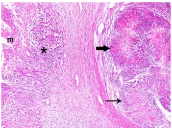
Figure 2. Conventional malignant melanoma (asterisk) just beneath the ulcerated oral mucosa (m) and neurosarcomatous differentiation area with nuclear palisading (thin arrow) and pseudopalisading structures with necrotic center (thick arrow), covered by a fibrous tissue. HE. X65.
Şekil 2. Ülserli oral mukozanın hemen altında klasik malign melanoma (yıldız) ile yoğun fibröz doku ile çevrili palisad (ince ok) ve nekrotik merkezli yalancı-palisad (kalın ok) bulunan nörosarkomatöz farklılaşma alanı. HE. X65.
Ankara Üniv Vet Fak Derg, 63, 2016 91
Figure 3. Nuclear palisading (Verocay bodies) accompanied by garland-like array of nuclei, including thin collagen fibres in the centre of palisades. HE. X120. Inset shows fine collagen fibres in center of a palisad. Masson Trichrome stain. X120.
Şekil 3. Merkezlerinde ince kollajen iplikçiklerin olduğu ve çekirdeklerin çelenk benzeri tarzda dizildiği nükleer palisadlar (Verocay cisimleri). HE. X120. Küçük resim bir palisad merkezinde ince kollajen iplikçikleri göstermektedir. Masson Trikrom boyama. X120.
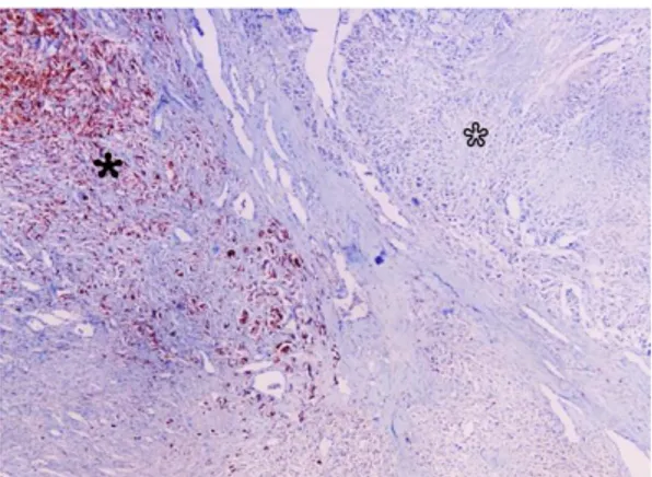
Figure 4. Melan-A immunoreactivity in the cellular components of conventional melanoma (black asterisk) in contrast to those of neurosarcomatous differentiation region (open asterisk). IHC, Hematoxylin counterstain. X65.
Şekil 4. Klasik melanom’daki hücrelerde Melan-A immunereaksiyonu (siyah yıldız) buna karşın nörosarkomatöz farklılaşma bölgesinde negative reaksiyon (beyaz yıldız). IHC, Hematoksilen karşıt boyama. X65.
At 1.5 months after the initial presentation, the dog
was resubmitted with bilateral enlargement of
retropharyngeal lymph nodes and total, bilateral lymphadenectomy was performed. Microscopic examination of the lymph nodes indicated metastatic tumor tissues composed of fusiform- or spindle-shaped cells arranged in fascicles and sheaths and whorled structures in both cortex and medulla of nodes. Neoplastic cells were amelanotic and had hyperchromatic, oval to ellipsoid nuclei, and fine eosinophilic cytoplasm. Nuclear palisading or pesudopalisading pattern were not detected. Immunohistochemically, tumor cells in the lymph nodes
Figure 5. Strong GFAP immunoreactivity in both neurosarcomatous area including a pseuodopalisading structure with tiny cellular processes and necrotic centre (open asterisk) and conventional melanocytic tumor cells (black asterisk). IHC, Hematoxylin counterstain. X120.
Şekil 5. Nörosarkomatöz alandaki yalancı-palisadların nekrotik merkezleri ve ince hücresel uzantıların yanında (beyaz yıldız), klasik melanositik tumor dokusunda (siyah yıldız) güçlü GFAP immunreaksiyonu. IHC, Hematoksilen karşıt boyama. X120.
were strongly positive for Melan-A and vimentin, whereas negative for other all antibodies used. The dog died approximately 1 year after initial presentation, but the owner declined a necropsy.
Histologic architectures with distinctive features, which are palisades and pseudopalisades special to some tumors, have been documented comprehensively in a review of neuropathology (12). Palisades, so-called Verocay bodies, are stacked arrangements of elongated palisading nuclei alternating with anuclear zones containing cell processes and are typical of schwannomas. Other varieties of tumors, including peripheral nerve sheath tumors (PNSTs), central nervous system (CNS) tumors, soft-tissue tumors, melanocytic tumors and even carcinomas, may display palisades (12). In contrast to palisades, pseudopalisades have been characterized by a garland-like arrangement of hypercellular tumor nuclei lining up around irregular foci of tumor necrosis containing pyknotic nuclei, and are frequently observed in glioblastoma (1,12). Pseuodopalisading pattern of tumor cells has been reported in peripheral primitive neuroectodermal tumors (PNET) (6) as well as in malignant PNST (3) in dogs. In the present case, nuclear palisading and pseudopalisading structures in various sizes were striking findings, representing neurosarcomatous differentiation. A diagnosis of malignant melanotic schwannoma was ruled out, because in only two canine cases (8), there was no an association of a spinal nerve roots and no melanin pigmentation in components displaying neurosarcomatous differentiation.
Melanocytic neoplasms are usually positive for vimentin, S100, NSE, HMB-45, and Melan-A, but negative for cytokeratin (9). Retrospective studies of canine oral melanomas showed that Melan-A is a specific
Mustafa Yavuz Gülbahar - Ahmet Özak - Yonca Betil Kabak - Mehmet Önder Karayiğit - Ayhan Gacar 92
and sensitive marker for canine melanomas with 100% vimentin positivity and lower positivity for S100 and NSE in all cases (2,9). In the present study, conventional melanocytic tumor cells in the primary site were strongly positive for vimentin, Melan-A, NSE and GFAP, but negative for two clones of S100 antibody, despite various antigen retrieval methods. Moreover, HMB-45 antibody also failed to detect melanocytic cells. Palisading and pseudopalisading structures were positive for vimentin, NSE, GFAP, suggesting neural differentiation, but
negative for the melanocytic marker Melan-A.
Interestingly, GFAP immunoreactivity was a constant finding in all components in the primary site, suggesting neurosarcomatous differentiation. GFAP and NSE expression in some tumors are regarded as neural differentiation, and variable GFAP and NSE expression is reported in canine PNSTs. Immunoreactivity for S100 is highly suggestive for canine Schwann cell tumors in contrast to low diagnostic importance of myelin basic protein (MBP) in diagnosis of canine PNSTs (3). In other studies, however, a lower percentage of S100 immunoreacitivity has been described in canine oral melanomas compared to Melan-A immunoreactivity. The lack of immunostaining for S100 has been explained by the fixation time of tissues in canine oral melanomas (9), or, depending on the degree of differentiation of the cells, given two S100 antibodies with different clones were used in the present study.
The melanocytic tumors may have different histologic features in metastatic sites than primary tumor. The primary canine oral melanoma may be pigmented and epitheloid, but metastatic sites may acquire an amelanotic spindle-shaped variant (10). In the present case, the primary tumor comprised slightly melanotic epitheloid or spindle shaped cells, whereas lymph node metastasis was amelanotic, and only fusiform- or spindle-shaped cells arranged in fascicles and sheaths or whorled bundles, were immunolabeled with only
vimentin and Melan-A. Higher percentage of
immunoreactivity of Melan-A has been reported in both primary tumors and their metastases of canine melanomas (9). However, there are no well-documented cases regarding preservation or loss of melanoma antigens or aberrant antigen expressions between a primary melanoma and metastases reported in animals.
In conclusion, on the basis of microscopy and immunohistochemical features of both conventional melanocytic and neurosarcomatous components in the primary site, with different features in a lymph node metastasis, verified that the tumor was malignant oral melanoma with a neural transformation fulfilling the criteria for diagnosis of coexistent melanocytic and neural differentiations given the cells of common origin from neural crest. The possibility of unusual histological variants and aberrant immunophenotypic features should
be considered in melanocytic tumors when regarding their inconsistent nature.
References
1. Brat DJ, Castellano-Sanchez AA, Hunter SB, Pecot M, Cohen C, Hammond EH, Devi SN, Kaur B, Van Meir EG (2004): Pseudopalisades in glioblastoma are hypoxic, express extracellular matrix proteases, and are formed by an actively migrating cell population. Cancer Res, 64, 920-927.
2. Brown CC, Baker DC, Barker IK (2007): Alimentary system, 29-30. In: Maxie MG (eds.), Jubb, Kennedy and Palmer’s Pathology of Domestic Animals, 5th
Edit., Vol 2, Philadelphia, Saunders Elsevier, USA.
3. Chijiwa K, Uchida K, Tateyama S (2004): Immunohistochemical evaluation of canine peripheral nerve sheath tumors and other soft tissue sarcomas. Vet Pathol, 41, 307-318.
4. Goldschmidt MH and Hendrick MJ (2002): Tumors of the skin and soft tisuues, 78-84. In: Meuten DJ (ed.), Tumors in Domestic Animals, 4th Edit., Iowa State Press, USA.
5. Head KW, Else RW, Dubielzig RR (2002): Tumors of the alimentary tract, 401-430. In: Meuten DJ (ed.), Tumors in Domestic Animals, 4th Edit., Iowa State Press, USA. 6. Katayama KI, Kuroki K, Uchida K, Nakayama H,
Sakai M, Mochizuki M, Nishimura R, Sasaki N, Doi K (2001): A case of canine primitive neuroectodermal tumor (PNET). J Vet Med Sci, 63, 103-105.
7. Maize JC, Foster G (1979): Age-related changes in melanocytic naevi. Clin Exp Dermatol, 4, 49-58.
8. Patnaik AK, Erlandson RA, Lieberman PH (1984): Canine malignant melanotic schwannomas: a light and electron microscopic study of two cases. Vet Pathol, 21, 483-488.
9. Ramos-Vara JA, Beissenherz ME, Miller MA, Johnson GC, Pace LW Fard A, Kottler SJ (2000): Retrospective study of 338 canine oral melanomas with clinical, histologic, and immunohistochemical review of 129 cases. Vet Pathol, 37, 597-608.
10. Smith SH, Goldschmidt MH, McManus PM (2002): A comparative review of melanocytic neoplasms. Vet Pathol, 39, 651-678.
11. Summers BA (2011): Melanocytic nevi, melanoma and malignant peripheral nerve sheath tumours: not in the textbook. 62nd Annual Meeting of the American College of Veterinary Pathologists and 46th Annual Meeting of the American Society for Veterinary Clinical Pathology, Nashville Tennessee, USA.
12. Wippold FJ 2nd, Lämmle M, Anatelli F, Lennerz J, Perry A (2006): Neuropathology for the neuroradiologist: palisades and pseudopalisades. Am J Neuroradiol, 27, 2037-2041.
Geliş tarihi: 21.10.2014 / Kabul tarihi: 21.01.2015 Address for correspondence:
Dr. MustafaYavuz Gülbahar Ondokuz Mayıs University, Faculty of Veterinary Medicine, Department of Pathology, Kurupelit 55139 Samsun-Turkey