Rare Clinical Variants of Pilomatricoma
Pilomatrikomanın Nadir Klinik VaryantlarıNesrin Tan Baser, Ünzile Balci Akbuga, Ali Cemal Yılmaz, Serdar Gökrem, Gurcan Aslan
S.B. Ankara Eğitim ve Araştırma Hastanesi, 2. Plastik Cerrahi Kliniği
Received: 12.09.2007 • Accepted: 09.11.2007 Corresponding author
Nesrin Tan Baser
S.B. Ankara Eğitim ve Araştırma Hastanesi, 2. Plastik Cerrahi Kliniği Phone : +90 (312) 595 36 62
Fax :
E-mail address : [email protected]
Pilomatricoma (calcifi ying epithelioma of Malherbe ) is a benign adnexal tumor of hair matrix cells. Pilomatricomas are usually observed as solitary lesions. Pilomatricoma mostly makes peaks in the fi rst and sixth decades. Female to male ratio is 3/2. More than 50% of the lesions are found on head and neck region, and 25-30% of the lesions are found on the upper extremities. The rest occurs in the trunk and rarely in the lower extremities. Pilomatricoma is not rare entity. On the con-trary, multiple and perforating forms of pilomatricoma are rare entities. Although pilomatricoma is not hereditary as a rule, there are limited familial pilomatricoma cases which are published in the literature. The patients, who come to our clinic during three years and histologically diagno-sed as having the pilomatricoma were analydiagno-sed. Rare clinic variants of pilomatricoma that familial multiple pilomatricoma, perforating pilomatricoma are reported. In spite of the small number of the patients in our series, we believe that familial multiple pilomatricoma and perforating pilo-matricoma cases will supply the literature.
Key Words: pilomatricoma, familial, multiple, perforating
Pilomatrikoma (Malherbe ’nin kalsifi ye epitelyoması saç matriks hücrelerinden gelişen benign ad-neksal bir tümördür. Pilomatrikoma genellikle solid kitleler olarak izlenir. Sıklıkla yaşamın 1. ve 6. dekadında pik yapar. Kadın/erkek görülme oranı 3/2 ’dir. Lezyonların %50 ’den fazlası baş boyun bölgesinde, %25-30 ’luk kısmı üst ekstremitede lokalizedir. Pilomatrikoma nadir değildir. Bunun yanında ailesel multiple pilomatrikoma ve perfore pilomatrikomanın ise nadir görüldüğü bildi-rilmektedir. Kural olarak pilomatrikomanın herediter olmadığı söylensede literatürde bildirilmiş sınırlı sayıda ailesel pilomatrikoma vakası vardır. Burada 3 yıllık bir peryodda kliniğimize başvuran ve histopatolojik olarak pilomatrikoma tanısı alan 10 vaka incelendi. Literatürde nadir rastlandığı bildirilen ailesel multiple pilomatrikoma ve pilomatrikomanın nadir bir klinik varyantı olan perfore pilomatrikoma vakaları sunuldu. Bu vakalar literatür eşliğinde gözden geçirildi. Serimizdeki hasta sayısının az olmasına rağmen ailesel multiple pilomatrikoma ve perfore pilomatrikoma olgularımı-zın literatüre katkısı olacağını düşünüyoruz.
Anahtar Kelimeler: pilomatrikoma, ailesel, multiple, perfore
Pilomatricoma is a benign adnexal tumour of hair matrix cells. It is most frequently seen in head and neck region and upper limbs (1). Malherbe and Chenantais defined the
“calcifying epithelioma” in 1880. In the beginning they thought this as a tumor of sebaceous glands. In 1905, this mistake was corrected by Malherbe and the entity was re-named as “calcifying epithelioma of Malherbe” (2). In 1949, Lever and Griesemer (3), claimed that
the tumor was taking origin from the hair matrix cells, and 1961, it was named as “pilomatrixoma by Forbis and Helwig” (4). Then, pi-lomatrixoma was changed to pilo-matricoma which is more etymo-logically accurate (2).
Pilomatricoma is not rare entity. When the literature is reviewed, incidance varies from one in 500 to 924 dermatologic specimens (5,6). On the contrary, multiple and perforating forms of
pilomat-Table 1: Patients information. ( L: left, R: right, lat.:laterale, month: mn., year: yr.)
Case Age (year)
Sex Localization Size (cm) Duration Clinical
diagnosis
Histopathollogical diagnosis
1 4 M L infraorbital 1,5x1x0,6 4 mn Organized hematom pilomatricoma 2 21 M L leg 3x2x1 2 yr. pilomatricoma pilomatricoma 3 21 F L right 1x0,7x0,7 18 mn pilomatricoma pilomatricoma 4 76 M R eyebrow 1,5x1x0,5 3 yr. epidermoid cyst pilomatricoma 5 47 F Scalp 0,5x0,5x0,3 3 yr epidermoid cyst pilomatricoma 6 59 M L pectoral, L subclaviculer 3x1,5x0,5
1x0,8x0,5
1 yr epidermoid cyst pilomatricoma
7 18 F L eyebrow 0,5x0,5x0,3 6 mn. epidermoid cyst pilomatricoma 8 66 E R temporal 0,8x0,7x0,3 6 mn. Basalcell carcinoma pilomatricoma 9 15 F Anterior cervicale 2,5x1,5x1 1 mn. epidermoid cyst pilomatricoma 10 17 F R lat. cantus L preauriculer Posterior cervicale 4x3x1,5 0,6x0,5x0,2 2x1,5x0,5 4 yr. pilomatricoma pilomatricoma
ricoma are rare entities (4,7-11). Although pilomatricoma is not he-reditary as a rule, there are limited familial pilomatricoma cases whi-ch are published in the literature (12-14).
In this article, we report 10 pilomat-ricoma cases operated in our cli-nic during three years and presen-ted three of them which are rarely seen in the literature (Table-1).
Case Reports
Case 2
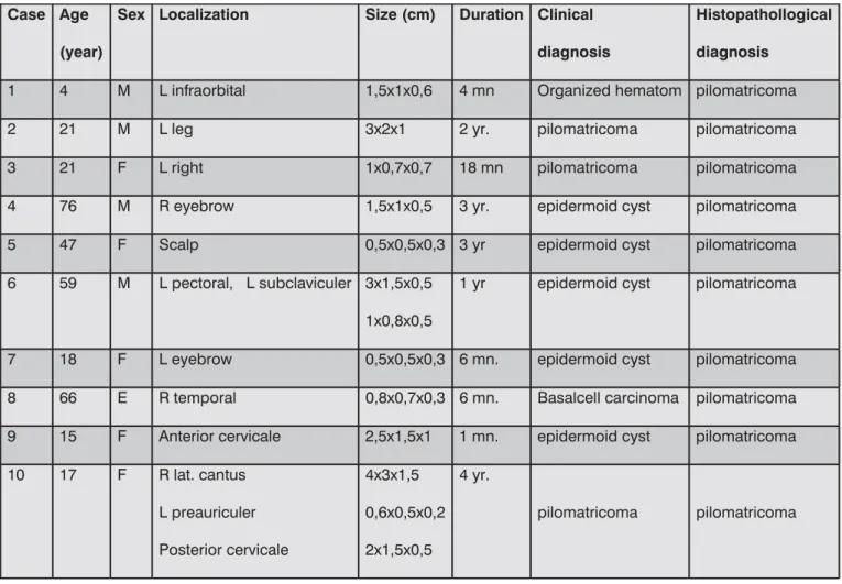
21 year old male patient presented with a 2 year history of painless mass on the left leg. Examinati-on showed mobile, well defined,
firm, 3x2x1 cm subcutaneous no-dule covered by normal skin. Local excision of nodule with overlying skin was performed (Figure 1.A-B). Histopathologic examination revealed that, tumor was consis-ted of island of epithelial cells. It was composed of basophilic and eosinophilic shadow cells. Areas of calsification and ossification were detection in lesion . Lesion was seperated from the neighbour skin with the regular border. Case 8
66 year old female patient presented to our clinic with the mass on her right temporal area. She had noti-ced the asymptomatic nodule with ulceration about 6 months previ-ously. Physical examination revea-led, bluish colored nodule with
ul-cer in center. That was 0,8x0,7x0,3 cm. size on the right temporal area. Lesion was exiced by clinical diagnoses of BCC (Figure 2.A-B). On microscopic examination, sha-dow cell groups, basophilic cells and cystic cavity were observed. There were also foreign body gi-ant cells. The epidermis adjacent to the tumor invaginated.
Case 10
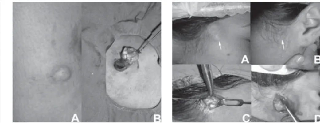
17 year old female patient refered to our clinic because of the masses in her face and neck since the age of 13 years. When she examined, the masses, which were painly, hard, mobile were found out and these masses did not cause colour change. The mass in the laterale of right eyebrow was 4x3x1,5 cm., the mass in the left preauriculer
area was 0,6x0,5x0,2 cm., and the mass in the posterior neck area was 2x10x0,5 cm. These three masses were surgically removed (Figure 3.A-C). On the histopato-logical observation, a few cells in the bazloid features around the lobuller occuring from the cell remnants which did not inclu-de nucleous in the fibroz stroma drew attention. In addition to this, calcification areas were found out. There were multinuclear giant cel-ls and lenfositier inflatration were seen adjacent.
After the pathologic diagnosis, sys-tematic illnesses, which could be seen with pilomatricoma, like myotonic distrofi, Gardner synd-rome, Reynaud phenomene were examined. However, none of the-se pathologies were detected. Moreover, it was learned that her sister, brother and uncle had the similiar masses. Her uncle’s mas-ses in his neck and left preauciliar areas were taken out 10 years ago. Its histopatological diagnosis was pilomatricoma. A hard mass was found in his brother’s right arm. Histopatological diagnoses of this mass came as pilomatricoma. Also, her sister had a painless mass. His-topatological examination of the mass could not be done because she was in abroad. These three ca-ses are not attached to the table.
Discussion
Pilomatricoma usually presents as firm to stone hard, asemptomatic nodule with dimensions ranging from 0,5 to 5 cm. The overlying skin is generally normal, but may show a blue- red discolaration (1).
Etiology of pilomatricoma is not known. Microscobic, biochemi-cal enzymatic, polarischopic, and immunohistochemical properti-es reveals that it originatproperti-es from the hair matrix cells (15). Normal hair matrix is characterized with anogen, telogen and catagen cyc-lus (16). This cyccyc-lus is control-led with programmed cell death (apoptosis) (17). It’s thought that it is a defect occuring in progress of the normal anogen phase in pi-lomatricomas (18). “bcl-2”, which is a proto-oncogen, suppresses the apoptosis in both a benign and malign tumor. In addition, both apoptosis and “bcl-2” are the critical factors in the normal hair follicule progressing. Farrier and his friends(17) showed that pilo-matricomas are painted strongly with “bcl-2” in their immunohis-tochemical study. They pointed out that this raised “bcl-2” pain-ting causes the wrong apoptosis
suppression. This contributes to the pathogenesis of pilomatrico-ma. In some studies it was shown that ß catenin mutation also play a role in this pathogenesis (19,20). ß catenin misregulation is respon-sible for the anormal proliferation of hair matrix cells (19). ß catenin takes a role in cell-cell adhesion rather than celluler proliferation in pilomatricoma tumorogenesis (20).
Pilomatricoma mostly makes peaks in the first and sixth decades. %40 of the patients are in the first de-cade. Female to male ratio is 3/2 (3,7,9). More than %50 of the le-sions are found on head and neck region, and %25-30 of the lesions are found on the upper extremi-ties. The rest occurs in the trunk and rarely in the lower extremities (2-12). Pain and tense seen in % 20 to 35 of cases (21-22).
In our study, female to male ratio was 3/2. Except the four whose age range was between fifth and eighth decades the age of other patients were in the first and se-cond decades. In two of our cases the masses were painfull (%20). Although in most of our cases lesi-ons were located in the head and neck region, one of them was lo-cated in the leg (Table 1).
Figure 1: Case 2 A) Firm, nodular lesion on
the leg. B) Peroperative view of the lesion Figure 2: Case 8 A) A nodular lesion on the temporal region with ulceration. B) Intraopera-tive view of the lesion
Figure 3: Case 10 A-B) Preoperative lateral
vi-ewes of masses. C-D) Peroperative vivi-ewes of masses
REFERENCES
1. Elder D, Elentistas R, Ragsdale BD. Tumors of epidermal appendages. In: Elder D, Elantistas R, Jaworsky C, Johnson B Jr, editors. Lever’s his-topatology of the skin, 8th ed. Phi-ladelphia:Lippincott-Raven.; 1997. p.747.
2. Fernandes R, Holmes, Mullenix C. Giant pilomatricoma ( epithelioma
of Melherbe): Report of a case and review of literature. J Oral Maxillofac Surg 2003;61:634-6.
3. Fetil E, Özkan S, Ilknur T, et.al. Mul-tiple pilomatricoma with perforati-on. Int J Dermatol 2002;41:892-3. 4. Forbis R, Helwig EB. Pilomatrixoma
(calcifying epithelioma). Arch. Der-matol 1961; 83: 606-8.
5. Margogi A, Wick MR, Dehner LP. Pi-lomatrical neoplasm in childern and young adults. Am J Dermatopathol 1992; 87:14.
6. Moehlenbeck, F. W. Pilomatrixoma (calcifying epithelioma): A statisti-cal study. Arch. Dermatol 1973; 108: 532-4.
7. Migirov L, Fridman Y, Talmi YP. Pilo-matrixoma of the retroauriculerarea
Pilomatricomas are usually obser-ved as solitary lesions. 4 clinical variances of were defined; eruptif type, perfore type, myotonic dis-trofi which occurs with or without familial type and recurrent invaziv nonmetastatic pilomatrix carsino-ma (2).
Although most of the articles report the occurence rate of multiple le-sions as %2-3,5 (2,3,7-12), in the article of Yoskimoto this was re-ported as %26 (23). When they compared this high rate with ot-her broader series (24,25), they conclude that this may be due to small number of cases in their report or racial factors (23). Mul-tiple pilomatricoma may be asso-ciated myotonic distrofi, sarkoi-dozis, Gardner syndrome, Turner syndrome and Reynaud pheno-menon. Multiple pilomatricoma is rare without myotonic distrofi. The associtation of multiple pilo-matricoma and myotonic distrofi is reported as %75 (2,3,9,12-14). Multiple pilomatricoma was seen in two of our patients .
The familial pilomatricoma is the most rarely seen type. Up to now, only 10 familial cases have been reported (7,11-14). Of these some were soliter and some were mul-tiple. Four familial pilomatricoma cases associated with miyotonic distrofi were multiple (11,14). In our familial case although both our patient and her brother not have myotonic distrofi.
Perforated pilomatricoma is seen rarely. When literature is exami-ned, it was seen that up to now, only 10 cases have been published (3,10,11). In our series, only one patient had perforated pilomatri-coma.
Clinic diagnosis of pilomatricoma is difficult. The wrong diagnosis rate ranges from %71 to % 79 (21,25). Pilomatricoma can be frequently confused with epidermoid cyst, calcifying lymf node, organized hematoma, foreign body reaction, dermoid cyst or parotis gland tu-mours. When perforation is seen, it may be misdiagnosed as in skin cancer (2-12,21-26). In our study, one lesion was misdiagnosed as organized hematom and one le-sion was misdiagnosed as BBC. Four lesions were misdiagnosed as epidermoid cyst (Table-1). Spontoneus regression has never
been observed. The treatment of choice is surgical excision. In-complete resections have been followed by local recurrence. After the resection local recurrence ra-tes were about % 2-6 (4,21). Diagnosis of pilomatricoma is
usual-ly made with histopatological exa-mination. Lesion is usually located in the lower dermis and subcuta-neous fat. It is usually surrounded by connective tissue capsule and, sharply demarced from skin and subcutaneous. Irregularly shaped islands of either basophilic or sha-dow epithelial cells are seen.
Ba-sophilic cells are usually arranged periphery of the tumor islands. From periphery to the center they lose their nucleuses step by step, and in the center of island sha-dow cells are seen. They are dyed eozinophlic (1,18). As the lesion age the number of basophilic cel-ls decreases (3). Calcium deposits may occur in 75 percent of lesi-ons and occification areas can be seen in 15-20 percent of lessions (2,3,12). When we observed the histophatologic investigation of our patients, the ossification are-as were approximately %50 in our five cases.
Conclusion
When the literature was reviewed, it was seen that familial pilomat-ricoma and perfore pilomatrico-ma, which are the clinic variances of pilomatricoma, have been in-formed rarely. In spite of the less number of the patients in our se-ries, we believe that familial mul-tiple pilomatricoma and perfore pilomatricoma cases will supply the literature.
and arm. J Pediat Surg 2002;8: 1. 8. Sarı A, Yavuzer R, Isık I, et al.
Aty-pical presentation of pilomatrico-ma: A case report . Dermatol Surg 2002;28:603-5.
9. Aslan G, Erdogan B, Akoz T, et.al. Multiple occurrence of pilomatrixo-ma. Plast Reconstr Surg 1996; 98: 510-3.
10. Onishi T, Nakamura Y, Watanebe S. Perforating pilomatricoma in a pro-cess of total elimination. J Am Acad Dermatol 2003;49:146-7.
11. Alli N, Gungor E, Artuz F. Perforating pilomatricoma. J Am Acad Dermatol 1996;35: 116-8.
12. Grabczynska SA, Budny P, Calonjet E, et al. Case 3. Clin Exp Dermatol 2002;27 :343-4.
13. Hills RJ, Ive FA. Familyal pilomatrixo-mas. Br J Dermatol 1992;127:194-5. 14. Graells J, Servitje O, Badell A.
Multip-le familial pilomatricomas associated with myotonic dystrophy. Int J
Der-matol 1996; 35: 732-3.
15. Haskimoto K, Nelson RG, Lewer WF. Calcifiying epithelioma of Malher-be: histochemical and electronmic-roscopic studies. J Invest Dermatol 1996;46:391-408.
16. Kligman AM. The human hair cycle. J Invest Dermatol 1959;33:307-16. 17. Farrier S, Morgan M. bcl-2
expressi-on in pilomatricoma. Am J Dermato-pathol 1997; 19: 254-7.
18. Headington JT. Tumors of hair folli-culer diferantiation. In: Farmer ER, Hood AF, editors. Pathology of the skin. Norwalk, Conn. & San Meteo, Calif.: Appleton & Lange.; 1990. p.596-614.
19. Chan EF, Gat U, McNiff JM, et.al. A common human skin tumour is ca-used by activating mutations in beta-catenin. Nat Genet 1999; 21: 410-3. 20. Park SW, Suh KS, Wang HY. ß catenin
expression in the transitional cell zone of pilomatrixoma. Br J
Derma-tol 2001;145:624-9.
21. Pirouzmanes A, Reinisch J, Gonzolez-Gomez I, et al Pilomatrixoma: A revi-ew of 346 cases. Plast Reconstr Surg 2003;112:1784-89.
22. Duflo S, Nicollas R, Roman S. Pilo-matrixoma of the head and neck in children: a study of 38 cases and a review of the literature. Arch Otolary-ngol Head Neck Surg 1998 Nov; 124: 1239-42.
23. Yoshimoto S, Ichinose M, Uda-gawa A, et al. Are Multiple pilomat-ricomas rare? Plast Reconstr Surg 2002;109:816-17.
24. Julian CG, Bowers PW. A clinical revi-ew of 209 pilomatricomas. J Am Acad Dermatol 1998 Aug; 39: 191-5. 25. Hernandez-Perez E, Cestoni-Parducci
RF. Pilomatricoma (calcifying epithe-lioma): a study of 100 cases in El Sal-vador. Int J Dermatol 1981 Sep; 20: 491-4.