177 Ankara Üniversitesi Tıp Fakültesi Mecmuası 2009, 62(4) MEDICAL SCIENCES/ DAHİLİ BİLİMLER
Original Imaging / Orjinal Görünteleme
Corresponding author Prof. Dr. Faruk Erzengin
I.U. Istanbul Medical Faculty Çapa, Istanbul, TURKEY Gsm : +90 532 453 51 79 E-mail Address : farukerzengin@ gmail.com.
Currently, Multislice Computed Tomography (MSCT) is a very useful and an important tool for the noninvasive evaluation and during the intervention of coronary arterial pathology(1-13). We present the assessment of coronary artery disease in a 82-year-old male and another 57-year old male using a MSCT coronary angiography with 64-slice technology which was first de-scribed by Leschka S et al (5). First patient was admitted to the Cardiology Department with exercise dispnea, and palpitation from time to time for about one month. ECG and Exercise ECG ( Maxi-mal effort test) were normal. The patient who was a medical doctor denied directly conventional coro-nary angiography and 64-slice multi-detector CT technique was performed (Fig.1). Coronary artery plaque leading to severe coronary artery stenosis
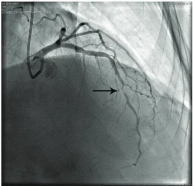
(%95) at the middle segment of LAD was detected and served as a guide for doing coronary angiography and for determining type and size of the stent. Single coronary lesion (LAD) was detected by coronary angiography (Fig.2) and the result was completely parallel to MSCT. Percutaneous coro-nary intervention was performed for LAD lesion and a drug-eluting stent was implanted after predilatation.The patient was exam-inved routinely every three mounths. He was asymptomatic at the end of the three years after the procedure.
Second patient was admited with trivial sore throat together with minimal diaphoresis and was admited to the Cardiology Department with elevated of cardiac enzymes. His ECG (Fig.3) showed slightly prominent and non sig-nificant T waves on V2-3. In the noninvasive technique (MSCT-Fig.4), com-pletely total occlusion in the proximal segment in the left circumflex artery and critical stenosis(%98) at the middle segment of LAD were detected and invasive coronary an-giography was done and the result was completely parallel to MSCT (Fig.5). This patient had a subacute silent posterior
myocar-Istanbul University, Faculty of Medicine, Department of Cardiology, Çapa, Istanbul, TURKEY.
Importance of Multislice Cardiac Computed Tomography For
The Diagnosis and Evaluation of Silent Ischemia and Myocardial
Infarction: Two Cases.
Sessiz İskemi ve Miyokart İnfaktüsü Tanısı ve Değerlendirmesinde Multislice Kardiyak Komputerize Tomografinin Önemi
Faruk Erzengin, Mustafa Özcan, Erhan Teker , Kıvanç Yalın, Güneş Hüseynova, Derya Baykız,
Kamil Adalet
Figure 2: Significant stenosis in the LAD of Case 1 with coronary
angiography.
Figure 1: The adventitial atherosclerotic plaques with minimal
calci-fications (thick arrow) on the proximal segment of LAD artery without stenosis and a vulnerable soft plaque (thin arrow) on the mid LAD artery with significant stenosis.
178 Importance of Multislice Cardiac Computed Tomography For The Diagnosis and Evaluation of Silent Ischemia and Myocardial Infarction: Two Cases. Ankara Üniversitesi Tıp Fakültesi Mecmuası 2009, 62(4)
dial infarction. Percutaneous coronary intervention and predila-tation was performed firstly for the total occluded circumflex artery and was opened successfully, and then to the critical lescion of LAD. Drug-eluting stents were implanted in these two lesions. The patient was examined routinely in every three mounths.This patient was also asymptomatic at the end of the two
years and four months after the procedure.
In conclusion the 64-slice multi-detector cardiac computed tomography is a very important device for the diagnosis, evaluation and the guidance for the treatment of silent ischemia and myocardial infarction.
Figure 4: MSCT of Case 2. Cx totaly occluded (thick arrow), a
signifi-cant stenosis of LAD (thin arrow).
Figure 5: Total occlusion of Cx artery (thick arrow), significant
steno-sis of LAD artery (thin arrow) with standart coronary angiography of Case 2.
Figure 3: Normal ECG of Case 2.
179
Faruk Erzengin, Mustafa Özcan, Erhan Teker , Kıvanç Yalın, Güneş Hüseynova, Derya Baykız, Kamil Adalet
Journal Of Ankara University Faculty of Medicine 2009, 62(4) ReFeRences
1. Wilson GT, Gopalakrishnon P, Tak T. Non-invasive cardiac imaging with computed to-mography. Clin Med Res. 2007 Oct; 5 (3): 165-71.
2. Saia F, Schaar J, Regar E, Rodriguez G, De Feyter PJ, Mastik F, Marzocchi A, Marro-zzini C, Ortolani P, Palmarini T, Branzi A, van der Steen AF, Serrays PW. Clinical imag-ing of the vulnerable plague in the coronary arteries: new intracoronary diagnostic meth-ods. J Cardiovasc Med (Hagerstown) 2006; 7:21-28.
3. Schroeder S, Kopp AF, Baumbach A, Meis-ner C, KuettMeis-ner A, Georg C, Ohnesorge B, Herdeg C, Claussen CD, Karsch KR. Non-invasive detection and evaluation of athero-sclerotic coronary plaques with multislice computed tomography. J Am Coll Cardiol 2001; 37:1430-1435
4. Hoffmann U, Ferencik M, Cury RC, Pena AJ. Coronary CT angiography. J Nucl Med. 2006; 47:797-806.
5. Leschha S, Alkadhi H, Plass A, Desbiolles L,Grunenfelder J, Merincek B,Wildermuth S.Accuracy of MSCT coronary an-giograghy with 64-slice technology: First ex-perience. Eur Heart J 2005;26: 1482-1487.
6. Hendel RC, Patel MR, Kramer CM, Poon M, Hendel RC, Carr JC, Gerstad NA, Gil-lam LD, Hodgson JM, Kim RJ, Kramer CM, Lesser JR, Martin ET, Messer JV, Red-berg RF, Rubin GD, Rumsfeld JS, Taylor AJ, Weigold WG, Woodard PK, Brindis RG, Hendel RC, Douglas PS, Peterson ED, Wolk MJ, Allen JM, Patel MR; American College of Cardiology Foundation Quality Strategic Directions Committee Appropri-ateness Criteria Working Group; American College of Radiology; Society for Cardiovas-cular Magnetic Resonance; American
Soci-ety of Nuclear Cardiology; North American Society for Cardiac Imaging; Society for Car-diovascular Angiography and Interventions; Society of Interventional Radiology. ACCF/ ACR/SCCT/SCMR/ASNC/NASCI/SCAI/ SIR 2006 appropriateness criteria for car-diac computed tomography and carcar-diac magnetic resonance imaging: a report of the American College of Cardiology Foundation Quality Strategic Directions Committee Apporpriateness Criteria Working Group, American College of Radiology, Society of Cardiovascular Computed Tomography, So-ciety for Crdiovascular Magnetic Resonance, American Society of Nuclear Cardiology, North American Society for Cardiac Imag-ing, Society for Cardiovascular Angiography and Interventions, and Society of Interven-tional Radiology. J Am Coll Cardiol 2006; 48:1475-1497.
7. Budoff MJ, Achenbach S, Blumenthal RS, Carr JJ, Goldin JG, Greenland P Guerci AD, Lima JA, Rader DJ, Rubin GD, Shaw LJ, Wiegers SE; American Heart Association Committee on Cardiovascular Imaging and Intervention; American Heart Association Council on Cardiovascular Imaging and Intervention; American Heart Association Committee on Cardiovascular Imaging, Council on Clinical Cardiology. Assess-ment of coronary artery disease by cardiac computed tomography. A scientific state-ment from the American Heart Association Committee on Cardiovascular Imaging and Intervention, Council on Cardiovascular Radiology and Intervention, and Commit-tee on Cardiac Imaging, Council on Clinical Cardiology. Circulation 2006; 114:1761-1791.
8. Achenbach S, Moselewski F, Ropers D, Fe-rencik M, Hoffmann U, MacNeill B, Pohle K, Baum U, Anders K, Jang IK, Daniel WG, Brady TJ. Detection of calcified and
noncalcified coronary atherosclerotic plaque by contrast-enhanced, submillimeter mul-tidetector spiral computed tomography: a segment-based comparison with intravscular ultrasound. Circulation 2004; 109:14-17. 9. Kopp AF, Schroeder S, Baumbach A,
Kue-ttner A, Georg C, Ohnesorge B, Heuschmid M, Kuzo R, Claussen CD. Noninvasive characterisation of coronary lesion morphol-ogy and composition by multislice CT: first results in comparison with intracoronary ul-trasound. Eur Radiol 2001; 11:1607-1611. 10. Dirksen MS, Bax JJ, de Ross A, Jukema
JW, van der Geest RJ, Geleijns K, Boersma E, van der Wall EE, Lamb HJ. Usefulness of dynamic multislice computed tomogra-phy of left ventricular function in unstable angina pectoris and comparison with echo-cardiography. Am J Cardiol 2002; 90:1157-1160.
11. Erzengin F, Büyüköztürk K. Cardiac Im-aging, Internal Medicine (Clinical Text Book, 3000 pages.) in part 2. Edit: Prof. Dr.K.Büyüköztürk. Nobel Tıp LTD. Pag-es.1663-1686; 2007.
12. Filippo Cademartiri, Erica Maffei, Nico R.Mollet. Is dual-source CT coronary angi-ography ready fort the real world? Depart-ment of Radiology and Cardiology, Erasmus Medical Center, Rotterdam, The Nether-lands; Department of Radiology and Car-diology, Azienda Ospedaliero-Universitaria/ University Hospital, Parma, Italy. Eur Heart Journal (2008) 29, 701-703.
13. Erzengin F. Adventitial calcification: Sur-prise location and a new pathway. The 59th The European Cardiovasculer-Cardiology Congress, 18-23 April, İzmir, TURKEY. 14. ) Erzengin F. The 7 8th EAS(European
Ath-erosclerosis Society) Congress 19-24 th June 2010/ Hamburg - Germany (Accepted).