Development and Evaluation of Epigenetic Regulation of Sucrose Metabolism in
Saccharomyces cerevisiae by Using COBRA Technique and Selected CpG Islands in
HXT10 and SUC2 genes
BURAK ALAYLAR 1*, MEDİNE GÜLLÜCE2 and MEHMET KARADAYI2
1Ağrı İbrahim Çeçen University, Faculty of Science and Arts, Department of Molecular Biology and Genetics, Agri, Turkey, 2Atatürk University, Faculty of Science, Department of Biology, Erzurum, Turkey
Abstract
Epigenetics mostly focuses on heritable changes in gene expression that does not contain changes to the underlying primary DNA sequences; an alteration in phenotype without an alteration in genotype. Among various cellular mechanisms, especially DNA methylation plays a central role in many epigenetic processes. Therefore, it has been the most studied epigenetic mechanism and many special techniques for assessment of changes in DNA methylation patterns have been developed. Combined bisulfite restriction analysis (COBRA) is one of these commonly used techniques. Although many studies have been conducted to get information about epigenetic regulation mechanisms in mammalian cell by using COBRA, there is still limited information on the the epigenetic programs and related pathways in other organisms. In the present study;we aimed to detect in silico applications for recognition sites of Hinf I and Taq I restriction enzyme and methylation profiles of specific gene region of HXT10 and SUC2 of Saccharomyces
cerevisiae depending on sucrose, glucose and sucrose and
glucose conditions with COBRA analysis. According to results; methylation status of selected CpG islands in the HXT10 and SUC2 gene regions were not altered by the presence of the sucrose, glucose and glucose+sucrose media.
Keywords: COBRA, DNA methylation,
Saccharomyces cerevisiae.
Received: 28.03.2019 Revised: 11.06.2019 Accepted:18.06.2019
Corresponding author: Burak ALAYLAR, PhD
Ağrı İbrahim Çeçen University, Faculty of Science and Arts, Department of Molecular Biology and Genetics, Agri, Turkey, E-mail: [email protected]
Cite this article as: B. Alaylar, M. Güllüce and M. Karadayı, Development and Evaluation of Epigenetic Regulation of Sucrose Metabolism in Saccharomyces cerevisiae by Using COBRA Technique and Selected CpG Islands in HXT10 and SUC2 genes, Eastern Anatolian Journal of Science, Vol. 5, Issue 1, 50-55,2019.
Introduction
Epigenetics, initially described in 1942 by Conrad Waddington, is the one of the most popular discipline that become a center of scientific attraction in the 20th century. Fundamentally the term of epigenetics refers to “beyond, alongside, above or outside conventional genetics”. This new discipline is mostly focus on heritable changes in gene expression that does not contain changes to the underlying primary DNA sequences; an alteration in phenotype without an alteration in genotype. This alteration in DNA sequences is stem from several factors; environment, lifestyle, embryonic development, nutrition and formation of wide range of diseases such as obesity, diabetes, cancer, variety of human disorders and fatal diseases. Each organism has to face constantly changing environment and respond to the intrinsic and extrinsic factors in throughout lifetime [1-5].
In this manner, epigenetic mechanisms of the cell play vital role. Epigenetic mechanisms cover a number of cellular mechanisms including S-nitrosylation, ubiquitination, SUMOylation, proline isomerization, ADP ribosylation, acetylation, phosphorylation, and methylation events. Among all of these mechanisms, DNA methylation is the most studied and well– known type of major epigenetic modification of the genome. DNA methylation plays a key role in the regulation of gene expression in several biological phenomena such as genomic imprinting, bookmarking, paramutation, gene silencing, X chromosome inactivation, position effect variegation, reprogramming, transvection, infection agent like prions, maternal conditioning, RNA interference, non-coding RNA, small RNAs, and chromatin modifications [2,3, 6-9 ].
Methylation of DNA at CpG dinucleotides is the best studied and highly charecterized epigenetic modification. DNA methylation in humans is based on transfer of a methyl group to the 5’- position of cytosine residues. In the DNA methylation, a methyl group from the universal methyl donor S-adenosylmethionine (SAM) is transferred to the 5th carbon position of a cytosine pyridine ring by a DNA
methyltransferases (DNMTs) catalyzed reaction. This epigenetic event occurs at cytosine residues in a cytosine phosphorylated guanine dinucleotide (CpG) [2,3].
CpG dinucleotides are predominantly clustered in promoter region of genes called CpG islands. Mostly metylation of CpG islands represses the initiation of transcription in the somatic cell of eukaryote. Additionally, some studies propose that environmental factor such as exposure to several dietary factors can change DNA methylation and histone acetylation patterns for this reason, affecting the gene regulation and the phenotypic expression of a gene [6,7]. Thus, exploring the relationship between epigenetics and nutrition remains as a milestone for understanding of many biological processes that response the environment and shape the phenotype from the gene. In this regard, the aim of the present study was designated as the determination of the changes in the methylation profiles of sucrose metabolism related gene regions HXT10 and SUC2 of Saccharomyces
cerevisiae by using combined bisulfite restriction
analysis (COBRA) determined.
Materials and Methods
Chemicals
Magnesium sulphate (MgSO4.7H2O), Glucose (C6H12O6), Potassium phosphate (KH2PO4) and Sodium chlorure (NaCI) were provided from Merck (Hohenbrunn, Germany). All other compounds such as; phenol-chloroform-isoamylalcohol, , isopropanol, glycerol, ethylenediamine tetraacetic acid (EDTA), Triton X-100, Direct Load TM wide range DNA marker and ethanol were purchased from Sigma-Aldrich (St. Louis, USA). Ammonium sulphate (NH4)2SO4), sucrose (C12H22O11) chloroform-isoamylalcohol and yeast extract were obtained from Fluka (Steinheim, Germany). The restriction enzymes for the COBRA analysis, Taq I and Hinf I were obtained Thermo-Scientific (Waltham, MA USA). Wizard genomic DNA purification kit and Methyledge bisulfite conversion system were purchased from Promega (Madison, USA). Expand high fidelity PCR system was acquired from Roche (Mannheim, Germany). Potato Dextrose Agar, bacteriological peptone and agarose acquired from Oxoid (Hampshire, England), LAB M (Lancashire, England) and Lonza (Rockland, USA) respectively.
Saccharomyces cerevisiae strain
Saccharomyces cerevisiae (ATTC® Number: 9763)
strain was provided by the American Type Culture Collection Microbiologiscs Incorporation –U.S.A.
Media
Medium Modified BMC was used as described in detail elsewhere. This media is including several solution such as; salt solution, sugar solution (Glucose, sucrose, glucose+sucrose), vitamin solution, trace metal solution and reproduction encouragement solution. These all solution was prepared in sterile cabin and all this solutions were combined with respectively [10].
Preparation of Test Samples
Stock culture was solubilized at room temperature, then 40 ml MBMC-sucrose 40 ml MBMC-glucose, 40 ml MBMC-sucrose+glucose broth mediums were prepared, then for each broth mediums 30 ml stock culture was added. Yeast cells were grown in sampling tubes 40 ml of MBMC solutions. These specimens were incubated for 24 h, 48 h and 72 h at 28 °C with shaking at 150 rpm. Sampling were performed every 24 h, then DNA isolation, PCR, sodium bisulfite treatment, high fidelity PCR implementation, restriction enzymatic digestion and visualizing applications were realized respectively [11].
Genomic DNA isolation
Genomic DNA was isolated from Saccharomyces
cerevisiae. DNA isolation protocol was explained as
follows.
Cultures were grown in MBMC media, and then centrifuged at 18.000 rpm for 10 minutes at 4 °C. 0.3 gr glass bead was added on the pellets which were inside of eppendorf tubes. 200 µl Lysis buffer was added and vortexed at the maximum speed for 2 minutes. 200 µl phenol-cloroform-isoamylalcohol (25:25:1) was added and overturned 5-10 times for mixing well. 200 TE buffer was added and vortexed again at the maximum speed for 2 minutes, then centrifuged 16.000 rpm for 10 minutes at +4 °C. After the centrifuge process transferred to the 400 µl supernatant containing DNA sterile 2 ml eppendorf tubes. Added to eppendorf tubes 1000 µl % 99.5 pure alcohol and overturned several times, then centrifuged at 16.000 rpm for 10 minutes at 4 °C after this process, Protein pellets were remained in the original eppendorf
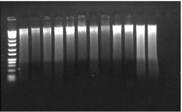
tubes and removed the liquid part of eppendorf tubes. Drained the tubes on clean blotting paper was allowed the pellet air-dry for 5 minutes. 300 µl Nuclei lysis solution (Promega) was added to DNA pellets inside of the eppendorf tubes and overturned 1-2 times. 100 µl Protein precipitation solution (Promega) was added to eppendorf tubes and overturned 1-2 times again after overturning of samples were vortexed 1-2 minutes. Samples were sat on the ice or at -20 °C for 5 minutes and then samples were centrifuged at 16.000 rpm for 10 minutes at 4 °C. 350 µl supernatant containing DNA samples were transferred to steril 2 ml eppendorf tubes and 300 µl isopropanol (-20 °C) was added to cell pellets and overturned 5-10 times. Supernatants were removed gently and pellets were remained in eppendorf tubes, blotting papers was allowed to the pellet air-dry for 1 minute. 300 µl % 70 ethanol (-20 °C) was added and centrifuged at 16.000 rpm for 6 minutes at 4 °C. After centifuruge process, samples were waited inside of the incubator for evaporated to ethanols inside of the cell pellets at 40 °C for 60 minutes. 50 µl DNA rehydration solution (Promega) was added to eppendorf tubes. 1.5 µl RNAase was added to the purified DNA samples and incubated at 37 °C for 15 minutes. Finally, after the incubation of DNA samples were waited in incubator at 65 °C for 60 minutes and samples were stored at 4 °C [11]. The results of DNA isolation were shown in Figure 1.
Figure 1 DNA isolation results of S. cerevisiae
samples
Sodium bisulfite treatment
Many bisulfite conversion kits have been advanced for bisulfite treatment because it is hard to success and optimal balance between complete conversion and minimal degradation of DNA. In this present study, DNA treatment with sodium bisulfite was performed using the Methyledge™ bisulfite conversion kit
(Promega, San Luis Obispo-USA) according to manufacturer’s protocol. Also specific primer sets were designed after the bisulfite treament for SUC2 and HXT10 gene region. Detailed information of primer sets for related genes were shown in Table 1 and Table 2.
Table 1. Primer set for SUC2 gene region
Forward CGGCTGCCAAATCACAAGAC
Reverse CTAGAGCGTTACCGGTGGTC
Product length
969 base pair
Table 2. Primer set for HXT10 gene region
PCR protocol
Samples were prepared for as a volume of 25 PCR reactions for each reaction. Each PCR reaction was contained 17.75 µl distilled water, 2.5 µl expand high fidelity buffer (10X concentrated without MgCI2), 1 µl DMSO, 1.5 µl MgCI2 solution (25mM), 0.25 µl F primer, 0.25 µl R primer, 0.5 µl dNTPs (deoxynucleotide triphosphates: dATP, dGTP, dCTP, dTTP), 0.5 µl Expand high fidelity enzyme mix and 1 µl bisulfite treated DNA.
PCR conditions were as follows: 94 °C for 3 minutes, 40 cycles of denaturing at 94 °C for 1 minute, 1 minute annealing at 61.5 °C (This step should prepare for each primer Tm temperature), 1.5 minutes and 7 minutes extension steps at 72 °C [11].
Restriction enzymatic digestion of PCR products
RestrictionMapper program is used to determine the restriction sites in the products to be obtained after the amplification of SUC2 and HXT10 gene regions with the designed specific primer sets. Restriction sites for PCR product of SUC2 and HXT10 gene informations were given in Table 3 and Table 4. Restriction enzymatic digestion of PCR product was performed with Hinf I and Taq I. These restriction enzymes were provided from Thermo-Scientific (Waltham, MA USA). According to manufacturer’s protocol was used as follows:
Hinf I: 10 µl PCR products, 18 µl distilled water, 2 µl
10X buffer R Hinf I and Hinf I enzyme were added a PCR tube respectively. Gently mixed and incubated for 1-16 h, stopping to restriction enzyme reaction was
Forward GTGTTTTGCGTGGGCAATCT
Reverse CTCCGGACTTCCATGGCTTT
Product length
stayed at 65 °C 20 minutes in heat block and products were stored at +4 °C.
Taq I: 10 µl PCR products, 18 µl distilled water, 2 µl
10X buffer R Taq I and Taq I enzyme were added a PCR tube respectively. Gently mixed and incubated for 1-16 h, stoping to restriction enzyme reaction was stayed at 80 °C 20 minutes in heat block and products were stored at +4 °C.
Table 3. Restriction sites of the PCR product of the
SUC2 gene region
Table 4. Restriction sites of the PCR product of the
HXT10 gene region
Analysis of Digested PCR Products
Digested PCR products were analyzed by QIAxcel advanced analysis system.
Results
According to the research findings; it was seen that methylation status of selected CpG islands in the
HXT10 and SUC2 gene regions were not changed by
the presence of the sucrose. Methylation profiles of
HXT10 and SUC2 regions of S. cerevisiae were not
changed methylation status depending on the growth in sucrose, glucose and glucose+sucrose media. In addition, using of capillary gel electrophoresis technique for such as sensitive applications were emerged as the most efficient method of the present methods. The method is convenience for separation of both small and large molecules. Accordingly, using of gel electrophoresis technique have many factors that make it very crucial and useful. These features are such as; broad analytical variability, high mass sensitivity, high efficiency separation, working opportunities with very low volume, short analysis time, using consumables with minimum level and basic device fiction.
When our study findings were considered; after the bisulfite treatment had caused high product loss. It was clearly demonstrated that using these products with capillary electrophoresis system that it had many advantages for analysis sensitively.
Discussion and Conclusions
In the past decades, epigenetics advances have been one of the most exciting discoveries among the scientific fields. It has been demonstrated to the scientists to have made many discoveries possible. Recent advances have highlighted important roles of epigenetic that focus on human diseases. Currently, many serious diseases such as; cancer, aging, inflammation, cardiovascular diseases, immune and neurocognitive disorders, diabetes and obesity associated with epigenetic alterations, histone modifications and DNA methylation [3,5, 12-15]. According to the present studies in the literature was demonstrated that understanding of basic epigenetics mechanisms and related rules about many cellular processes such as; quiescence, proliferation, migration, differentiation, etc., and that it enables development of new technologies for various areas especially including biology, agriculture, medicine, biotechnology. From this reason; advances in understanding to the basic mechanisms of unknown etiology of diseases field of epigenetics the most significant expectations [3,8,11].
However, many environmental effects may change epigenetic pattern of organisms and related gene expression levels. One of the most common environmental effects is nutrition. Nutrition can cause heritable alteration in phenotype without any changes in DNA sequences. It is now clear epigenetic processes can be transferred in organisms from one generation to another. For this reason, many scientists focus on the epigenetics-associated studies have become crucial in the nutritional field to research active nutrition compounds to understand their effects on organisms. This new field is called nutritional epigenetics [4,3,11]. In the present study, we determined the changes in the methylation profiles of sucrose metabolism related specific gene region HXT10 and SUC2 of S. cerevisiae by using COBRA technique. COBRA is a quantitative analysis method and it is based on the amplification of bisulfite-converted DNA using gene-specific primers and subsequent restriction digestion of PCR products in small quantity of biological samples [16, 3,11]. Previous studies have mentioned that the methylation levels of specific genes may play a key role in many Restriction
Enzyme Sequence Cutting sites
HinfI GANTC 69
TaqI TCGA 583, 597
Restriction
Enzyme Sequence Cutting sites
HinfI GANTC 50, 261, 350
serious disease process such as; cancer, diabetes etc. COBRA used in laboratories due to suitable application based techniques are commonly, analyzing large number of samples and cost effectiveness [16-19].
According to the literature; it has many different applications about COBRA in eukaryotic organisms. This study is the one of the prestudy for gene specific methylation analysis in eukaryotic organisms to understand their related epigenetics processes with capillary gel electrophoresis in Combined bisulfite restriction analysis.
In the light of these experiments, we examined in silico applications for recognition sites of Hinf I and Taq I restriction enzyme and methylation profiles of specific gene region of HXT10 and SUC2 of S. cerevisiae depending on sucrose, glucose and sucrose and glucose conditions with COBRA analysis.
ACKNOWLEDGEMENT
This study was supported by Atatürk University Scientific Research Projects Coordination Unit. Project Number: BAP-2015/349
References
[1] FREİDBERG, J. (2007). Plasma Physics and Fusion Energy. Cambridge university press. 25-30.
[2] HO S, TANG W, 2007. Techiques used in studies of epigenome dysregulation due to aberrant DNA methylation: An emphasis on fetal-based adult diseases. Reproductive Toxicology. 23: 267-282.
[3] DALTON VS, KOLSHUS E, MCLOUGHLIN DM, 2014. Epigenetics and depression: return of the repressed. Journal of Affective Disorders. 155: 1-12.
[4] GULLUCE M, ALAYLAR B, KOC TY, KARADAYI M, 2014. Epigenetics: An Innovative Approach for Biotechnology and Food Science. Internetional Journal of Bioscience, Biochemistry and Bioinformatics. 4(3): 195-199.
[5] CHOI SW, FRISO S, 2010. Epigenetics: A New Bridge between Nutrition and Health. Advances in Nutrition An International Review Journal. 1: 8-16.
[6] TOLLEFSBOL TO, 2011. Chapter 1:Epigenetics: The New Science of Genetics. Handbook of Epigenetics: The New Molecular and Medical Genetics, Ed: T.O. Tollefsbol. Academic Press, San Diego. 1-6.
[7] BIRD A, 2002. DNA methylation patterns and epigenetic memory. Cold Spring Harbor Laboratory Press. 6-21.
[8] HOLLIDAY R, 2006. Epigenetics: A Historical overview. Epigenetics, 1: 76-80.
[9] İZMIRLI M, 2013. Epigenetic Mechanisms and Approaches in Cancer Treatments. Van Medical Journal. 20(1): 48-51.
[10] JANITZ K, JANITZ M, 2011. Chapter 12: Assessing Epigenetic Information. Handbook of Epigenetics: The New Molecular and Medical Genetics, Ed: T.O. Tollefsbol. Academic Press, San Diego. 173-181. [11] TAHERZADEH MJ, FOX M, HJORTH H,
EDEBO L, 2003. Production of mycelium biomass and ethanol from paper pulp sulfite liquor by Rhizopus oryzae. Bioresource Technology. 88: 167-177.
[12] ALAYLAR B, 2014. Determination of epigenetic changes depending on presence of sucrose and glucuse in related gene regions of
Saccharomyces cerevisiae. Master Thesis,
Atatürk University Institute of Natural and Applied Sciences. 1-58.
[13] EGGER G, LIANG G, APARICIO A, JONES PA, 2004. Epigenetics in human disease and prospects for epigenetic therapy. Nature. 429: 457-463.
[14] TAYLOR KH, KRAMER RS, DAVIS JW, GUO J, DUFF DJ, XU D, CALDWELL CW, SHI H, 2007. Ultradeep Bisulfite Sequencing Analysis of DNA Methylation Patterns in Multiple Gene Promoters by 454 Sequencing. Cancer Research. 67: 8511-8518.
[15] KRISTENSEN LS, HANSEN LL, 2009. PCR-Based Methods for Detecting Single-Locus DNA Methylation Biomarkers in Cancer Diagnostics, Prognostics and Response to Treatment. Clinical Chemistry. 55(8): 1471-1483.
[16] KARPINSKI P, SZMIDA E, MISIAK B, RAMSEY D, LESZCZYNSKI P, BEBENEK M, SEDZIAK T, GRZEBIENIAK Z, JONKISZ A, LEBIODA A, SASIADEK MM, 2012. Assessment of Three Epigenotypes in Colorectal Cancer by Combined Bisulfite Restriction Analysis. Molecular Carcinogenesis. 51: 1003-1008. [17] XIONG Z, LAIRD PW, 1997. COBRA: a
sensitive and quantitative DNA methylation assay. Nucleic Acids Research. 25(12): 2532-2534.
[18] EADS CA, LAIRD PW, 2002. Chapter 7: Combined Bisülfite Restriction Analysis (COBRA). Methods in Molecular Biology: DNA Methylation Protocols, Ed: K.I.Mills
and B.H. Ramsahoye. Humana Press Inc. 200: 71-85.
[19] FRAGA MF, ESTELLER M, 2002. DNA Methylation: A profile of Methods and Applications. Biotechniques. 33: 632-649. [20] BRENA RM, AUER H, KORNACKER K,
HACKANSON B, RAVAL A, BYRD JC, PLASS C, 2006. Accurate quantification of DNA methylation using combined bisulfite restriction analysis coupled with the Agilent 2100 Bioanalyser platform. Nucleic Acid Research. 34(3): 1-8.