Original Article
Oncol Res Treat 2017;40:523–527 DOI: 10.1159/000476037
Adjuvant Small Pelvic Radiotherapy in Patients with
Cervical Cancer Having Intermediate Risk Factors Only –
Is It Sufficient?
Sezin Yuce Sari
aOzan C. Guler
bMelis Gultekin
aHuseyin C. Onal
cFerah Yildiz
aa Department of Radiation Oncology, Hacettepe University, Faculty of Medicine, Ankara, Turkey;
b Department of Radiation Oncology, Faculty of Medicine, Karadeniz Technical University, Trabzon, Turkey; c Department of Radiation Oncology, Dr Turgut Noyan Research and Treatment Center,
Baskent University Faculty of Medicine, Adana, Turkey
Introduction
Cervical cancer is the most common gynecologic cancer world-wide [1]. In International Federation of Gynecology and Obstetrics (FIGO; revised 2009) stages I–IIA, either surgery or radiotherapy (RT) can be administered with similar rates of disease control, sur-vival, and toxicity. However, the combination of surgery and RT increases the complication rates [2–4].
A number of risk factors have been described for recurrence after surgery, i.e., parametrial invasion, compromised surgical margins, regional lymph node (LN) metastasis, unfavorable histol-ogy, large tumor, deep (> 50%) stromal invasion (DSI), and lym-phovascular space involvement (LVSI) [5–8]. In this case, adjuvant RT or chemoradiotherapy can be beneficial [2, 9]. However, post-operative RT fields and treatment volumes are still a matter of de-bate. The Radiation Therapy Oncology Group (RTOG) recom-mends delineating the uterine surgical bed, bilateral parametria, upper third of the vagina, paravaginal soft tissues, and common, external, and internal iliac lymph nodes, and presacral lymphatics [10]. However, this field encompasses a large portion of the intesti-nal segments, and can cause increased toxicity. Therefore, several attempts to reduce the RT field have been made. In a pilot study by Kridelka et al. [11] ‘small pelvic’ RT produced high local control rates with less toxicity. Based on this trial, in 2001, we began treat-ing all cervical cancer patients with intermediate risk factors ustreat-ing small pelvic external beam RT (EBRT) including the surgical bed, parametria, upper vagina, and presacral lymphatics.
In this bi-institutional study, we retrospectively evaluated the treatment outcomes and prognostic factors for survival and disease control in cervical cancer patients treated with postoperative small pelvic EBRT. The toxicity of this treatment field was also evaluated.
Keywords
Cervical cancer · Prognostic factor · External beam radiotherapy
Summary
Background: We sought to determine the outcomes of
adjuvant small pelvic external beam radiotherapy (EBRT) and prognostic factors for survival and disease control.
Patients and Methods: We retrospectively evaluated 113
cervical cancer patients treated with postoperative me-dian 50.4-Gy small pelvic EBRT. We treated the surgical bed, bilateral parametria, paravaginal soft tissues, upper third of the vagina, and presacral lymphatics. Results: Median follow-up of all patients and survivors was 58 and 67 months, respectively. The 2- and 5-year overall survival (OS) and disease-free survival rates were 91 and 82%, and 85 and 74%, respectively. The locoregional fail-ure rate was 10%. Age was a significant predictor for OS and distant metastasis-free survival (DMFS) on univari-ate analysis. The number of dissected lymph nodes being < 30 negatively affected the pelvic recurrence-free survival. The only independent predictor on multivariate analysis was older age for DMFS. Although no severe acute toxicity was observed, late grade ˰ 3 toxicity de-veloped in 8 patients. Conclusion: Small pelvic EBRT produces satisfactory survival and locoregional control with acceptable toxicity, and can be an alternative to whole pelvic EBRT in selected cervical cancer patients.
© 2017 S. Karger GmbH, Freiburg
Received: March 03, 2017 Accepted: April 25, 2017 Published online: August 21, 2017
Ferah Yildiz, M.D.
Department of Radiation Oncology © 2017 S. Karger GmbH, Freiburg
Patients and Methods
The medical charts of 113 patients with early-stage intermediate-risk cervi-cal cancer treated between 2001 and 2014 in 2 departments were retrospectively evaluated. All patients underwent pretreatment staging involving history, phys-ical examination, vaginal examination under general anesthesia, hematologic and biochemical parameters, chest X-ray, and computed tomography (CT) or magnetic resonance imaging (MRI) of the pelvis with/without cystoscopy, rec-toscopy, and intravenous pyelogram. Patients were eligible if adequate complete surgical staging was performed. Adequate LN dissection was described as the bilateral dissection of ˰ 10 obturator and external iliac LNs [12]. Patients with ˰ 2 intermediate risk factors (i.e., adenocarcinoma, DSI, LVSI, and > 4 cm tumor) or DSI alone were included in the study. All patients were treated with small pelvic EBRT. 39 (35%) patients received additional brachytherapy, and 44 (39%) received concurrent chemotherapy. Patients with distant metastasis or high-risk factors (i.e., positive surgical margins, LNs and/or parametria) and patients that received chemotherapy prior to RT were excluded from the study. This study was approved by the institutional review board.
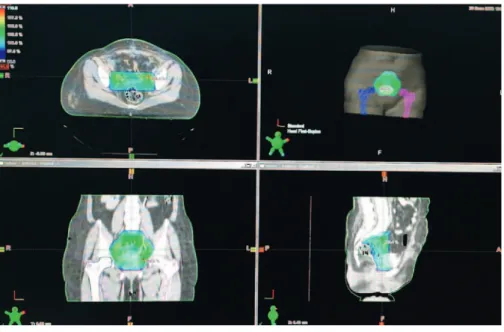
All patients received either 2-dimensional (2D) or 3D conformal EBRT in supine position with a comfortably full urinary bladder and an empty rectum according to RTOG recommendations (RTOG 0418: A Phase II Study of IMRT to the Pelvis +/- Chemotherapy for Post-operative Patients with either Endo-metrial or Cervical Carcinoma; 2011). EBRT was administered via Philips SL25 (Philips Healthcare, Andover, MA, USA), Elekta Synergy Platform (Elekta AB, Stockholm, Sweden), or Varian Clinac DHX High Performance (Varian Medi-cal Systems, Inc. Palo Alto, CA). In 2D EBRT, the superior border of the RT field was the lumbar (L)5-sacral (S)1 vertebral interspace, the inferior border was inferior to the obturator foramina, and the lateral borders were adjacent to the medial border of iliac bones. For lateral fields, the anterior border was pos-terior to the symphysis pubis, and the pospos-terior border was the S3–4 vertebral interspace. In 3D EBRT, the surgical bed and presacral lymphatics were deline-ated according to the RTOG guidelines [10]. Common, internal, and external iliac LNs as well as obturator LNs were excluded. Organs at risk were detected in related slices, and were also contoured (fig. 1). In both 2D and 3D EBRT, a pelvic box technique with 4 fields (i.e., antero-posterior, postero-anterior and 2 laterals) was applied.
Brachytherapy was performed when positive or close (< 5 mm) surgical margins on the vagina were reported. A 2D and 3D remote after-loading tech-nique of high-dose-rate (HDR) brachytherapy was administered via microSe-lectron® (Elekta AB), GammaMed Plus® (Varian Medical Systems Inc.), or VariSource® (Varian Medical Systems Inc.). All patients were treated in supine position with an empty urinary bladder and rectum. A vaginal cylinder cover-ing the whole vagina was placed. The dose was prescribed at a 5-mm distance
from the mucosa. In 3D brachytherapy, a planning CT with a slice thickness of 2.5 mm was taken, and the clinical target volume (CTV) was contoured encom-passing the upper third (length) and whole thickness of the vagina.
A radiation oncologist followed all patients during the course of RT and, in association with a gynecologic oncologist, monitored patients every 3 months for the first 2 years, every 6 months until year 5, and annually thereafter. Fol-low-up information was obtained from department charts, referring doctors, and patients and/or next of kin. RTOG/European Organization for the Treat-ment of Cancer (EORTC) toxicity criteria were used to score RT-related acute and late toxicities [13].
All statistical analyses were performed using the Statistical Package for the Social Sciences (SPSS) version 18.0 (SPSS Inc., IBM Corp., Armonk, NY, USA). The primary end point was pelvic recurrence-free survival (PRFS). Secondary end points included overall survival (OS), disease-free survival (DFS), distant metastasis-free survival (DMFS), and treatment toxicity. Time to progression was calculated as the period from the date of diagnosis to the date of death or disease recurrence. Survival analyses were carried out using the Kaplan-Meier method and compared using the log-rank test. Multivariate analysis was performed using the Cox proportional hazards model, including all factors found significant by univariate analysis. p < 0.05 was considered statistically significant.
Results
Patient and treatment characteristics are presented in table 1. The median age was 54 years (range 34–84 years). Tumors were mostly squamous cell carcinoma. The median number of dissected total, pelvic, and para-aortic LNs was 41 (range 12–120), 30 (range 12–97), and 8 (range 0–36), respectively. 61 (54%) patients received 2D EBRT, whereas 52 (46%) received 3D EBRT. The median EBRT dose was 50.4 Gy (range 45–50.4 Gy) in median 1.8-Gy (range 1.8–2 Gy) daily fractions. 39 (35%) patients underwent HDR brachyther-apy with a median 15 Gy (range 12–28 Gy) in 5-Gy (range 4–7 Gy) daily fractions. 44 (39%) patients received concurrent 40 mg/m2
weekly cisplatin with a median of 5 (range 3–6) cycles.
The median follow-up times for all patients and for those that survived were 58 months (range 2–177 months) and 67 months (range 4–177 months), respectively. In total, 6 patients harbored disease (local, locoregional, or distant), including 2 with distant
Fig. 1. Dose-color wash images of 3-dimentional radiotherapy plan in a cervical cancer patient with postoperative intermediate risk factors. Right upper image: right and left femur depicted in blue and pink, respectively. Dose-color wash images are seen in transverse, coronal, and sagittal slices in the upper left, lower left, and lower right boxes, respec-tively.
metastasis, 2 with pelvic recurrence, and 2 with both. At the final follow-up, 91 (81%) patients were alive (9 with disease), and 22 (19%) patients had died (6 of their disease, 17 of other causes).
The 2- and 5-year OS rate was 91 and 82%, respectively. In uni-variate analysis, age was the only significant factor (table 2). In mul-tivariate analysis, no statistically significant factor was observed. The 2- and 5-year DFS rate was 85 and 74%, respectively. In uni-variate analysis, a trend for DFS was observed in patients ˯ 50 years (p = 0.09) and in patients with ˰ 30 LNs dissected (p = 0.06). No statistically significant factor was found in multivariate analysis. The 2- and 5-year PRFS rate was 86 and 75%, respectively. The lo-coregional control rate was 92% in 2 years and 89% in 5 years. The
2- and 5-year PRFS rates were 74 and 66%, and 92 and 79% in pa-tients with < 30 and ˰ 30 dissected LNs, respectively (p = 0.04). Multivariate analysis was not performed as there was only 1 statisti-cally significant factor in univariate analysis. The 2- and 5-year DMFS rate was 88 and 79%, respectively. The distant metastasis rate was 6% for the entire cohort. The only statistically significant factor in univariate analysis was age. The 2- and 5-year DMFS rate was 90 and 88% in patients ˯ 50 years compared to 87 and 73% in patients > 50 years, respectively (p = 0.02). Although not statistically signifi-cant, positive/close surgical margins on the vagina resulted in a de-creased DMFS rate (2- and 5-year DMFS rate 79 and 67% vs. 92 and 83% in patients with negative margins; p = 0.075). Older age (> 50 years) was the only significant factor in multivariate analysis (haz-ard ratio = 2.77, 95% confidence interval 1.02–7.56; p = 0.04).
None of the patients had toxicities that necessitated the discon-tinuation of RT. No grade ˰ 3 adverse events occurred during EBRT or brachytherapy. 45 (40%) patients developed grade 1–2 acute toxicity (cystitis, n = 24; proctitis, n = 24; diarrhea, n = 21; dermatitis, n = 7). Late serious complications were observed in 8 (7%) patients: 1 patient each with proctitis, enteritis, lymphedema, rectovaginal fistula, rectovesical fistula, enterovesical fistula, femo-ral aseptic necrosis, and femofemo-ral aseptic necrosis with a possibly RT-related sarcoma of the iliac bone. The rate of late toxicity in patients treated with 2D EBRT was significantly higher compared to that in patients treated with 3D EBRT (p = 0.04). However, only 1 patient with a rectovaginal fistula was treated with 3D EBRT. The patient with late proctitis had received 4×7 Gy brachytherapy, and also developed grade 2 proctitis during EBRT. Other patients with late toxicity had not received brachytherapy, and none of them de-veloped acute toxicity.
Discussion
The results of the present study revealed that omitting the iliac and obturator lymphatics during EBRT in early-stage cervical can-cer patients with intermediate risk factors does not negatively im-pact survival and disease control. Additionally, we did not find any significant prognosticators for survival or local control, except for
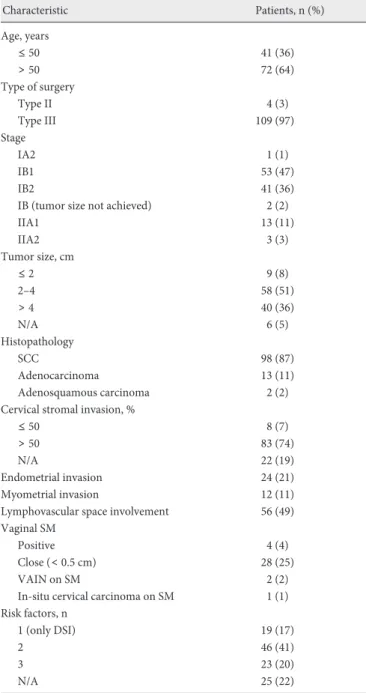
Table 1. Patient and treatment characteristics
Characteristic Patients, n (%) Age, years ˯ 50 41 (36) > 50 72 (64) Type of surgery Type II 4 (3) Type III 109 (97) Stage IA2 1 (1) IB1 53 (47) IB2 41 (36)
IB (tumor size not achieved) 2 (2)
IIA1 13 (11) IIA2 3 (3) Tumor size, cm ˯ 2 9 (8) 2–4 58 (51) > 4 40 (36) N/A 6 (5) Histopathology SCC 98 (87) Adenocarcinoma 13 (11) Adenosquamous carcinoma 2 (2)
Cervical stromal invasion, %
˯ 50 8 (7)
> 50 83 (74)
N/A 22 (19)
Endometrial invasion 24 (21)
Myometrial invasion 12 (11)
Lymphovascular space involvement 56 (49)
Vaginal SM
Positive 4 (4)
Close (< 0.5 cm) 28 (25)
VAIN on SM 2 (2)
In-situ cervical carcinoma on SM 1 (1) Risk factors, n
1 (only DSI) 19 (17)
2 46 (41)
3 23 (20)
N/A 25 (22)
N/A = Not available; SCC = squamous cell carcinoma; SM = surgical margin; VAIN = vaginal intraepithelial neoplasia; DSI = deep stromal invasion.
Table 2. Prognostic factors in univariate analysis for overall survival 2-year OS, % 5-year OS, % p value
Age, years 0.04a ˯ 50 90 90 > 50 92 78 Dissected LNs, n 0.08 < 30 75 75 ˰ 30 92 83 SM status 0.09 Positive/close 82 70 Negative 92 86 aStatistically significant.
age for OS and DMFS. Older patients in our study received a quite similar treatment schedule with high compliance rates. We can only assume that the reason for the worse outcome in patients > 50 years may be somewhat aggressive tumor factors other than the al-ready known.
Postoperative adjuvant RT improves local control in selected cases. In the studies on postoperative RT, cervical cancer patients mostly have high and intermediate risk factors [14, 15]. In a phase III study of patients with intermediate risk factors alone, Sedlis et al. [2] treated 277 patients with 46–50.4 Gy EBRT that encom-passed the obturator and iliac LNs. With a median 10-year follow-up, the authors reported DFS rates of 82.5% in patients treated with adjuvant RT and 69.3% for no further treatment [16]. The lo-coregional recurrence rate was 13.9 and 20.7% in the respective pa-tients. The overall pelvic disease control rate in our study was 90.3% which is quite similar to the above study with whole pelvic EBRT.
A number of studies did not recommend adjuvant RT in early-stage cervical cancer with intermediate risk factors as it did not in-crease disease control and survival rates [17, 18]. Nevertheless, the majority of studies has come up with opposite results. DSI and tumor size were demonstrated as strong prognostic factors for OS, DFS, and DMFS [9, 19, 20]. In the Gynecologic Oncology Group (GOG)-49 study, Delgado et al. [21] demonstrated that tumor size, LVSI, and DSI were independent predictors for DFS in stage IB disease. In the GOG-92 study with 277 patients having ˰ 2 inter-mediate risk factors, compared to surgery alone, adjuvant RT in-cluding iliac and obturator LNs reduced the recurrence and mor-tality risk by 44 and 36%, respectively [2]. In the update of this study with a median 10-year follow-up, local and distant failures were still higher in the surgery-only arm compared to the adjuvant RT arm (20.7 vs. 13.9%, and 8.6 vs. 2.9%) with similar OS rates (70 vs. 81%) [16]. Based on these findings, we began delivering adju-vant RT to all patients with DSI alone or ˰ 2 intermediate risk fac-tors. However, we found none of the intermediate risk factors to be a significant prognostic factor in the current study. The routine use of adjuvant RT may have precluded these risk factors and hence the determination of their effect on local and distant recurrence.
According to the American College of Radiology (ACR) Appro-priateness Criteria, patients with early-stage cervical cancer are di-vided into 3 risk groups following radical surgery, and patients with LVSI, DSI, and/or large tumor constitute the intermediate-risk group [22]. Adjuvant RT is indicated in this group as the re-currence risk is approximately 30%. For adjuvant RT, ACR recom-mends that the CTV should encompass the paracervical, parame-trial, and paravaginal soft tissues, the upper vagina, the ligaments supporting the cervix, and all LNs at high risk of involvement [22]. In 2D simulation, the superior border is the L5-S1 vertebral inter-space, the inferior border is inferior to the iliac bones, and the lat-eral borders are 2 cm latlat-eral to the iliac foramina. In our small
pel-vic field, the superior border is the L5-S1 vertebral interspace, but the inferior border is just inferior to the iliac foramina and the lat-eral borders are latlat-eral to these. Hence, our field is relatively smaller than conventional fields. Nevertheless, only 10% of patients experienced pelvic recurrence in a median follow-up of 58 months. Our results are comparable with those of Kriedelka et al. [11] who used small pelvic fields in 25 patients with intermediate risk factors of whom only 1 patient recurred.
RTOG published a consensus guideline for IMRT in postopera-tive endometrial and cervical cancer [10]. They recommend delin-eating common, external, and internal iliac LNs as well as presacral LNs together with the uterine and cervical surgical bed, bilateral parametria, upper third of the vagina, and paravaginal soft tissues. We strictly follow this guideline when determining targets. How-ever, we irradiate presacral lymphatics alone in patients with ade-quate LN dissection. We give an at least 1.5-cm margin for the planning target volume based on organ motion according to our previous study with sequential CT scans at different time periods of treatment [23].
None of our patients experienced toxicity that necessitated the discontinuation of RT. Only 8 patients developed considerable late toxicity. In studies on cervical cancer patients with high and inter-mediate risk factors treated with adjuvant low pelvic field EBRT (i.e., exclusion of common iliac LNs), local and distant control rates were found to be similar to those in patients treated with whole pelvic RT with much lower rates of gastrointestinal compli-cations [15, 24, 25]. In our study, delineating only the presacral LNs resulted in an excellent pelvic control rate of 90.3% compared to studies with whole pelvic EBRT [14, 26]. A 5-year OS rate of 82% and the absence of grade ˰ 3 acute toxicity also reveal that adjuvant small pelvic EBRT is an adequate and sufficient treatment for patients with intermediate risk factors.
The main limitations of our study are that it is retrospective in nature, and it only presents the results of small pelvic EBRT with-out comparing it to whole pelvic EBRT. However, this study is im-portant in that it offers a somewhat innovative technique to radia-tion oncologists which leads to excellent disease control with negli-gible toxicity. Although there are previously published studies pre-senting acceptable outcomes with small pelvic EBRT, our study is the only one that applied a true small pelvic field. Besides the iden-tified intermediate risk factors, older age and < 30 LN dissection deteriorated the prognosis. On the other hand, prospective trials analyzing the treatment outcomes and prognostic factors for small and whole pelvic field irradiation are required for accurate decision making.
Disclosure Statement
References
1 2012 GLOBOCAN Cancer Incidence and Mortality Worldwide. IARC CancerBase, 2010; globocan.iarc.fr/ Pages/fact_sheets_cancer.aspx.
2 Sedlis A, Bundy BN, Rotman MZ, Lentz SS, Muder-spach LI, Zaino RJ: A randomized trial of pelvic radia-tion therapy versus no further therapy in selected pa-tients with stage IB carcinoma of the cervix after radi-cal hysterectomy and pelvic lymphadenectomy: a Gy-necologic Oncology Group Study. Gynecol Oncol 1999;73:177–183.
3 Landoni F, Maneo A, Colombo A, Placa F, Milani R, Perego P, Favini G, Ferri L, Mangioni C: Randomised study of radical surgery versus radiotherapy for stage Ib-IIa cervical cancer. Lancet 1997;350:535–540. 4 Gray HJ: Primary management of early stage cervical
cancer (IA1-IB) and appropriate selection of adjuvant therapy. J Natl Compr Canc Netw 2008;6:47–52. 5 Horn LC, Fischer U, Raptis G, Bilek K, Hentschel B:
Tumor size is of prognostic value in surgically treated FIGO stage II cervical cancer. Gynecol Oncol 2007; 107:310–315.
6 Aoki Y, Sasaki M, Watanabe M, Sato T, Tsuneki I, Aida H, Tanaka K: High-risk group in node-positive patients with stage IB, IIA, and IIB cervical carcinoma after radical hysterectomy and postoperative pelvic irradiation. Gynecol Oncol 2000;77:305–309. 7 Atahan IL, Yildiz F, Ozyar E, Pehlivan B, Genc M,
Kose MF, Tulunay G, Ayhan A, Yuce K, Guler N, Kucukali T: Radiotherapy in the adjuvant setting of cervical carcinoma: treatment, results, and prognostic factors. Int J Gynecol Cancer 2007;17:813–820. 8 Nahhas WA, Sharkey FE, Whitney CW, Husseinzadeh
N, Chung CK, Mortel R: The prognostic significance of vascular channel involvement and deep stromal pene-tration in early cervical carcinoma. Am J Clin Oncol 1983;6:259–264.
9 Peters WA 3rd, Liu PY, Barrett RJ 2nd, Stock RJ, Monk BJ, Berek JS, Souhami L, Grigsby P, Gordon W Jr, Al-berts DS: Concurrent chemotherapy and pelvic radia-tion therapy compared with pelvic radiaradia-tion therapy alone as adjuvant therapy after radical surgery in high-risk early-stage cancer of the cervix. J Clin Oncol 2000; 18:1606–1613.
10 Small W Jr, Mell LK, Anderson P, Creutzberg C, De Los Santos J, Gaffney D, Jhingran A, Portelance L, Schefter T, Iyer R, Varia M, Winter K, Mundt AJ: Con-sensus guidelines for delineation of clinical target vol-ume for intensity-modulated pelvic radiotherapy in postoperative treatment of endometrial and cervical cancer. Int J Radiat Oncol Biol Phys 2008;71:428–434. 11 Kridelka FJ, Berg DO, Neuman M, Edwards LS,
Rob-ertson G, Grant PT, Hacker NF: Adjuvant small field pelvic radiation for patients with high risk, stage IB lymph node negative cervix carcinoma after radical hysterectomy and pelvic lymph node dissection. A pilot study. Cancer 1999;86:2059–2065.
12 Nijman HW, Khalifa M, Covens A: What is the num-ber of lymph nodes required for an ‘adequate’ pelvic lymphadenectomy? Eur J Gynaecol Oncol 2004;25: 87–89.
13 Cox JD, Stetz J, Pajak TF: Toxicity criteria of the Radi-ation Therapy Oncology Group (RTOG) and the Euro-pean Organization for Research and Treatment of Cancer (EORTC). Int J Radiat Oncol Biol Phys 1995; 31:1341–1346.
14 Yeh SA, Wan Leung S, Wang CJ, Chen HC: Postopera-tive radiotherapy in early stage carcinoma of the uter-ine cervix: treatment results and prognostic factors. Gynecol Oncol 1999;72:10–15.
15 Hong JH, Tsai CS, Lai CH, Chang TC, Wang CC, Lee SP, Tseng CJ, Hsueh S: Postoperative low-pelvic irra-diation for stage I-IIA cervical cancer patients with risk factors other than pelvic lymph node metastasis. Int J Radiat Oncol Biol Phys 2002;53:1284–1290. 16 Rotman M, Sedlis A, Piedmonte MR, Bundy B, Lentz
SS, Muderspach LI, Zaino RJ: A phase III randomized trial of postoperative pelvic irradiation in Stage IB cer-vical carcinoma with poor prognostic features: follow-up of a gynecologic oncology grofollow-up study. Int J Radiat Oncol Biol Phys 2006;65:169–176.
17 Sartori E, Tisi G, Chiudinelli F, La Face B, Franzini R, Pecorelli S: Early stage cervical cancer: adjuvant treat-ment in negative lymph node cases. Gynecol Oncol 2007;107:S170–174.
18 Van der Velden J, Samlal R, Schilthuis MS, Gonzalez DG, ten Kate FJ, Lammes FB: A limited role for adju-vant radiotherapy after the Wertheim/Okabayashi radical hysterectomy for cervical cancer confined to the cervix. Gynecol Oncol 1999;75:233–237.
19 Chen SW, Liang JA, Yang SN, Lin FJ: Early stage cervi-cal cancer with negative pelvic lymph nodes: pattern of failure and complication following radical hysterec-tomy and adjuvant radiotherapy. Eur J Gynaecol Oncol 2004;25:81–86.
20 Gadducci A, Fabrini MG, Bonuccelli A, Fanucchi A, Perutelli A, Facchini V: Recurrence patterns in patients with early stage cervical cancer treated with radical hysterectomy and external pelvic irradiation. Antican-cer Res 1995;15:1071–1077.
21 Delgado G, Bundy BN, Fowler WC Jr, Stehman FB, Sevin B, Creasman WT, Major F, DiSaia P, Zaino R: A prospective surgical pathological study of stage I squa-mous carcinoma of the cervix: a Gynecologic Oncol-ogy Group Study. Gynecol Oncol 1989;35:314–320. 22 Wolfson AH, Varia MA, Moore D, Rao GG, Gaffney
DK, Erickson-Wittmann BA, Jhingran A, Mayr NA, Puthawala AA, Small W Jr, Yashar CM, Yuh W, Card-enes HR; American College of Radiology (ACR): ACR Appropriateness Criteria(R) role of adjuvant therapy in the management of early stage cervical cancer. Gy-necol Oncol 2012;125:256–262.
23 Yilmaz C, Gultekin M, Eren G, Yuce D, Yildiz F: De-termination of optimal planning target volume mar-gins in patients with gynecological cancer. Phys Med 2015;31:708–713.
24 Ohara K, Tsunoda H, Satoh T, Oki A, Sugahara S, Yoshikawa H: Use of the small pelvic field instead of the classic whole pelvic field in postoperative radio-therapy for cervical cancer: reduction of adverse events. Int J Radiat Oncol Biol Phys 2004;60:258–264. 25 De Jong RA, Pras E, Boezen HM, van der Zee AG,
Mourits MJ, Arts HJ, Aalders JG, Slot A, Timmer PR, Hollema H, Nijman HW: Less gastrointestinal toxicity after adjuvant radiotherapy on a small pelvic field compared to a standard pelvic field in patients with endometrial carcinoma. Int J Gynecol Cancer 2012;22: 1177–1186.
26 Tsai CS, Lai CH, Wang CC, Chang JT, Chang TC, Tseng CJ, Hong JH: The prognostic factors for patients with early cervical cancer treated by radical hysterec-tomy and postoperative radiotherapy. Gynecol Oncol 1999;75:328–333.