Comparison of intravitreal bevacizumab and
triamcinolone acetonide theraphies for diffuse diabetic
macular edema
窑Clinical Research窑
1Department of Ophthalmology, Fatih Sultan Mehmet Training and Research Hospital, Istanbul 34452, Turkey 2Department of Ophthalmology, Faculty of Medicine, Baskent University, Ankara 06490, Turkey
3Department of Biostatistics, Faculty of Medicine, Baskent University, Ankara 06490, Turkey
Correspondence to: Sibel Aksoy. Fatih Sultan Mehmet Training and Research Hospital, E-5 Karayolu Uzeri 34452, Bostanci, Istanbul, Turkey. [email protected] Received: 2014-02-26 Accepted: 2014-07-21
Abstract
·
AIM: To compare therapeutic effects of intravitreal triamcinolone acetonide (IVTA) versus intravitreal bevacizumab (IVB) injections for bilateral diffuse diabetic macular edema (DDME).·
METHODS: Forty eyes of 20 patients with bilateral DDME participated in this study. For each patient, 4 mg/ 0.1 mL IVTA was injected to one eye and 2.5 mg/0.1 mL IVB was injected to the other eye. The effects of injection for diabetic macular edema (DME) were evaluated using best -corrected visual acuity (BCVA), central macular thickness (CMT) by optical coherence tomography (OCT) and intraocular pressure (IOP) by applanation tonometer. Patients underwent eye examinations, including BCVA, CMT, and IOP at pre-injection, 1, 4, 8, 12 and 24wk after injection. During the follow -up, second injections were performed to eyes which have CMT greater than 400 滋m at 12wk for salvage therapy.·
RESULTS: BCVA (logarithm of the minimum angle of resolution) at pre -injection, 1, 4, 8, 12 and 24wk after injection was 0.71依0.19, 0.62依0.23, 0.63依0.12, 0.63 依0.13, 0.63依0.14 and 0.61依0.24 in the IVTA group and 0.68依0.25, 0.61依0.22, 0.60依0.24, 0.62依0.25, 0.65依0.26 and 0.59依0.25 in the IVB group, respectively. CMT (滋m) at pre-injection, 1, 4, 8, 12 and 24wk after injection was 544依125, 383依96, 335依87, 323依87, 333 依92, 335依61 in the IVTA group and 514依100, 431依86, 428依107, 442依106, 478依112, 430依88 in the IVB group respectively. Reduction ratios of mean CMT were 29% at 1wk, 38% at 4wk, 40% at 8wk, 38% at 12wk, and 38% at 24wk in the IVTA group. Second IVTA injections were performed to the 6 eyes (30%) at 12wk. Reduction ratios of mean CMT were 16% at 1wk, 17% at4wk, 14% at 8wk, 7% at 12wk, and 16% at 24wk in the IVB group. Second IVB injections were performed to the 15 eyes (75%) at 12wk.
·
CONCLUSION: This study showed earlier and more frequent macular edema recurrences in the eyes treated with bevacizumab compared with the ones treated with triamcinolone acetonide. Triamcinolone acetonide was found to provide more efficient and long-standing effect in terms of reducing CMT compared with the bevacizumab.·
KEYWORDS: bevacizumab; diabetic macular edema; triamcinolone acetonideDOI:10.3980/j.issn.2222-3959.2015.03.20
Aksoy S, Yilmaz G, Akkoyun I, Yazici AC Comparison of intravitreal bevacizumab and triamcinolone acetonide theraphies for diffuse diabetic macular edema. 2015;8(3):550-555
INTRODUCTION
M
acular edema is the main cause of visual loss in patients with diabetic retinopathy, which may occur at any stage of diabetic retinopathy and often bilaterally [1]. The Early Treatment of Diabetic Retinopathy Study (ETDRS) indicates that focal/grid laser photocoagulation for clinically significant macular edema effectively reduces the risk of moderate vision loss [2]. Later studies showed that grid laser photocoagulation has limited efficacy, and may cause decreased vision because of progressive macular scar and subretinal fibrosis [3-5]. At present, there have been several therapies for the treatment of diabetic macular edema (DME) such as intravitreal steroids and anti-vascular endothelial growth factor (VEGF) injections. Ranibizumab and bevacizumab are two main anti-VEGF agents for DME. Although ranibizumab has been approved by the United States Food and Drug Administration for the treatment ofDME, bevacizumab, which costs much less than
ranibizumab, is commonly used as an off-label therapeutic option in treating DME. Many studies have indicated intravitreal bevacizumab (IVB) was effective for reducing DME[6-8]. Triamcinolone acetonide (TA), one of corticosteroids, has the effect of anti-inflammatory and anti-angiogenic. Many reports have demonstrated the usefulness of intravitreal
栽藻造押8629原愿圆圆源缘员苑圆 8629-82210956 耘皂葬蚤造押ijopress岳员远猿援糟燥皂
TA (IVTA) in patients with DME[9,10]. With the increasing use of IVB and IVTA, it is of interest to confirm which agent is more effective and safe. In the literature apart from one study, efficacy of IVTA and IVB therapies have been compared by applying them on only one eye of different patients. The purpose of this prospective, clinical controlled study to compare the efficacy of IVTA versus IVB for bilateral diffuse DME (DDME), and also their possible side effects in both eyes of the same patient.
SUBJECTS AND METHODS
Subjects Forty eyes of 20 patients with bilateral DDME were included in this study. The study was approved by the local ethic committee of Baskent University and written inform concent was taken from all patients according to the Declaration of Helsinki. Ophthalmological examination findings were included; best-corrected visual acuity (BCVA) which was measured with the Snellen chart and then converted to logMAR, intraocular pressure (IOP) by Goldmann applanation tonometry and biomicroscopic anterior-posterior segment findings. The diagnosis of diabetic retinopathy and macular edema was made with slit-lamp fundus examination and also by fundus fluorescein angiography (FFA). Retinal thickness was measured by CirrusTMHD-OCT (Carl Zeiss Inc. MEDITEC., Dublin, CA, USA), 512伊128 cube map using the macular scan pattern. Patients were included if they had: 1) retinal thickening of two or more disk areas involving some portion of the foveal avascular zone and diffuse fluorescein leakage involving the fovea and most of the macular area on fluorescein angiography; 2) logMAR BCVA of 0.5 or worse; and 3) central macular thickness (CMT) greater than 300 滋m on OCT. Exclusion criterias were: 1) previous therapies for macular edema, including grid-laser treatment, intravitreal injection of any drugs, and/or vitreous surgery; 2) macular ischemia on fluorescein angiography; 3) aphakic or 1 eye phakic and other eye pseudophakic patients; 4) glycosylated haemoglobin (HbA1c) level above 10% ; 5) history of glaucoma or ocular hypertension; 6) panretinal photocoagulation history shorter than 3mo; 7) an ocular condition other than diabetes mellitus (DM) that, the investigator thinks that might affect macular edema or alter visual acuity during the course of the study such as retinal vein occlusion, uveitis or other ocular inflammatory disease, epiretinal membrane, macular degeneration, vitreomacular traction; 8) systemic corticosteroid therapy; 9) history of thromboembolic event or current use of anticoagulative medication other than acetylsalicylic acid; 10) uncontrolled hypertension.
Intravitreal injections were performed in the outpatient clinic, under sterile conditions. The 2.5 mg/0.1 mL of bevacizumab was injected to one eye (Avastin, Genentech, Inc., San Francisco, California, USA), and 4 mg/0.1 mL of TA
(Kenacort-A, 40 mg/mL, Bristol-Myers Squibb, New Jersey, USA) was injected to the other eye for every patient with 1d interval. All patients were examined for control 1d after each injection. After the injection 0.3% ofloxacin drops were prescribed four times per day for a week.
According to the CMT values determined by OCT, TA was injected to the eye with thicker CMT and bevacizumab was injected to the other eye in first patient and subsequently in the next patient bevacizumab was injected into the eye with thicker CMT and TA was injected to the other eye. TA and bevacizumab were injected into the eye with thicker CMT of consecutive patients by turn to make possible equal conditions of macular edema between groups. Reduction ratios for CMT values were calculated by the formula; final CMT-initial CMT/initial CMT at 1, 4, 8, 12 and 24wk. Patients' visual acuity, CMT, IOP data were evaluated at 1, 4, 8, 12 and 24wk. During the follow-up, same injections were repeated to patients who have CMT greater than 400滋m at 12wk for the salvage therapy.
Statistical Analysis Statistical analysis of continuous variables with normal distribution was checked for compliance Shapiro-Wilk test. Variances were analyzed with the homogeneity of the Levene test. Parametric tests on pre-conditions are not fulfilled; in order to compare the two drug groups Mann-Whitney test was used. Friedman test and then with the period between six different multiple comparison methods were compared with Bonferroni-Dunn test. Results were expressed as mean依standard deviation. Data set SPSS program (SPSS version 15.0, SPSS Inc., Chicago, IL, USA) were analyzed using.
RESULTS
Forty eyes of 20 patients were included in this study. There were 10 (50%) female and 10 (50%) male patients, with mean age of 62 (range 53-74)y. All patients had type 2 DM, and the duration of DM from 10 to 20y. Ten (50%) patients had comorbide hypertension and were taking oral antihypertensive drugs. Thirteen (65% ) patients had pan-retinal photocoagulation in both eyes. Patients had no ocular disease except refractive errors or cataract. There wasn't concurrent retinal or optic nerve disorder other than diabetic retinopathy. Four (20%) patients had bilateral pseudophakic, the remaining 16 (80%) patients had phakic.
BCVA was 0.71依0.19 logMAR in the IVTA group and 0.68依0.25 logMAR in the IVB group at baseline and there were not significant differences between these groups ( =0.237). BCVA improved at one week after the injection (0.62依0.23 logMAR, <0.001) in the IVTA injected eyes and remained at the same level up to 24wk (0.61依0.24 logMAR, <0.001). Visual acuity in the bevacizumab injected eyes statistically significant improved at 1, 4, and 24wk (0.61依0.22, 0.60依0.24, 0.59依0.25 =0.001). However, at 12wk, visual acuity returned to the initial level (0.65依0.26). Between these
two groups, visual acuity improvement was clinically significant in favor of triamcinolone acetonide only at first week ( =0.055). Other time points of 4, 8, 12 and 24wk there were not statistically significant differences ( =0.247,
=0.142, =0.294, =0.206; Table 1).
Before the administration of the drugs baseline average CMT value was found 544依125 滋m in the IVTA group, 514依100 滋m in the IVB group, and there was no statistically significant difference between the two groups ( =0.715). One week after IVTA injection, CMT values were significantly decreased (383依96 滋m, =0.001) and these decrease continued up to 24wk. Although, triamcinolone acetonide injection was administered at 12wk second time to 6 (30%) eyes in which CMT values greater than 400 滋m. Average CMT value was 335依61 滋m at 24wk and decrease was statistically significant compared with baseline and 12wk values ( =0.001). After intravitreal injection of bevacizumab CMT values statistically significant decreased (431依86 滋m, =0.001) at 1wk and decrease continued until to 8wk. Average CMT value increased to 442依112 滋m at 12wk and this change statistically insignificant compared with basale CMT. Second injection was administered at 12wk to 15 (75% ) eyes in which CMT greater than 400 滋m. At 24wk, the decrease of the CMT values were statistically significant compared with the baseline and 12wk values (430依88 滋m, =0.001). Between these two groups, there was a statistically significant difference of CMT in favor for IVTA at all time points ( =0.045, =0.007, =0.001, =0.001, =0.001; Table 2, Figure 1).
Reduction ratios of mean CMT were 29% at 1wk, 38% at 4wk, 40% at 8wk, 38% at 12wk, and 38% at 24wk in the IVTA group. And the reduction ratios of mean CMT were 16% at 1wk, 17% at 4wk, 14% at 8wk, 7% at 12wk, and 16%
at 24wk in the IVB group.
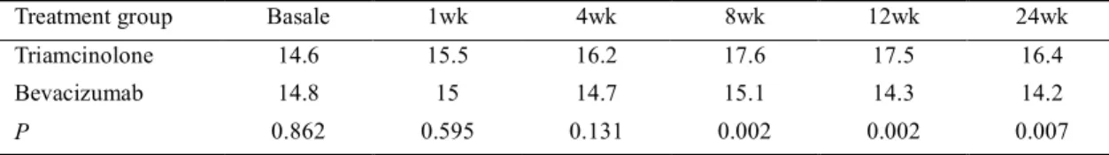
Average IOP values at baseline 14.6 mm Hg in the IVTA group, and 14.8 mm Hg in the IVB group and difference were not significant between the groups ( =0.862). One month after the injection, IOP in the IVTA group showed a statistically significant increase and then gradually increased with time ( =0.001). Four (20%) patients whose IOP values were higher than 21 mm Hg, treated with topical antiglaucomatous theraphy. In contrast, IOP in the bevacizumab-treated eyes did not show a statistically significant change during the clinical course. At 8, 12 and 24wk IOP in the IVTA group was significantly higher than the IVB group ( =0.002, =0.002, =0.007; Table 3). None of the patients who received IVB or IVTA, displayed a systemic side effect or an ocular complication associated with the applied invasive procedure. The development or progression of lens opacities was occurred in 4 (25%) eyes in the IVTA group. Two of four (50%) eyes that had second
Table 1 Alteration of logMAR visual acuity after each treatment in patients with bilateral DDME
Treatment group Basale 1wk 4wk 8wk 12wk 24wk Triamcinolone 0.71±0.19 0.62±0.23 0.63±0.12 0.63±0.13 0.63±0.14 0.61±0.24 Bevacizumab 0.68±0.25 0.61±0.22 0.60±0.24 0.62±0.25 0.65±0.26 0.59±0.25
P 0.237 0.055 0.247 0.142 0.294 0.206
Table 2 Alteration of central macular thickness after each treatment in patients with bilateral DDME
Treatment group Basale 1wk 4wk 8wk 12wk 24wk Triamcinolone 544±125 383±96 335±87 323±87 333±92 335±61 Bevacizumab 514±100 431±86 428±107 442±106 478±112 430±88
P 0.715 0.045 0.007 0.001 0.001 0.001
Table 3 Alteration of intraocular pressure after each treatment in patients with bilateral DDME
Treatment group Basale 1wk 4wk 8wk 12wk 24wk Triamcinolone 14.6 15.5 16.2 17.6 17.5 16.4 Bevacizumab 14.8 15 14.7 15.1 14.3 14.2
P 0.862 0.595 0.131 0.002 0.002 0.007
Figure 1 Comparison of the clinical course of central macular thickness between the triamcinolone acetonide injected eyes (dark line) and the bevacizumab injected eyes (light line) in patients with bilateral diffuse diabetic macular edema Each square and vertical bar indicates mean central macular thickness依 standard deviation (SD) of the mean.
栽藻造押8629原愿圆圆源缘员苑圆 8629-82210956 耘皂葬蚤造押ijopress岳员远猿援糟燥皂
IVTA injections developed lens opacities. No cataract surgery was performed during the follow-up period. Cataract progression has not been observed in the IVB group.
DISCUSSION
Breakdown of inner blood-retinal barrier due to retinal infiltration of inflammatory cells and biochemical alterations arising from chronic hyperglycemia, plays an important part in the pathogenesis of diabetic macular edema [11-13]. Focal macular edema can be controlled by laser photocoagulation, however, eyes with diffuse macular edema generally demonstrate a resistance against therapy. Among treatment strategies towards the pathogenesis of the disease, TA and bevacizumab, alone or in combination with laser photocoagulation, have produced successful results in the treatment of DME [ 6 , 7 , 14, 15]. In the studies by Chakrabarti [16]and Marey and Ellakwa [17], the response to therapy with bevacizumab showed superiority compared with triamcinolone for DME. However, these studies differed from that of Paccola [18], Isaac [19]and Lim [20], Song [21]who demonstrated that intravitreal triamcinolone was more efficient in reducing DME relative to bevacizumab. And in the other study by Rensch [22], IVTA and IVB did not differ markedly in term of their effects in improving VA and reducing macular thickness. Which treatment is more effective remains controversial. In these studies, efficacy of IVTA and IVB therapies have been compared by applying them on only one eye of different patients. Thus, in our study, by considering that the influence of systemic risk factors such as glycemic level, blood pressure, and nephropathy may alter the efficacy of the drugs in every individual, both eyes of the each patient received intravitreal treatment of both drugs with equal volume were compared. One of the potential limitation of this study is that, we used only the macular scan pattern of the HD-OCT. As we used just macular pattern of the HD-OCT instead of full field 3-D SD-OCT, extrafoveal vitreous tractions including vitreopapillary traction and extrafoveal traction membranes which is a common fenomen in DDME, might not be detected, as recently re-described by Ophir [23]. Thus, the therapeutic effect of the two medications could be different if all tractional components were excluded.
The most remarkable problem encountered in cases treated with intravitreal injections is the transient nature of the therapeutic effect and frequent recurrences. Pharmacokinetic data suggest a single intravitreal injection of 1.25 mg bevacizumab is effective for 6-7wk [24]. Kreutzer [25] suggested that a single triamcinolone injection may be as effective as a 3 injections of bevacizumab for the treatment of DME. Less number of injections of triamcinolone reduces injection-related complications and improve the patient compliance. In a Meta-analysis including 6 study which
compare the efficacy of the IVTA versus IVB in the treatment of DME shows IVTA is superior in improving VA at earlier follow-up (1mo and 3mo) and in reducing CMT at later follow -up (6mo) for DME [26]. In the current study, visual acuity was observed to rise at 1 and 4wk and remain stable at 8, 12, and 24wk in the IVTA group. Reduction ratios for mean CMT values were 29% at 1wk, 38% at 4wk, 40% at 8wk, 38% at 12wk, and 38% at 24wk. Thus, CMT demonstrated a tendency to increase starting from the 12wk and therefore 6 (30%) eyes received a second IVTA injection in the IVTA group.
In multicenter trial of Pan-American Collaborative Retina Study Group (PACORES), 139 eyes of 115 DDME patients received 1.25 mg and 2.5 mg IVB injection and the patients were followed-up for a mean period of 24mo. No difference was determined between the 1.25 and 2.5 mg doses with regard to efficacy. While 95% of patients required a second injection, 84% required a third injection, 36% required 6 injections, and 11% required 9 injections [6]. In the present study, visual acuity in the IVB group increased at 1 and 4wk, restarted to decrease at 8 and 12wk, and returned to the initial levels at 24wk. The reduction ratios of mean CMT were 16.4% at 1wk, 16.8% at 4wk, 14% at 8wk, 7% at 12wk, and 16% at 24wk. Thus, CMT demonstrated a tendency to increase starting from the 8th week and 15 (75%) eyes were subjected to second bevacizumab injection at 12wk. According to the literature and our results, the reduction in CMT after intravitreal bevacizumab injection, can be maintained only for about 1mo even with 2.5 mg injection, macular thickness begins to reincrease and require a second injection after 1mo.
A crossover effect of bevacizumab in the contralateral eye on proliferative diabetic retinopathy has been reported [7]. However pharmacokinetic studies of intravitreal bevacizumab injection revealed that only a very small amounts of bevacizumab or no changes on VEGF levels were detected in the contralateral eye [27,28]. And also in a PET/CT study on small animals with I-124 radiolabeled bevacizumab and ranibizumab showed that there were no significant escape of bevacizumab and ranibizumab from the vitreous cavity after intravitreal injection [29]. Although, it may be concluded that bevacizumab may have some or little effect on the contralateral eye, we did not establish a significant effect primary eye. This study actually showed that IVB does not have a beneficial effect in DDME and such edema may be would naturally adversely affect the posterior macula over time to irreversible changes.
The most common side effect observed after IVTA injection is, raised IOP and its incidence ranges between 20%-80% in various studies [30]. Considerable IOP increases are reported even in small case series. On the other hand, clinical studies
performed with bevacizumab have shown insignificant alterations in IOP levels. In the present study, IOP levels at 4, 8, and 12wk exhibited a statistically significant elevation in the IVTA group and remained stable at 24wk. However, IOP levels in the IVB group demonstrated no significant change. At routine doses of both drugs, bevacizumab may be more advantageous than triamcinolone with regard to IOP stability. There is only one study in the literature which compares triamcinolone acetonide and bevacizumab therapies in the same patients. In that study, Shimura [31]applied single dose of bevacizumab 1.25 mg one eye of each of 14 patients with bilateral persistant DME, while delivering single dose of triamcinolone acetonide 4 mg on the contralateral eye, after which the patients were followed-up for 24wk. Triamcinolone acetonide was found to provide more efficient and long-standing effect in terms of reducing CMT and increasing visual acuity compared with the bevacizumab. Bevacizumab the VEGF inhibitor which was used in our study as well, provided a reduction in macular edema 4wk after the injection, however, its effect was not as efficient as triamcinolone acetonide and macular edema relapsed at 12wk. While the reduction in CMT one week after the injection was 16% for the IVB group, it was 29% for the IVTA group. CMT reduction rate was 7% in the IVB group 12wk after the injection which is an evidence indicating a decrease in the effect of bevacizumab. On the other hand, CMT reduction rate among triamcinolone acetonide group was still 38% which shows the continuation of the influence of triamcinolone injection even after 12wk. At 12wk, 75% of IVB group and 30% of IVTA group received a second injection due to recurrence of macular edema.
Our results showed earlier and more frequent recurrences in the eyes treated with bevacizumab compared with the ones treated with triamcinolone. Therefore, in order to prolong the reductive effect, multiple injections of bevacizumab required. DME is not only associated with VEGF, but also with many growth factors and inflammatory cytokines. Bevacizumab reduces vascular permeability by blocking the influence of
VEGF. Triamcinolone acetonide decreases VEGF
expression, prevents fluid accumulation in the extracellular area, and inhibits release of edema-inducing inflammatory mediators. In this regard, triamcinolone is a multipotent drug and has more advantages when compared with bevacizumab which only reduces intraocular VEGF levels. In the current study, 6mo follow-up results of each drug regimen with the same volume and same patient were presented, however, studies with longer duration and larger sample size are required in order to further evaluate multiple dose-response relationship and safety of the drugs.
ACKNOWLEDGEMENTS
Conflicts of Interest: Aksoy S, None; Yilmaz G, None; Akkoyun I, None; Yazici AC, None.
REFERENCES
1 Klein R, Klein B, Moss S, Cruickshanks KC. The Wisconsin Epidemiologic Study of Diabetic Retinopathy. The long-term incidence of
macular edema. 1995;102(1):7-16
2 Ferris FL 3rd. The Early Treatment Diabetic Retinopathy Study Research
Group. Treatment techniques and clinical guidelines for photocoagulation of diabetic macular edema.
1987;94(7):761-774
3 Fong DS, Ferris FL 3rd, Davis MD, Chew EY. Causes of severe visual loss in the early treatment diabetic retinopathy study: ETDRS report no. 24. Early Treatment Diabetic Retinopathy Study Research Group.
1999;127(2):137-141
4 Striph GG, Hart WM Jr, Olk RJ. Modified grid laser photocoagulation for diabetic macular edema. The effect on the central visual field.
1988;95(12):1673-1679
5 Rutledge BK, Wallow IHL, Poulsen GL. Sub-pigment epithelial membrans after photocoagulation for diabetic macular edema.
1993;111(5):608-613
6 Arevalo JF, Sanchez JG, Wu L, Maia M, Alezzandrini AA, Brito M,
Bonafonte S, Lujan S, Diaz-Llopis M, Restrepo N, Rodr侏guez FJ,
Udaondo-Mirete P; Pan-American Collaborative Retina Study Group. Primary intravitreal bevacizumab for diffuse diabetic macular edema: The Pan-American Collaborative Retina Study Group at 24 months.
2009;116(8):1488-1497, 1497.e1
7 Avery RL, Pearlman J, Pieramici DJ, Rabena MD, Castellarin AA, Nasir MA, Giust MJ, Wendel R, Patel A. Intravitreal bevacizumab (Avastin) in
the treatment of proliferative diabetic retinopathy. 2006;113
(10):1695.e1-15
8 Haritoglou C, Kook D, Neubauer A, Wolf A, Priglinger S, Strauss R, Gandorfer A, Ulbig M, Kampik A. Intravitreal Bevacizumab (Avastin)
Therapy for Persistent Diffuse Diabetic Macular Edema. 2006;26(9):
999-1005
9 Jonas JB, Akkoyun I, Kreissig I, Degenring RF. Diffuse diabetic macular edema treated by IVTA: a comparative, non-randomised study.
2005;89(3):321-326
10 Jonas JB, Harder B, Kamppeter BA. Inter-eye difference in diabetic macular edema after unilateral injection of triamcinolone acetonide.
2004;138(6):970-977
11 Antcliff RJ, Marshall J. The pathogenesis of edema in diabetic
maculopathy. 1999;14(4):223-232
12 Joussen AM, Murata T, Tsujikawa A, Kirchhof B, Bursell SE, Adamis AP. Leukocyte-mediated endothelial cell injury and death in the diabetic
retin. 2001;158(1):147-152
13 Mizutani M, Kern TS, Lorenzi M. Accelerated death of retinal microvascular cells in human and experimental diabetic retinopathy.
1996;97(12):2883-2890
14 Martidis A, Duker JS, Greenberg PB, Rogers AH, Puliafito CA, Reichel E, Baumal C. Intravitreal Triamcinolone for Refractory Diabetic Macular
Edema. 2002;109(5):920-927
15 Sutter FK, Simpson JM, Gillies MC. Intravitreal triamcinolone for diabetic macular edema that persists after laser treatment: three month
栽藻造押8629原愿圆圆源缘员苑圆 8629-82210956 耘皂葬蚤造押ijopress岳员远猿援糟燥皂
efficacy and safety results of a prospective, randomized, double-masked,
placebo-controlled clinical trial. 2004;111(11): 2044-2049
16 Chakrabarti M, John SR, Chakrabarti A. Intravitreal monotherapy with bevacizumab (IVB) and triamcinolone acetonide (IVTA) versus combination therapy (IVB and IVTA) for recalcitrant diabetic macular edema.
2009;21(2):139-148
17 Marey HM, Ellakwa AF. Intravitreal bevacizumab alone or combined with triamcinolone acetonide as the primary treatment for diabetic macular
edema. 2011;5:1011-1016
18 Paccola L, Costa RA, Folgosa MS, Barbosa JC, Scott IU, Jorge R. Intravitreal triamcinolone versus bevacizumab for treatment of refractory
diabetic macular oedema (IBEME study). 2008;92 (1):
76-80
19 Isaac DL, Abud MB, Frantz KA, Rassi AR, Avila M. Comparing IVTA and bevacizumab injections for the treatment of diabetic macular oedema: a
randomized double-blind study. 2012;90(1):56-60
20 Lim JW, Lee HK, Shin MC. Comparison of intravitreal bevacizumab alone or combined with triamcinolone versus triamcinolone in diabetic
macular edema: a randomized clinical trial. 2012;227(2):
100-106
21 Song HJ, Lee JJ, Lee SJ. Comparison of the Short-Term Effects of IVTA and Bevacizumab Injection for Diabetic Macular Edema.
2011;25(3):156-160
22 Rensch F, Spandau UH, Wickenhauser A, Jonas JB. Diffuse diabetic macular oedema treated with intravitreal bevacizumab or triamcinolone
acetonide. 2010;88(2):e36-37
23 Ophir A. Full-field 3-D optical coherence tomography imaging and treatment decision in diffuse diabetic macular edema.
2014;55(5):3052-3053
24 Zhu Q, Ziemssen F, Henke-Fahle S, Tatar O, Szurman P, Aisenbrey S, Schneiderhan-Marra N, Xu X, Grisanti S. Vitreous levels of bevacizumab and vascular endothelial growth factor-A in patients with choroidal
neovascularization. 2008;115(10):1750-1755
25 Kreutzer TC, Al Saeidi R, Kook D, Wolf A, Ulbig MW, Neubauer AS, Haritoglou C. Comparison of intravitreal bevacizumab versus triamcinolone for the treatment of diffuse diabetic macular edema.
2010;224(4):258-264
26 Zhang XL, Chen J, Zhang RJ, Wang WJ, Zhou Q, Qin XY. Intravitreal triamcinolone versus intravitreal bevacizumab for diabetic macular edema:
a meta-analysis. 2013;6(4):546-552
27 Bakri SJ, Snyder MR, Reid JM, Pulido JS, Singh RJ. Pharmacokinetics
of intravitreal bevacizumab (Avastin). 2007;114 (5):
855-859
28 Sawada O, Kawamura H, Kakinoki M, Ohji M. Vascular endothelial growth factor in fellow eyes of eyes injected with intravitreal bevacizumab.
2008 Oct;246(10):1379-1381 29 Christoforidis JB, Carlton MM, Knopp MV, Hinkle GH. PET/CT imaging of I-124 radiolabeled bevacizumab and ranibizumab after intravitreal
injection in a rabbit model. 2011;52 (8):
5899-5903
30 Kiddee W, Trope GE, Sheng L, Beltran-Agullo L, Smith M, Strungaru MH, Baath J, Buys YM. Intraocular pressure monitoring post-intravitreal
steroids:a systematic review. 2013;58(4):291-310
31 Shimura M, Nakazawa T, Yasuda K, Shiono T, Lida T, Sakamoto T, Nishida K. Comparative therapy evaluation of intravitreal bevacizumab and triamcinolone acetonide on persistent diffuse diabetic macular edema.