Address for correspondence: Dr. Mehmet Vedat Çaldır, Başkent Üniversitesi Konya Hastanesi Kardiyoloji Bölümü. Hocacihan Mah., Saray Cad., No: 1, Selçuklu, Konya-Türkiye
Phone: +90 332 257 06 06/2116 E-mail: [email protected] Accepted Date: 09.11.2016 Available Online Date: 01.12.2016
©Copyright 2017 by Turkish Society of Cardiology - Available online at www.anatoljcardiol.com DOI:10.14744/AnatolJCardiol.2016.7425
Mehmet Vedat Çaldır, Güner Koyuncu Çelik
1, Özgür Çiftçi
2, İbrahim Haldun Müderrisoğlu*
Department of Cardiology, Faculty of Medicine, Başkent University, Konya; Ankara*-Turkey
1Department of Norology, Beyhekim State Hospital; Konya-Turkey 2Department of Cardiology, Koru Private Hospital; Ankara-Turkey
The effect of high-dose steroid treatment used for the treatment of
acute demyelinating diseases on endothelial and cardiac functions
Introduction
Multiple sclerosis (MS) and neuromyelitis optica (NMO) are inflammatory and demyelinating central nervous system (CNS) diseases characterized by acute exacerbations and remissions. They frequently affect young adults, and their disability rates are high (1). The brain, brain stem, cerebellum, spinal cord, and optic nerves can be involved either individually or combined, accor- ding to the subtypes of the disease. High-dose steroid treatment is frequently preferred in the acute exacerbation periods of the diseases regardless of the subtypes (2, 3). Recently, steroids have been successfully used not only in neurological diseases but also in acute exacerbations of renal diseases, musculoskele- tal system disorders, collagen vascular diseases, and dermato-logical and hematodermato-logical diseases (4, 5). Some early side effects are fluid retention, weight gain, high blood pressure, and mood changes (6). In long-term steroid use, cataracts, bone changes
(osteoporosis or osteonecrosis), hypertension, lipid abnormali-ties, diabetes and adrenal suppression, and diabetes suscepti-bility can develop (7–9).
It is known that vascular function is deteriorated and that premature atherosclerosis is triggered after long-term steroid treatment for some chronic inflammatory diseases such as rheu-matoid arthritis (10, 11). Furthermore, it has also been reported that sinus bradycardia, sinus tachycardia, and rhythm problems such as conduction system blocks or atrial fibrillation in the acute period can develop with high-dose steroid treatment (12, 13). Accelerated atherosclerosis and cardiovascular mortality increase in Cushing’s disease patients exposed to long-term high-dose steroid treatment. However, it has been shown that short-term high-dose steroid treatment does not lead to the de-terioration of endothelial function or increase in the carotid in-tima–media thickness (cIMT) in healthy adults (14, 15). There are a limited number of studies in which the cardiovascular effects
Objective: The cardiovascular effects of short-term high-dose steroid treatment (pulse steroid treatment) have not yet been clarified. We exa- mined the short- and long-term effects of pulse steroid treatment in demyelinating diseases on endothelial and cardiac functions.
Methods: In this prospective study, we included 35 patients (20 females and 15 males; mean age, 32.8±9.3 years) who were not treated with steroids and who were previously diagnosed with multiple sclerosis or neuromyelitis optica. Patients were evaluated before, 1 week after, and 3 months after the steroid treatment. Brachial artery flow-mediated relaxation and cardiac systolic/diastolic function were evaluated using echocardiography to assess physical examination results, carotid intima–media thickness, and endothelial function.
Results: There was no difference between biochemical values, systolic function, left ventricular dimensions, and carotid intima–media thick-nesses in the three evaluation periods. There were significant increases in the body mass index, body weight, and systolic/diastolic blood pressure measurements at 1 week and 3 months after treatment (p<0.001). There was a significant decrease in brachial artery flow-mediated relaxation at 1 week and 3 months (1 versus 2, p=0.042; 1 versus 3, p=0.003). In Doppler measurements at 1 week and 3 months, there was an increase in mitral A velocity, IVRT, and EDT values and a decrease in the E/A ratio in line with diastolic dysfunction.
Conclusion: Pulse steroid therapy used for demyelinating diseases deteriorated endothelial and left ventricular diastolic functions in the early and late periods. Future studies are needed to evaluate the development of cardiovascular mortality and morbidity in patients receiving this type of treatment. (Anatol J Cardiol 2017; 17: 392-7)
Keywords: demyelinating diseases, diastolic function, endothelial dysfunction, pulse steroid
A
BSTRACTof short-term high-dose steroid treatment have been examined. Therefore, in this study, we examined the effects of high-dose steroid treatment in acute demyelinating diseases on endothe-lial and cardiac functions in the early and late periods.
Methods
Study design
This was a single-center, prospectively designed, observa-tional study. This study was approved by Clinical Research and Ethical Committee (project no: KA15/351).
Study group
In total, 35 consecutive patients (20 females and 15 males; mean age, 32.8±9.3 years) who did not receive any steroid treatment for MS according to the McDonald criteria (16) or for NMO according to the Wingerchuk criteria (17) were included in 6 months. Patients who had hypertension, diabetes mellitus, hyperlipidemia, chro- nic renal failure, cardiac insufficiency, collagen vascular dis-ease, congenital heart disdis-ease, acquired valve disdis-ease, or atrial fibrillation were excluded. Furthermore, patients who smoked, who were obese [body mass index (BMI) >30], who were preg-nant, who may be pregpreg-nant, who were using contraceptives, who were in the menstruation or luteal phase, who were not in the age group of 18 and 50 years, and who previously received steroids were excluded.
Study protocol
Patients were evaluated in three different periods: before high-dose steroid treatment (for 5 days, everyday; 1 g of methyl- prednisolone and 2 h of intravenous infusion in 5% dextrose so-lution), 1 week after the end of treatment (early phase), and 3 months after the end of treatment (late phase). In each evalu-ation period, physical examinevalu-ations were performed, and the blood pressures of patients were recorded. Furthermore, height and weight measurements were determined, and the BMIs of patients were calculated. Venous blood samples were collected after a 12-h starving period to measure the blood glucose, li- pid, creatinine, high-sensitivity C-reactive protein (hs-CRP), and whole blood levels. The brachial artery flow-mediated dilatation (FMD) test was performed to indirectly evaluate endothelial func-tion. cIMT was assessed using Doppler ultrasound. The systolic and diastolic functions of the heart were echocardiographically evaluated. All these measurements were performed by the same cardiologist who was unaware of the measurement period and laboratory findings of patients. High-dose steroid treatment was given to patients after basal evaluation. This steroid treatment was not performed before the two following evaluations.
Brachial artery FMD and cIMT measurement
Dilatation after transient ischemia of the brachial artery was ensured as it was previously defined (18). Measurements were performed using a 12-MHz linear vascular probe of a General
Electric Vivid S6 (GE Healthcare, Chicago, USA) model echocar-diography device. The brachial artery was longitudinally visu-alized 3–5 cm above the antecubital fossa. The mean of three separate measurements of the basal systolic and diastolic diameters of the inner vessel were recorded. Then, a sphyg-momanometer cuff was placed proximal to the section of the brachial artery and was inflated to 50 mm Hg higher than the systolic blood pressure. Systolic and diastolic diameters of the brachial artery were measured again 30 and 60 s after remo- ving the cuff at the end of the 5 min of ischemia. The difference between the baseline and hyperemic values were calculated as a percentage change.
Carotid Doppler ultrasound was performed as it was pre- viously described (19). Measurements were performed from the posterior wall and 10 mm proximal to the left and right carotid bifurcation. Both carotid measurements were repeated three times. The mean of the measurements was recorded as IMT.
Echocardiographic examination
All patients were examined using 3V2c transthoracic probe of a Sequoia C256 (Siemens Healthcare GmbH, Germany) echo-cardiography device. Measurements were performed from apical parasternal windows in the lateral decubitus position. Two-dimensional, M-mode, pulsed-wave (PW) Doppler and tis-sue Doppler methods were used. Systolic and diastolic inter-ventricular septum (IVS) thicknesses, posterior wall thickness, left ventricular end-systolic and end-diastolic diameters, aor-tic systolic and diastolic diameters, and diastolic diameter of the left atrium were measured from the parasternal long axis using the M-mode. Ejection fraction (EF) was calculated u- sing the Teicholz method (20), and the left ventricular mass was calculated using the Devereux formula (21). The left ventricular mass index was calculated by dividing the left ventricular mass with the body surface area. The early diastolic flow velocity (E), late diastolic flow velocity (A), E/A ratio, E wave decelera-tion time (EDT), and isovolumetric relaxadecelera-tion time (IVRT) were measured by placing the pulse Doppler sample volume on the tip of the mitral valve. We switched to the tissue Doppler mode to measure the regional myocardial velocity, and the gain was reduced to a minimum optimal level. The Doppler velocity range was set within the range of –30 and 30cm/s. The PW sample volume was placed at the level of the lateral wall mitral annulus and in the basal IVS in the apical four-chamber window. Early (Em) and late (Am) diastolic flow velocities and IVRT of both areas were calculated.
Statistical analysis
SPSS 15.0 (SPSS for Windows 15.0, Chicago, IL) was used for all statistical analyses. According to the power analysis, the minimum patient number was 15 (with 95% power). The variables were shown as mean±standard deviation. Pairwise comparisons of basal, 1-week, and 3-month findings were performed using a paired t-test. p<0.05 was accepted to be statistically significant.
Results
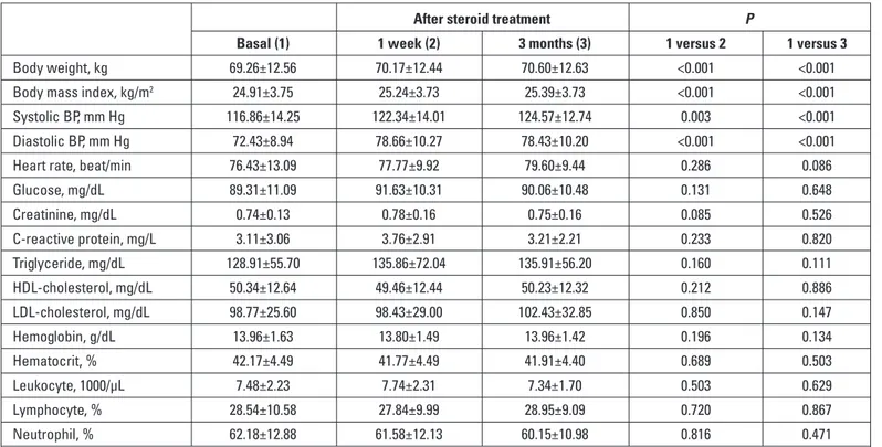
The general characteristics and laboratory findings of pa-tients during the evaluation periods can be seen in Table 1. There was no difference between the heart rate; glucose, creatinine, hs-CRP, and serum lipid levels; and complete blood counts of pa-tients. The basal body weight and mean BMI of patients were 69.26±12.56 and 24.91±3.75, respectively. However, these values were 70.17±12.44 kg and 25.24±3.73, kg/m2 respectively, at 1 week
after treatment and 70.60±12.63 and 25.39±3.73, respectively, at 3 months after. The increase in the basal body weight and mean BMI at 1 week and 3 months after was significant (p<0.001) (Fig. 1a). Basal systolic and diastolic blood pressures were 116.86±14.25 mm Hg and 72.43±8.94. The systolic and diastolic blood pressures at 1 week after treatment were 122.34±14.01 and 78.66±10.27 mm Hg, respectively, and the corresponding pressures at 3 months after treatment were 124.57±12.74 and 78.43±10.20. According to the systolic and diastolic blood pres-sure meapres-surements, there was a significant increase in the val-ues at 1 week and 3 months after treatment compared to the basal values (for the systolic blood pressure: 1 versus 2, p=0.003 and 1 versus 3, p<0.001, respectively; for diastolic blood pres-sure, for both measurements, p<0.001) (Fig. 1b).
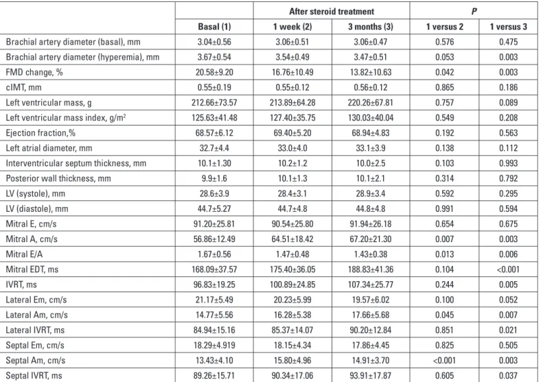
As a result of the comparison of the echocardiographic and Doppler findings at 1 week and 3 months after treatment with the basal values, we found that there was no significant decrease in the hyperemic brachial artery diameter at 1 week, whereas the reduction in the values at 3 months was significant (1 versus 2, p=0.576; 1 versus 3, p=0.003, respectively). On the other hand,
there was a significant decrease in the FMD in both values at 1 week and 3 months after treatment compared to basal values (1 versus 2, p=0.042; 1 versus 3, p=0.003, respectively) (Fig. 1c). There was no difference among the three measurements in terms of the cIMT (Fig. 1d), left ventricular mass, left ventricu-lar mass index, EF, cardiac chamber size, and wall thickness. According to the conventional and tissue Doppler evaluations, there was a significant increase in the mitral A velocities values at 1 week and 3 months after treatment (1 versus 2, p=0.007; 1 versus 3, p=0.003) (Fig. 2a), whereas there was a significant dec- rease in the mitral E/A ratio (Fig. 2b). There was a significant in-crease in the mitral E deceleration time and IVRT at 3 months after treatment (Fig. 2c, d). Similarly, we observed an increase in lateral Am and septal Am wave velocities. There was a sig-nificant prolongation particularly in the IVRT of both walls at 3 months (Table 2).
Discussion
To our knowledge, this study is the first in which the effects of high-dose steroid treatment on endothelial and cardiac func-tions were examined in demyelinating CNS diseases. In this study, we observed that high-dose steroid treatment negatively affected endothelial function and left ventricular diastolic func-tion in the early and late periods. However, there was no diffe- rence in the cIMT.
Steroids lead to an increase in angiotensin II type 1 receptors and α-1 receptors and a decrease in prostacyclin E2 synthesis in endothelial and vascular smooth muscle cells in experimental
Table 1. Demographic characteristics and laboratory results
After steroid treatment P
Basal (1) 1 week (2) 3 months (3) 1 versus 2 1 versus 3
Body weight, kg 69.26±12.56 70.17±12.44 70.60±12.63 <0.001 <0.001
Body mass index, kg/m2 24.91±3.75 25.24±3.73 25.39±3.73 <0.001 <0.001
Systolic BP, mm Hg 116.86±14.25 122.34±14.01 124.57±12.74 0.003 <0.001
Diastolic BP, mm Hg 72.43±8.94 78.66±10.27 78.43±10.20 <0.001 <0.001
Heart rate, beat/min 76.43±13.09 77.77±9.92 79.60±9.44 0.286 0.086
Glucose, mg/dL 89.31±11.09 91.63±10.31 90.06±10.48 0.131 0.648 Creatinine, mg/dL 0.74±0.13 0.78±0.16 0.75±0.16 0.085 0.526 C-reactive protein, mg/L 3.11±3.06 3.76±2.91 3.21±2.21 0.233 0.820 Triglyceride, mg/dL 128.91±55.70 135.86±72.04 135.91±56.20 0.160 0.111 HDL-cholesterol, mg/dL 50.34±12.64 49.46±12.44 50.23±12.32 0.212 0.886 LDL-cholesterol, mg/dL 98.77±25.60 98.43±29.00 102.43±32.85 0.850 0.147 Hemoglobin, g/dL 13.96±1.63 13.80±1.49 13.96±1.42 0.196 0.134 Hematocrit, % 42.17±4.49 41.77±4.49 41.91±4.40 0.689 0.503 Leukocyte, 1000/µL 7.48±2.23 7.74±2.31 7.34±1.70 0.503 0.629 Lymphocyte, % 28.54±10.58 27.84±9.99 28.95±9.09 0.720 0.867 Neutrophil, % 62.18±12.88 61.58±12.13 60.15±10.98 0.816 0.471
models. Consequently, the blood pressure increases (22). It has also been observed that steroids lead to the in vitro production of superoxide radicals in endothelial cells in addition to their well-known metabolic effects (23). Further, in our study, signifi-cant early and late weight gains and increased BMIs and blood
pressures were observed after high-dose steroid treatment. All these mechanisms may be related to the prominent deteriora-tion of brachial artery FMD, which is an important indicator of endothelial dysfunction detected after high-dose steroid treat-ment. Nevertheless, an improvement in FMD has been reported
Body mass index (kg/m
2)
Flow mediated dilatation (%)
Systolic BP (mm Hg) Carotid IMT (mn)
a
c
b
d
30.00 50.00 40.00 30.00 20.00 10.00 0.00 150.00 1.00 0.80 0.60 0.40 140.00 130.00 120.00 110.00 100.00 90.00 27.00 24.00 21.00 18.00 Basal Basal Basal Basal First week First week First week First week Time Time Time Time Third month Third month Third month Third month P<0.001 P:0.003 NS NS P<0.001 P<0.001 P:0.003 P:0.042Figure 1. Body mass index (a), systolic blood pressure (BP) (b), flow-mediated dilatation (c), carotid intima–media thickness (cIMT) (d), and basal, first week, and third month values
Mitral A v
elocity (cm/s)
Mitral E deceleration time (ms)
Mitral E/A
Isov
olumetric relaxation time (ms)
a
c
b
d
125.00 300.00 250.00 200.00 150.00 100.00 100.00 75.00 50.00 25.00 3.50 175.00 150.00 125.00 100.00 75.00 3.00 2.50 2.00 1.50 1.00 0.50 Basal Basal Basal Basal First week First week First week First week Time Time Time Time Third month Third month Third month Third month P:0.003 P:0.013 P:0.006 P:0.007 P<0.001 P:0.005 NS NSFigure 2. Mitral A velocity (a), mitral E/A ratio (b), mitral E deceleration time (c), mitral isovolumetric relaxation time (d), and basal, first week, and third month values
with steroid treatment in patients with inflammatory vascular diseases as giant cell arteritis (24).
Vascular endothelium homeostasis changes are observed in the development of atherosclerosis, which is a complex and multi-factorial process. It is accepted that the FMD of the deterio-rated brachial artery is an indicator of endothelial dysfunction. It is also an early sign of atherosclerosis because they have common pathophysiological processes (25, 26). In our study, we examined the cIMT to evaluate atherosclerosis development. However, we did not detect any changes in the early or late periods. We believe that this was because our study was performed in a relatively short time period. There should be long-term studies where we can eval-uate these patients in terms of atherosclerosis development.
It has been shown that there was deterioration in diastolic function and an increase in the left ventricular mass depending on increased blood steroid levels in Cushing's disease patients. Furthermore, it was related to increased cardiovascular morta- lity (27). In this study, there were findings in line with diastolic dysfunction in mitral flow and tissue Doppler velocities. Diastolic dysfunction is accepted as the earliest finding in heart failure,
and the myocardial response depends on the coronary artery disease. Heart failure with preserved EF and deteriorated dias- tolic function is similar to heart failure with low EF. A diagnosis of diastolic dysfunction is very important because it is difficult to diagnose and is very easy to be ignored (28). In line with these findings, it can be stated that undesirable major cardiovascular events can develop in patients after high-dose steroid treatment.
Study limitations
The most important limitation of this study is the low number of patients. It was difficult to find a greater number of patients who complied with the inclusion and exclusion criteria.
Patients were evaluated for a period of 3 months. However, more time may be required to examine the cIMT and left ventri- cular mass increase. Additional radiological examinations such as cardiac magnetic resonance imaging could provide more ac-curate results. Furthermore, studies with long follow-up periods of pulse steroid treatment might show no adverse effect on ED and diastolic dysfunction due to spontaneous remission.
Table 2. Echocardiographic and Doppler measurement results
After steroid treatment P
Basal (1) 1 week (2) 3 months (3) 1 versus 2 1 versus 3
Brachial artery diameter (basal), mm 3.04±0.56 3.06±0.51 3.06±0.47 0.576 0.475
Brachial artery diameter (hyperemia), mm 3.67±0.54 3.54±0.49 3.47±0.51 0.053 0.003
FMD change, % 20.58±9.20 16.76±10.49 13.82±10.63 0.042 0.003
cIMT, mm 0.55±0.19 0.55±0.12 0.56±0.12 0.865 0.186
Left ventricular mass, g 212.66±73.57 213.89±64.28 220.26±67.81 0.757 0.089
Left ventricular mass index, g/m2 125.63±41.48 127.40±35.75 130.03±40.04 0.549 0.208
Ejection fraction,% 68.57±6.12 69.40±5.20 68.94±4.83 0.192 0.563
Left atrial diameter, mm 32.7±4.4 33.0±4.0 33.1±3.9 0.138 0.112
Interventricular septum thickness, mm 10.1±1.30 10.2±1.2 10.0±2.5 0.103 0.993
Posterior wall thickness, mm 9.9±1.6 10.1±1.3 10.1±2.1 0.314 0.792
LV (systole), mm 28.6±3.9 28.4±3.1 28.9±3.4 0.592 0.295 LV (diastole), mm 44.7±5.27 44.7±4.8 44.8±4.8 0.991 0.594 Mitral E, cm/s 91.20±25.81 90.54±25.80 91.94±26.18 0.654 0.675 Mitral A, cm/s 56.86±12.49 64.51±18.42 67.20±21.30 0.007 0.003 Mitral E/A 1.67±0.56 1.47±0.48 1.43±0.38 0.013 0.006 Mitral EDT, ms 168.09±37.57 175.40±36.05 188.83±41.36 0.104 <0.001 IVRT, ms 96.83±19.25 100.89±24.85 107.34±25.77 0.244 0.005 Lateral Em, cm/s 21.17±5.49 20.23±5.99 19.57±6.02 0.100 0.052 Lateral Am, cm/s 14.77±5.56 16.28±5.38 17.66±5.68 0.045 0.007 Lateral IVRT, ms 84.94±15.16 85.37±14.07 90.20±12.84 0.851 0.021 Septal Em, cm/s 18.29±4.919 18.15±4.34 17.86±4.45 0.825 0.505 Septal Am, cm/s 13.43±4.10 15.80±4.96 14.91±3.70 <0.001 0.003 Septal IVRT, ms 89.26±15.71 90.34±17.06 93.91±17.87 0.605 0.037
EDT - E wave deceleration time; FMD - flow-mediated dilatation; cIMT - carotid intima–media thickness; IVRT - isovolumetric relaxation time; LV - left ventricle. A paired t-test was used for statistical analyses
Conclusion
In our study, we detected significant weight gain, increase in blood pressure, left ventricular diastolic dysfunction, and en-dothelial dysfunction as a result of pulse steroid treatment in patients who did not have cardiovascular risk factors. However, we did not detect an increase in the left ventricular mass and an increase in the cIMT that is accepted as an early indicator of atherosclerosis.
Consequently, patients who required pulse steroid adminis-tration should be closely monitored in terms of cardiovascular mortality and morbidity, and further investigations are required.
Conflict of interest: None declared. Peer-review: Externally peer-reviewed.
Authorship contributions: Concept – M.V.Ç.; Design – M.V.Ç., G.K.Ç., Ö.Ç.; Supervision – İ.H.M.; Fundings – G.K.Ç.; Materials – M.V.Ç., G.K.Ç.; Data collection &/or processing – M.V.Ç., G.K.Ç., Ö.Ç.; Analysis and/ or interpretation – M.V.Ç.; Literature review – M.V.Ç.; Writing- M.V.Ç., G.K.Ç.; Critical review – İ.H.M., Ö.Ç.
References
1. Murray TJ. Diagnosis and treatment of multiple sclerosis. BMJ 2006; 332: 525-7. [CrossRef]
2. Shaygannejad V, Ashtari F, Alinaghian M, Norouzi R, Salari M, Fate-hi F. Short-term safety of pulse steroid therapy in multiple sclerosis relapses. Clin Neuropharmacol 2013; 36: 1-3. [CrossRef]
3. Arnold AC. Evolving management of optic neuritis and multiple sclerosis. Am J Ophthalmol 2005; 139: 1101-8. [CrossRef]
4. Funauchi M, Ikoma S, Imada A, Kanamaru A. Combination of im-munoadsorption therapy and high-dose methylprednisolone in patients with lupus nephritis; possible indications in patients with early stage. J Clin Lab Immunol 1997; 49: 47-57.
5. Werth VP. Treatment of pemphigus vulgaris with brief, high-dose intravenous glucocorticoids. Arch Dermatol 1996; 132: 1435-9. 6. Moreland LW, O'Dell JR. Glucocorticoids and rheumatoid arthritis:
back to the future? Arthritis Rheum 2002; 46: 2553-63. [CrossRef]
7. Tyblova M, Kalincik T, Zikan V, Havrdova E. Impaired ambulation and steroid therapy impact negatively on bone health in multiple sclerosis. Eur J Neurol 2015; 22: 624-32. [CrossRef]
8. Zimmerman J, Fainaru M, Eisenberg S. The effects of prednisone therapy on plasma lipoproteins and apolipoproteins: a prospective study. Metabolism 1984; 33: 521-6. [CrossRef]
9. Reaven GM, Lithell H, Landsberg L. Hypertension and associated metabolic abnormalities: the role of insulin resistance and the sym-pathoadrenal system. N Engl J Med 1996; 334: 374-81. [CrossRef]
10. Raynauld JP. Cardiovascular mortality in rheumatoid arthritis: how harmful are corticosteroids? J Rheumatol 1997; 24: 415-6.
11. del Rincón I, O'Leary DH, Haas RW, Escalante A. Effect of glucocor-ticoids on the arteries in rheumatoid arthritis. Arthritis Rheum 2004; 50: 3813-22. [CrossRef]
12. Liu D, Ahmet A, Ward L, Krishnamoorthy P, Mandelcorn ED, Leigh R, et al. A practical guide to the monitoring and management of the
complications of systemic corticosteroid therapy. Allergy Asthma Clin Immunol 2013; 9: 30. [CrossRef]
13. Vasheghani-Farahani A, Sahraian MA, Darabi L, Aghsaie A, Minagar A.Incidence of various cardiac arrhythmias and conduc-tion disturbances due to high dose intravenous methylpredniso-lone in patients with multiple sclerosis. Neurol Sci 2011; 309: 75-8. 14. Faggiano A, Pivonello R, Spiezia S, De Martino MC, Filippella M, Di
Somma C, et al. Cardiovascular risk factors and common carotid artery caliber and stiffness in patients with Cushing’s disease dur-ing active disease and 1 year after disease remission. J Clin Endo-crinol Metab 2003; 88: 2527-33. [CrossRef]
15. Brotman DJ, Girod JP, Garcia MJ, Patel JV, Gupta M, Posch A, et al. Effects of short-term glucocorticoids on cardiovascular biomark-ers. J Clin Endocrinol Metab 2005; 90: 3202-8. [CrossRef]
16. McDonald WI, Compston A, Edan G, Goodkin D, Hartung HP, Lub-lin FD, et al. Recommended Diagnostic Criteria for MS. Ann Neurol 2001; 50: 121-7. [CrossRef]
17. Wingerchuk DM, Banwell B, Bennett JL, Cabre P, Carroll W, Chitnis T, et al. International consensus diagnostic criteria for neuromyeli-tis optica spectrum disorders. International Panel for NMO Diagno-sis. Neurology 2015; 85: 177-89. [CrossRef]
18. Corretti MC, Anderson TJ, Benjamin EJ, Celermajer D, Charbon-neau F, Creager MA, et al. Guideline for the ultrasound assessment of endothelium-dependent flow-mediated vasodilatation of the brachial artery. J Am Coll Cardiol 2002; 39: 257-65. [CrossRef]
19. O’Leary DH, Polak JF, Kronmal RA, Manolio TA, Burke GL, Wolfson Jr SK. Carotid artery intima and media thickness as a risk factor of myocardial infarction and stroke in older adults. N Eng J Med 1999; 340: 14-22. [CrossRef]
20. Teichholz LE, Kreulen T, Herman MV, Gorlin R. Problems in echo-cardiographic volume determinations: echoecho-cardiographic-angio- echocardiographic-angio-graphic correlations in the presence of absence of asynergy. Am J Cardiol 1976; 37: 7-11. [CrossRef]
21. Devereux RB, Reichek N. Echocardiographic determination of left ventricular mass in man. Anatomic validation of the method. Circu-lation 1977; 55: 613-8. [CrossRef]
22. Ullian ME. The role of corticosteriods in the regulation of vascular tone. Cardiovasc Res 1999; 41: 55-64. [CrossRef]
23. Iuchi T, Akaike M, Mitsui T, Ohshima Y, Shintani Y, Azuma H, et al. Glucocorticoid excess induces superoxide production in vascular endothelial cells and elicits vascular endothelial dysfunction. Circ Res 2003; 92: 81-7. [CrossRef]
24. Gonzalez-Juanatey C, Llorca J, Garcia-Porrua C, Sanchez-Andrade A, Martín J, Gonzalez-Gay MA. Steroid therapy improves endothe-lial function in patients with biopsy-proven giant cell arteritis. J Rheumatol 2006; 33: 74-8.
25. Heitzer T, Schlinzig T, Krohn K, Meinertz T, Münzel T. Endothelial dysfunction, oxidative stress, and risk of cardiovascular events in patients with coronary artery disease. Circulation 2001; 104: 2673-8. 26. Verma S, Buchanan MR, Anderson TJ. Endothelial function testing
as a biomarker of vascular disease. Circulation 2003; 108: 2054-9. 27. Muiesan ML, Lupia M, Salvetti M, Grigoletto C, Sonino N, Boscaro
M, et al. Left ventricular structural and functional characteristics in Cushing's syndrome. J Am Coll Cardiol 2003; 41: 2275-9. [CrossRef]
28. Steinberg BA, Zhao X, Heidenreich PA, Peterson ED, Bhatt DL, Can-non CP, et al. Get with the Guidelines Scientific Advisory Committee and Investigators. Trends in patients hospitalized with heart failure and preserved left ventricular ejection fraction: prevalence, thera-pies, and outcomes. Circulation 2012; 126 :65-75. [CrossRef]