157
Nerve conduction study plays a key role in the correct
diagnosis of HNPP
Reha Kuruoğlu1 , Kübra Mehel Metin2 , Bülent Cengiz1
1Department of Neurology and Clinical Neurophysiology, Gazi University School of Medicine, Ankara, Turkey 2Clinic of Neurology, Ankara Training and Research Hospital, Ankara, Turkey
Corresponding Author: Reha Kuruoğlu E-mail: [email protected] Submitted: 11 June 2017 Accepted: 20 July 2017
You may cite this article as: Kuruoğlu R, Mehel Metin K, Cengiz C. Nerve conduction study plays a key role in the correct diagnosis of HNPP. Neurol Sci Neurophyisol 2018; 35(4): 157-164.
Abstract
Objective: As nerve conduction study (NCS) is a commonly employed tool in the diagnosis of hereditary neuropathy with
liability to pressure palsies (HNPP), we describe the electrophysiological features of our patients, comparing the findings with those of healthy subjects.
Methods: The charts of 19 HNPP patients were reviewed and NCS including residual latency (RL) and terminal latency index
(TLI) were compared with the findings of 32 control subjects.
Results: Motor NCS revealed significant differences (p<0.05) in all variables except for the tibial distal latency, median
com-pound muscle action potential amplitude, ulnar and peroneal TLI, and tibial RL in the patient cohort compared with healthy controls. Tibial TLI was bigger in the patient cohort (p<0.05). Nerve conduction velocity (NCV) slowing of the knee-ankle seg-ments were more marked compared with the elbow-wrist segseg-ments (p<0.05). NCV of the ulnar nerve was significantly slower in the elbow segment, compared with the below elbow-wrist and axilla-above elbow segments (p=0.000). F-responses were pro-longed in the lower extremities and RL prolongations were especially prominent in the median nerve (p<0.05). TLI values were smaller in the median nerve, compared with all other nerves (p<0.05). Sensory NCS were notably abnormal in the patient cohort compared with the healthy controls (p<0.001). However, the sural compound nerve action potential amplitude was markedly enlarged compared with the upper extremity nerves (p<0.01).
Conclusion: Correct interpretation of the NCS findings plays an essential role in the diagnosis and rational use of mutation
analysis in suspected cases.
Keywords: Hereditary neuropathy with liability to pressure palsies, hereditary neuropathy, polyneuropathy, nerve conduction
studies
INTRODUCTION
Hereditary neuropathy with liability to pressure palsies (HNPP) is an autosomal dominant demyelinating neurop-athy, characterized by recurrent painless mononeuropathies at entrapment sites, following minor trauma or acute compression (1-5). While a heterozygous deletion is present at the chromosome site 17p11.2 encoding peripheral myelin protein (PMP22) gene in 85% of the affected individuals, a point mutation is responsible in approximately 15% (2, 3, 6-8). Nerve conduction studies demonstrate multifocal or segmental abnormalities at compression sites or distal segments of the peripheral nerves (1). In addition to electrodiagnostic investigation, histopathological and genetic tests that have an important place in diagnosis, sonographic and magnetic resonance imaging of compression sites may be of value as complementary diagnostic tools (2,5,7). The electrodiagnostic findings reveal a generalized slowing of the sensory nerve conduction velocities (NCV), reduction of the sensory nerve action po-tential amplitudes, prolongation of terminal latencies and focal slowing at multiple compression sites, while motor NCVs are mildly slowed (5, 7, 9, 10). Because the clinical picture and evolution of the disease is quite heterogeneous, atypical phenotypes including asymptomatic cases have been reported in the literature (1, 2, 7).
Although the diagnosis is important for prognosis and genetic counseling, it can remain elusive (1). Family history may not always be helpful due to the presence of sporadic cases. Affected individuals can be asymptomatic, with-out obvious neurological findings (1, 11). Additionally atypical forms may be encountered such as entrapment
158
and acute or chronic demyelinating neuropathies (2). Electro-diagnostic studies are important in this setting, being read-ily available, to exclude other possible causes, as well as to identify the asymptomatic and atypical cases (1). However a certain degree of experience is required to interpret the nerve conduction findings. In this report we aimed to present our experience and describe the clinical and electrophysiological features of our patients, comparing their electrophysiological findings with those of healthy subjects.
METHODS
Patients and Controls
The charts of 19 HNPP (11 female, 8 male) patients, ranging from 16 to 58 years of age with a mean age of 36.5 years, eval-uated at the EMG laboratory were retrieved and compared with the findings of 32 healthy control subjects (23 female, 9 male) ranging from 19-68 years of age with a mean age of 36.5 years. There was no statistically significant difference in terms of age (t=-0.009, p=0.993) and gender (chi-square=1.049, p=0.306) between the patient and control groups. Typical HNPP cases presented with acute compression neuropathies and brachial plexopathy. Chronic entrapment neuropathies in the form of carpal tunnel syndrome or ulnar neuropathy at the elbow, polyneuropathy either acute or chronic, fatigue, cramps, nonspecific symptoms such as pain and
asymptomat-ic individuals were considered as atypasymptomat-ical phenotypes. Other causes of peripheral neuropathy were ruled out by appropri-ate laboratory studies, including fasting blood glucose, blood urea nitrogen, creatinine levels, hepatic enzymes, fT3, fT4 and thyroid stimulating hormones, vitamin B12 and folate levels, urinalysis, serologic tests for HBV, HCV and brucella, erythro-cyte sedimentation rate, anti SSA, SSB antinuclear (ANA), anti double stranded DNA, anti-neutrophil cytoplasmic (ANCA) antibodies and cerebro spinal fluid analysis in 1 patient. Lo-cal ethics committee approved the study. The diagnosis was confirmed with mutation analysis, except for 3 patients, who were demonstrated to have tomaculous neuropathy on sural nerve biopsy.
Electrophysiologic Investigations
Nerve conduction studies were performed utilizing a Neu-ropack ∑, MEB-5504K (Nihon-Kohden, Tokyo, Japan) EMG ma-chine, employing standard techniques of surface stimulation and recording. Bipolar, felt-tip electrodes were employed for supramaximal stimulation with a pulse duration of 100 µs. Recordings were made with 7x4 mm disposable silver chlo-ride electrodes. Ground electrode was placed on the same limb, usually between the stimulation and recording sites. Belly tendon recordings were obtained for motor nerve con-duction studies with a sweep speed was 5 ms, sensitivity of 2 mV, and a bandpass filter of 20-10.000 Hz. Latencies were
Table 1. Clinical characteristics of the patient group
Age/Gender Symptoms and Findings Diagnosis Treatment Phenotype Consanguity
19/F Ulnar neuropathy at the elbow,
Carpal tunnel syndrome PMP22 deletion Surgical Atypical None 21/M Saturday night paralysis Tomacula on NB Conservative Typical None 16/M Acute median and ulnar
neurop-athy at the wrist PMP22 point mutation Conservative Typical Mother
54/M Ulnar neuropathy at the elbow PMP22 deletion Surgical Atypical Father and daughter 17/F Paresthesias at the left hand PMP22 deletion Preventive measures Atypical Mother
37/F Carpal tunnel syndrome PMP22 deletion Surgical Atypical None 18/M Ulnar neuropathy at the elbow PMP22 deletion Surgical Atypical None 31/F Ulnar neuropathy at the elbow PMP22 deletion Conservative Atypical None 58/F Ulnar neuropathy at the elbow PMP22 deletion Conservative Atypical None 25/F Diffuse pain PMP22 deletion Preventive measures Atypical None 19/M Brachial plexopathy PMP22 deletion Conservative Typical None 36/F Ulnar neuropathy at the elbow PMP22 deletion Surgical Atypical None 58/F Ulnar neuropathy at the elbow PMP22 deletion Conservative Atypical None 57/F Ulnar neuropathy at the elbow PMP22 deletion Conservative Atypical None 44/F Carpal tunnel syndrome PMP22 deletion Release operation Atypical Daughter 45/M Peroneal paralysis PMP22 deletion Conservative Typical None 36/M Generalized paresthesias Tomacula on NB Preventive measures Atypical None 49/M Carpal tunnel syndrome Tomacula on NB Surgical Atypical None 54/F Asymptomatic PMP22 deletion Preventive measures Atypical None
159
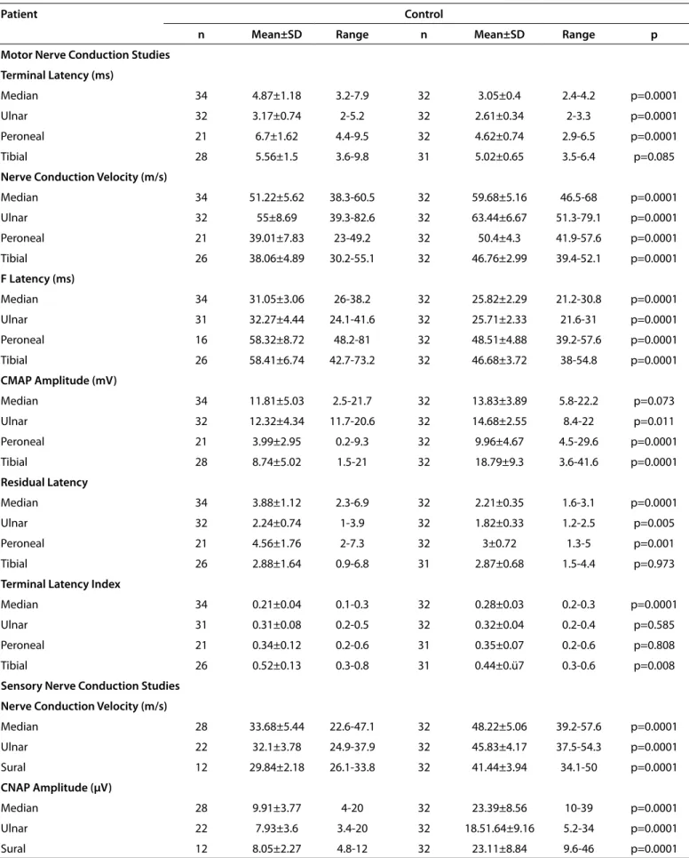
Table 2. Comparison of nerve conduction studies between the patient cohort and healthy controls
Patient Control
n Mean±SD Range n Mean±SD Range p Motor Nerve Conduction Studies
Terminal Latency (ms)
Median 34 4.87±1.18 3.2-7.9 32 3.05±0.4 2.4-4.2 p=0.0001
Ulnar 32 3.17±0.74 2-5.2 32 2.61±0.34 2-3.3 p=0.0001
Peroneal 21 6.7±1.62 4.4-9.5 32 4.62±0.74 2.9-6.5 p=0.0001
Tibial 28 5.56±1.5 3.6-9.8 31 5.02±0.65 3.5-6.4 p=0.085
Nerve Conduction Velocity (m/s)
Median 34 51.22±5.62 38.3-60.5 32 59.68±5.16 46.5-68 p=0.0001 Ulnar 32 55±8.69 39.3-82.6 32 63.44±6.67 51.3-79.1 p=0.0001 Peroneal 21 39.01±7.83 23-49.2 32 50.4±4.3 41.9-57.6 p=0.0001 Tibial 26 38.06±4.89 30.2-55.1 32 46.76±2.99 39.4-52.1 p=0.0001 F Latency (ms) Median 34 31.05±3.06 26-38.2 32 25.82±2.29 21.2-30.8 p=0.0001 Ulnar 31 32.27±4.44 24.1-41.6 32 25.71±2.33 21.6-31 p=0.0001 Peroneal 16 58.32±8.72 48.2-81 32 48.51±4.88 39.2-57.6 p=0.0001 Tibial 26 58.41±6.74 42.7-73.2 32 46.68±3.72 38-54.8 p=0.0001 CMAP Amplitude (mV) Median 34 11.81±5.03 2.5-21.7 32 13.83±3.89 5.8-22.2 p=0.073 Ulnar 32 12.32±4.34 11.7-20.6 32 14.68±2.55 8.4-22 p=0.011 Peroneal 21 3.99±2.95 0.2-9.3 32 9.96±4.67 4.5-29.6 p=0.0001 Tibial 28 8.74±5.02 1.5-21 32 18.79±9.3 3.6-41.6 p=0.0001 Residual Latency Median 34 3.88±1.12 2.3-6.9 32 2.21±0.35 1.6-3.1 p=0.0001 Ulnar 32 2.24±0.74 1-3.9 32 1.82±0.33 1.2-2.5 p=0.005 Peroneal 21 4.56±1.76 2-7.3 32 3±0.72 1.3-5 p=0.001 Tibial 26 2.88±1.64 0.9-6.8 31 2.87±0.68 1.5-4.4 p=0.973
Terminal Latency Index
Median 34 0.21±0.04 0.1-0.3 32 0.28±0.03 0.2-0.3 p=0.0001
Ulnar 31 0.31±0.08 0.2-0.5 32 0.32±0.04 0.2-0.4 p=0.585
Peroneal 21 0.34±0.12 0.2-0.6 31 0.35±0.07 0.2-0.6 p=0.808
Tibial 26 0.52±0.13 0.3-0.8 31 0.44±0.ü7 0.3-0.6 p=0.008
Sensory Nerve Conduction Studies Nerve Conduction Velocity (m/s)
Median 28 33.68±5.44 22.6-47.1 32 48.22±5.06 39.2-57.6 p=0.0001 Ulnar 22 32.1±3.78 24.9-37.9 32 45.83±4.17 37.5-54.3 p=0.0001 Sural 12 29.84±2.18 26.1-33.8 32 41.44±3.94 34.1-50 p=0.0001 CNAP Amplitude (µV) Median 28 9.91±3.77 4-20 32 23.39±8.56 10-39 p=0.0001 Ulnar 22 7.93±3.6 3.4-20 32 18.51.64±9.16 5.2-34 p=0.0001 Sural 12 8.05±2.27 4.8-12 32 23.11±8.84 9.6-46 p=0.0001
160
measured from the first negative deflection. Terminal latency distances were 5 cm for the median and ulnar nerves, 8 cm for the peroneal nerve and 10 cm for the tibial nerve. Motor nerve conduction velocities of the wrist-elbow segment of the median and ulnar nerves, below-above elbow and elbow-ax-illa segments of the ulnar nerve, knee-ankle segments of the peroneal and tibial nerves were analyzed. Compound muscle action potential (CMAP) was measured from peak to peak. Conduction block was defined as a 50% or more reduction of the CMAP amplitude of the proximal stimulation point, com-pared to the distal stimulation point, which was required to have a CMAP amplitude of at least 1 mV. Temporal dispersion was defined as greater than 30% prolongation of the negative peak duration of the CMAP obtained at the proximal stimula-tion point, compared with the distal stimulastimula-tion point. Mini-mum F-wave latencies were also obtained. Normal values of our laboratory were used to define nerve conduction abnor-malities (12). Motor nerve conduction studies of the median, ulnar, peroneal and tibial nerves were performed and residual latency (RL) and terminal latency index (TLI) for each motor nerve were calculated. Sensory nerve conduction studies of the digit II-wrist segment of the median, digit V-wrist segment of the ulnar and sural-ankle segment of the sural nerves were obtained by a sweep speed of 1 ms, sensitivity of 10 µV, utiliz-ing a bandpass filter of 20-2.000 Hz. While orthodromic tech-nique was utilized for median and ulnar nerves, sural nerve conduction study was performed antidromically. Latency measurements were made to the negative peak of the com-pound nerve action potential (CNAP), while amplitudes were measured from peak to peak. The skin temperature was mon-itored. When it fell below 31°C, terminal latencies (TL) were corrected by subtracting from the measured latency 0.2 ms for each degree centigrade and nerve conduction velocities (NCV) were multiplied by constant values defined for each
0.5°C according to DeJesus formula (13). No corrections were made on F-wave latencies.
Statistical Analysis
To check the normality of distribution of the measured vari-ables in patients and controls Kolmogorov-Smirnov test was performed. All tested variables distributed normally in the HNPP group. Parameters of the healthy controls also distrib-uted normally, except for the ulnar terminal latency and TLI of the ulnar and tibial nerves. Therefore comparisons of the means were analyzed by the Student’s t-test. Nerve conduc-tion variables in the patient group were expressed by dividing measured variables by the upper or lower limits of our lab-oratory and calculating the percentages. Group differences of individual nerves were analyzed by the Kruskal-Wallis test. Post-hoc analyses were performed by the Mann-Whitney U test. The significance level was defined as p<0.05.
RESULTS Clinical Findings
Family history was present 4 (21.1%) in the patient group, as 2 being mother and daughter and another 2 had first degree relatives with a history of acute compression neuropathies or chronic sensori-motor peripheral neuropathy of unidentified etiology. Fifteen patients carried deletion of the chromosome 17p22, while 1 patient was found to have a point mutation. Three patients displayed tomaculous changes in the sural nerve biopsy. Four (21%) patients presented with typical phenotypes in the form of acute compression neuropathies or brachial plexopathy, whereas the remaining majority had an atypical presentation including entrapment neuropathies, diffuse pain or paresthesias (Table 1). Treatment consisted of conservative rehabilitative measures or surgical exploration
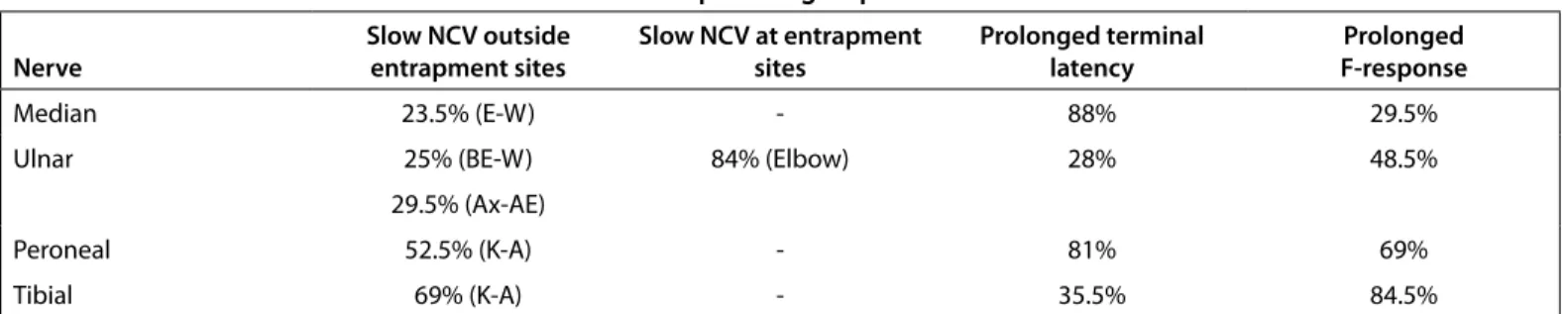
Table 3. Motor nerve conduction abnormalities in the patient group
Nerve Slow NCV outside entrapment sites Slow NCV at entrapment sites Prolonged terminal latency F-responseProlonged
Median 23.5% (E-W) - 88% 29.5%
Ulnar 25% (BE-W) 84% (Elbow) 28% 48.5%
29.5% (Ax-AE)
Peroneal 52.5% (K-A) - 81% 69%
Tibial 69% (K-A) - 35.5% 84.5%
A: ankle; Ax: axilla; AE: above elbow; BE: below elbow, K: knee, NCV: nerve conduction velocity; W: wrist
Table 4. Sensory nerve conduction abnormalities in the patient group
Nerve Segment Slow NCV Reduced CNAP Amplitude Absent CNAP Normal
Median Digit II-Wrist 85.5% 46.5% 17.5% 12%
Ulnar Digit V-Wrist 95.5% 32% 35.5% 3%
Sural Sura-Ankle 91.5% 8.5% 68.5% 2.5%
161
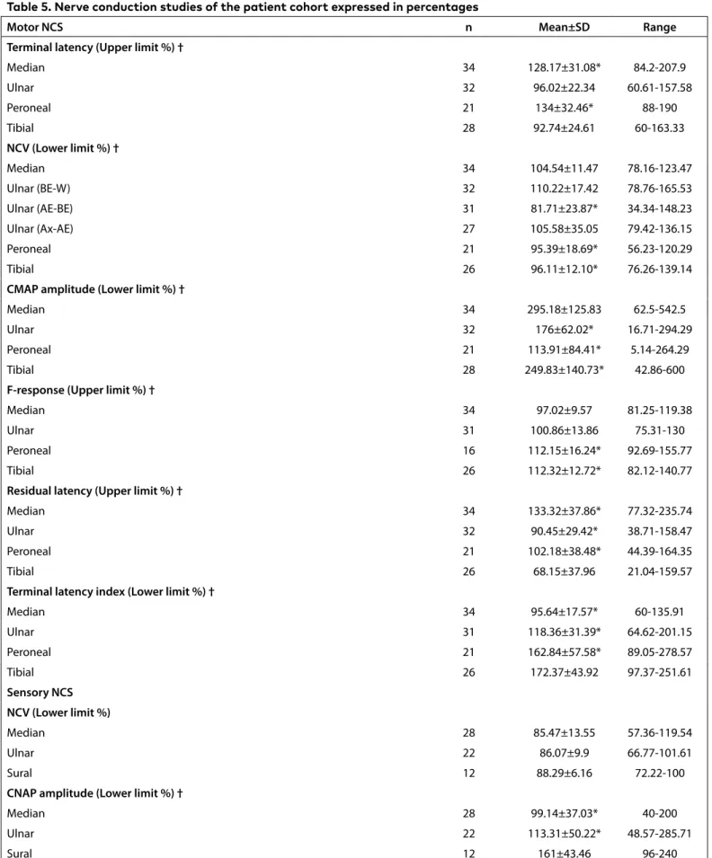
Table 5. Nerve conduction studies of the patient cohort expressed in percentages
Motor NCS n Mean±SD Range
Terminal latency (Upper limit %) †
Median 34 128.17±31.08* 84.2-207.9 Ulnar 32 96.02±22.34 60.61-157.58 Peroneal 21 134±32.46* 88-190 Tibial 28 92.74±24.61 60-163.33 NCV (Lower limit %) † Median 34 104.54±11.47 78.16-123.47 Ulnar (BE-W) 32 110.22±17.42 78.76-165.53 Ulnar (AE-BE) 31 81.71±23.87* 34.34-148.23 Ulnar (Ax-AE) 27 105.58±35.05 79.42-136.15 Peroneal 21 95.39±18.69* 56.23-120.29 Tibial 26 96.11±12.10* 76.26-139.14
CMAP amplitude (Lower limit %) †
Median 34 295.18±125.83 62.5-542.5
Ulnar 32 176±62.02* 16.71-294.29
Peroneal 21 113.91±84.41* 5.14-264.29
Tibial 28 249.83±140.73* 42.86-600
F-response (Upper limit %) †
Median 34 97.02±9.57 81.25-119.38
Ulnar 31 100.86±13.86 75.31-130
Peroneal 16 112.15±16.24* 92.69-155.77
Tibial 26 112.32±12.72* 82.12-140.77
Residual latency (Upper limit %) †
Median 34 133.32±37.86* 77.32-235.74
Ulnar 32 90.45±29.42* 38.71-158.47
Peroneal 21 102.18±38.48* 44.39-164.35
Tibial 26 68.15±37.96 21.04-159.57
Terminal latency index (Lower limit %) †
Median 34 95.64±17.57* 60-135.91 Ulnar 31 118.36±31.39* 64.62-201.15 Peroneal 21 162.84±57.58* 89.05-278.57 Tibial 26 172.37±43.92 97.37-251.61 Sensory NCS NCV (Lower limit %) Median 28 85.47±13.55 57.36-119.54 Ulnar 22 86.07±9.9 66.77-101.61 Sural 12 88.29±6.16 72.22-100
CNAP amplitude (Lower limit %) †
Median 28 99.14±37.03* 40-200
Ulnar 22 113.31±50.22* 48.57-285.71
Sural 12 161±43.46 96-240
AE-BE: above elbow-below elbow; Ax-AE: axilla-above elbow; BE-W: below elbow-wrist; CMAP: compound muscle action potential; CNAP: compound nerve action potential; n: number, NCS: nerve conduction study; NCV: nerve conduction velocity
† indicates p<0.01 Kruskal-Wallis test * indicates p<0.05 Mann-Whitney U test
162
and release in symptomatic patients, while asymptomatic individuals and patients with ill-defined symptoms received recommendations to prevent compression neuropathies.
Electrophysiological Findings
Motor nerve conduction studies revealed significant differ-ences (p<0.05) in all variables except for the tibial distal la-tency (t=1.772, p=0.085), median CMAP amplitude (t=-1.820, p=0.73), ulnar (t=-0.551, p=0.585) and peroneal TLI (t=-0.245, p=0.808), and tibial RL (t=0.034, p=0.973) in the patient co-hort compared with healthy controls. Tibial TLI value was actually higher in the patient cohort (Table 2). The details of motor NCV slowing in the patient cohort is demonstrated in Table 3. Prolongation of the terminal latencies were mainly observed in the median and peroneal nerves. The slowing of motor NCV, exceeding the lower limit of normal, was most prominent at the elbow segment of the ulnar nerve. NCV slowing and F-wave prolongation were more marked at the lower extremities (Table 3). Out of 31 elbow segments of the ulnar nerves tested, 2 right and 5 left sided conduction blocks (22.5%) was observed. Another patient displayed conduction
block at the knee-ankle segment of the left peroneal nerve. One patient with conduction block at the right elbow seg-ment of the ulnar nerve demonstrated conduction block at the knee-ankle segment of the right tibial nerve. Temporal dispersion was not observed.
Sensory nerve conductions in the patient cohort displayed significant abnormalities in all variables compared with the healthy controls (p<0.001, Table 2). Patients demonstrated absent sural CNAP in the 68.5% of the tested nerves. The de-tails of the sensory nerve conduction abnormalities are pre-sented in table 4.
Percentages of the nerve conduction variables according to the upper and lower limits are shown in Table 5 and Figure 1. All motor nerve variables demonstrated significant differences by the Kruskal-Wallis test among the nerves tested in the up-per and lower extremities. Post hoc analyses were up-performed by the Mann-Whitney test. In terms of terminal latencies, median and peroneal nerves demonstrated more significant prolongations (p<0.001). NCV slowing of the knee-ankle
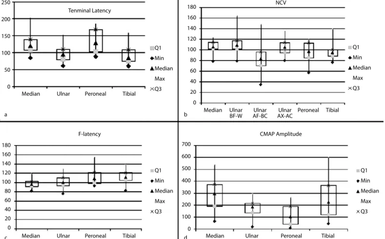
seg-Figure 1. a-d. Box plot graphics of the most relevant findings in nerve conduction variables expressed as percentages of the upper or lower
limits of normal and shown in Table 5. Terminal latency prolongations are most marked in the median and peroneal nerves (a), NCV slowing is prominent at the compression sites and knee-ankle segments of the lower extremity nerves (b), prominent F-wave prolongations are ob-served in the lower extremity nerves (c), peroneal CMAP amplitude is unproportionally reduced compared to other motor nerves (d) AE: above elbow; Ax: axilla; BE: below elbow; CMAP: compound muscle action potential; Q1 and Q3: first and third quartiles; Min and Max: minimum and maximum values; W: wrist
Q1 Min Median Max Q3 250 200 150 100 50 0 700 600 500 400 300 200 100 0 180 160 140 120 100 80 60 40 20 0 180 160 140 120 100 80 60 40 20 0 a c b d Median Ulnar Peroneal Tibial
Tenminal Latency
F-latency
NCV
CMAP Amplitude
Median Ulnar Peroneal Tibial Median Ulnar Peroneal Tibial Median Ulnar Ulnar Ulnar Peroneal Tibial
BF-W AF-BC AX-AC Q1 Min Median Max Q3 Q1 Min Median Max Q3 Q1 Min Median Max Q3
163
ments were more prominent compared with the elbow-wrist segments of the upper extremity nerves (p<0.05). NCV of the ulnar nerve was significantly slower at the elbow segment, compared with the below elbow-wrist and axilla-above elbow segments (p=0.0001). On the other hand, there was no signif-icant difference in the degree of slowing between the below elbow-wrist and axilla-above elbow segments (p=0.447). F-re-sponses were prolonged in the lower extremities (p<0.05). Re-sidual latency prolongations were especially prominent in the median nerve compared to all other nerves (p<0.05), while the residual latencies of all other nerves were prolonged compared to the tibial residual latency (p<0.01). Similarly, ter-minal latency index values were smaller in the median nerve compared with all other nerves (p<0.05), while the ulnar and peroneal nerve values were smaller in comparison with the tibial nerve terminal latency index (p<0.005). Peroneal CMAP amplitude was markedly reduced, compared with the CMAP amplitudes of all other nerves (p<0.05). NCV of the sensory nerves did not show a significant difference (p=0.72). How-ever, the sural nerve CNAP amplitude was markedly bigger compared with the upper extremity nerves (p<0.01).
DISCUSSION
Clinically atypical phenotypes were found to be more common and the family history was not conspicuous in our cohort. Lu-igetti et al. reported typical presentation in 44% of the cases, the most common phenotype being peroneal neuropathy at the level of the fibular head (2). Out of 36 of their atypical pa-tients 18 (50%) had either chronic ulnar neuropathy at the level of the elbow, or carpal tunnel syndrome. Similarly 11 (58%) of our 19 patients had either ulnar neuropathy at the elbow or carpal tunnel syndrome or both reflecting the common occur-rence of chronic compression neuropathies in HNPP, necessi-tating surgery as a therapeutic measure. However this issue re-mains controversial, as controlled studies concerning surgical decompression have not yet been conducted and some do not recommend surgery in HNPP at all (14). Other atypical cases present with chronic sensory neuropathy, fatigue and cramps, Charcot-Marie-Tooth like phenotype and acute onset of weak-ness resembling Guillain-Barré syndrome (2). Facial and trigem-inal neuropathies have also been reported (11). Non-specific pain and paresthesias were the presenting symptoms in 3 (16%) of our cases. Asymptomatic cases are also encountered, during an electrodiagnostic investigation performed for other reasons. In Luigetti et al. cohort of 73 cases, 32 (44%) had a fam-ily history (2). The incidence of sporadic cases is reported to be highly variable, ranging from 20 to 78% of the cases, explained by asymptomatic carriers and de novo mutations (1, 11). The frequent occurrence of atypical patients without a family histo-ry often creates a diagnostic dilemma and a need to resort to electrophysiologic methods. Additionally typical cases of acute compression neuropathies may present diagnostic challenge. In motor nerve conduction studies a prominent prolongation of the terminal latencies of the median and peroneal nerves
were noted as previously demonstrated (15). Interestingly, tibial terminal and residual latencies did not show a differ-ence compared to controls. Moreover the tibial nerve termi-nal latency index was actually higher than the control pop-ulation. Ulnar nerve terminal latency was also less involved. These findings obviously cannot be explained on the basis of a length dependent neuropathy or a distal myelinopathy. The most likely explanation is the susceptibility to compression of these individual nerves at the level of the wrist and ankle as suggested by Li et al. (15). Although RL and TLI measurements are convincingly abnormal in the median nerve, they do not have an overall diagnostic contribution, probably because slow NCVs influence calculations especially in lower extrem-ity nerves. Motor NCVs show non-homogenous slowing more prominent at the knee-ankle segments of the lower extremity nerves. However NCV slowing was markedly present at the el-bow segment of the ulnar nerve as previously demonstrated, being present in the 84% of the segments tested (1, 11). This finding has also been reported in the fibular head segment of the peroneal nerve, but this segment was not checked rou-tinely by us, one of the drawbacks of our study (1, 2). Slow motor NCV markedly present in the lower extremities had not been previously reported. In fact Infante et al. stated that the motor nerve conduction velocities were normal in more than 50% of the cases (11). F-wave prolongation was also more marked at the lower extremities. In Infante’s series peroneal F-wave prolongations were also more common compared to those of the upper extremity nerves, which was always in association with increased distal motor latency and motor nerve conduction slowing. This could explain the notable minimal F-wave abnormalities in the lower extremities in our series, due to the prominent motor nerve conduction slowing in peroneal and tibial nerves. Conduction block was almost non-existent except for the elbow segment of the ulnar nerve, which is a common entrapment site. This finding was also ob-served in other studies (15). It should be noted that temporal dispersion was never observed, unlike acquired demyelinat-ing neuropathies.
Sensory nerve conduction velocities were markedly slow and CNAP amplitudes low both in the upper and lower extrem-ities, as previously demonstrated (1, 15). However sensory nerve conduction velocity slowing calculated as the percent-ages of the lower limit of normal was uniform compared to previous studies (1). Absent sural CNAP was a notable finding in our study as previously reported by Li et al. (15). CNAP am-plitudes calculated as the percentages of the lower limit of normal showed marked reductions in the median and ulnar nerves, whereas the sural nerve amplitudes did not show a comparable reduction, which does not support the presence of a length dependent neuropathy.
In conclusion HNPP patients display marked prolongation of the median and peroneal terminal latencies, as well as pero-neal and tibial minimal F-wave latencies. Motor NCV slowing
164
is prominent at compression sites and lower extremity nerves. Peroneal CMAP amplitude is reduced out of proportion in com-parison to other motor nerves. Conduction block is usually ob-served at sites of compression and hardly present outside these segments. Temporal dispersion is never seen. On the other hand sensory NCV demonstrate diffuse slowing. This combination of findings seems to have a greater diagnostic contribution rath-er than individual abnormalities of nrath-erve conduction. Previous studies demonstrated that no significant difference exists in terms of nerve conduction studies between typical and asymp-tomatic cases (11). Therefore, typical nerve conduction studies as described above may guide the clinician to order mutation analysis when confronted with atypical cases.
Ethics Committee Approval: Ethics committee approval was
re-ceived for this study from the ethics committee of Ankara Numune Training and Research Hospital (335/2014).
Informed Consent: Due to the retrospective chart review of the
study, informed consent was not taken.
Peer-review: Externally peer-reviewed.
Author Contributions: Concept – K.M.M., B.C., R.K.; Design – R.K.;
Supervision – R.K., B.C.; Resources – R.K., B.C.; Materials – R.K., B.C.; Data Collection and/or Processing - K.M.M., B.C., R.K.; Analysis and/or Interpretation – R.K.; Literature Search – K.M.M., R.K., Writing Manu-script – R.K.; Critical Review – R.K., K.M.M., B.C.
Conflict of Interest: The authors have no conflicts of interest to
declare.
Financial Disclosure: The authors declared that this study has
re-ceived no financial support. REFERENCES
1. Hong YH, Kim M, Kim HJ, Sung JJ, Kim SH, Lee KW. Clinical and electrophysiologic features of HNPP patients with 17p11.2 dele-tion. Acta Neurol Scand 2003; 108: 352-358. [CrossRef]
2. Luigetti M, Del Grande A, Conte A, et al. Clinical, neurophysio-logical and pathoneurophysio-logical findings of HNPP patients with 17p12 deletion: A single-centre experience. J Neurol Sci 2004; 341:
46-50. [CrossRef]
3. Beydoun SR, Cho J. Hereditary neuropathy with liability to pres-sure palsy: Two cases of difficult diagnosis. J Clin Neuromuscul Dis 2013; 15: 28-33. [CrossRef]
4. Koehler PJ, Baas F. Hereditary neuropathy with liability to pres-sure palsies. Diagnosis in the first family (1947) confirmed. J Pe-ripher Nerv Syst 2012; 17: 412-413. [CrossRef]
5. Hooper DR, Lawson W, Smith L, Baker SK. Sonographic features in hereditary neuropathy with liability to pressure palsies. Mus-cle Nerve 2011; 44: 862-867. [CrossRef]
6. Russo M, Laura M, Polke JM, et al. Variable phenotypes are as-sociated with PMP22 missense mutations. Neuromuscul Disord 2011; 21: 106-114. [CrossRef]
7. Yurrebaso I, Casado OL, Barcena J, Perez de Nanclares G, Aguirre U. Clinical, electrophysiological and magnetic resonance find-ings in a family with hereditary neuropathy with liability to pres-sure palsies caused by a novel PMP22 mutation. Neuromuscul Disord 2014; 24: 56-62. [CrossRef]
8. He Y, Wu Q, Xu Z, et al. Hereditary neuropathy with liability to pressure palsy: An investigation in a rare and large Chinese fam-ily. Eur Neurol 2012; 68: 322-328. [CrossRef]
9. Casasnovas C, Banchs I, De Jorge L, et al. A novel small deletion in PMP22 causes a mild hereditary neuropathy with liability to pressure palsies phenotype. Muscle Nerve 2012; 45: 135-138.
[CrossRef]
10. Ginanneschi F, Filippou G, Giannini F, et al. Sonographic and electrodiagnostic features of hereditary neuropathy with liabil-ity to pressure palsies. J Peripher Nerv Syst 2012; 17: 391-398.
[CrossRef]
11. Infante J, Garcia A, Combarros O, et al. Diagnostic strategy for fa-milial and sporadic cases of neuropathy associated with 17p11.2 deletion. Muscle Nerve 2001; 24: 1149-1155. [CrossRef]
12. Baysal Aİ, Kuruoğlu R, Beyazova M, et al. Nerve conduction val-ues in normal population. Aegean J Neurol Sci 1989; 6: 9-15. 13. Oh SJ. Clinical Electromyography: Nerve Conduction Studies.
Philadelphia: Lippincott Williams & Wilkins; 2003.
14. van Paassen BW, van der Kooi AJ, van Spaendonck-Zwarts KY, Verhamme C, Baas F, de Visser M. PMP22 related neuropathies: Charcot-Marie-Tooth disease type 1A and hereditary neuropa-thies with liability to pressure palsies. Orphanet J Rare Dis 2014; 9: 38. [CrossRef]
15. Li J, Krajewski K, Shy ME, Lewis RA. Hereditary neuropathy with liability to pressure palsy: the electrophysiology fits the name. Neurology 2002; 58: 1769-1773. [CrossRef]